Advanced Veterinary Dental Procedures: Periodontal Surgery, Endodontics, Restorations, and Orthodontics

13 Advanced Veterinary Dental Procedures


Periodontal Surgery, Endodontics, Restorations, and Orthodontics


At one time, veterinary dentistry meant either scraping calculus from the teeth or extracting them if they could not be saved by conservative techniques. Fortunately for the patient, dentistry has made significant advances. Although not all practices have the resources to perform advanced procedures, the veterinary staff should have an idea of the range of procedures that can be performed to save teeth, what each procedure entails, and the type of equipment necessary. Some practices may perform many advanced dental procedures, and this chapter briefly addresses the most common. Prior to using any of these techniques, training by a board-certified veterinary dentist is recommended.


Periodontal Surgical Techniques


Periodontal surgical techniques are employed after more conservative measures, such as closed periodontal debridement, have been attempted without success or have been ruled out as impossible. In periodontal surgery, flaps are created to expose the tooth root and associated bone. The bone may be reshaped or augmented, and the gingiva may be sutured back to the initial position. Alternatively, the gingival height may be changed apically by gingival or bone surgery to decrease the pocket or coronally with guided tissue regeneration to increase the height of attachment.


Instruments and Materials


Periodontal surgery requires various instruments and materials (Box 13-1). The No. 3 handle is the standard type for scalpels. A variety of different blades may be used, including special periodontal knives. The No. 15c blade is extremely fine and therefore useful in periodontal surgical procedures. In addition, small tissue scissors, such as LaGrange scissors, are helpful in trimming periodontal tissue. Periosteal elevators are used to lift the gingiva away from the bone. Several types are available. The Molt elevator is one type; the Molt No. 9 is particularly popular. Many practitioners also like the ST-No. 7 instruments. Having a variety of periodontal surgical instruments available makes treating various anatomic and pathologic conditions less difficult.


Solutions for irrigating the tissue are also important in periodontal surgery. Sterile saline solution and chlorhexidine are most common. Chlorhexidine is used in a 0.1% to 0.2% solution and is available in two forms: diacetate and gluconate. Gluconate is preferred.


Tissue forceps and needle holders are necessary for suturing. Although expensive, the best is the spring-locking Castroviejo-type needle holder. For the sutures themselves, 4-0 or 5-0 absorbable material with a reverse cutting FS-2 needle is used. The high-speed handpiece is used in the removal of bone, and burs may be added for the removal of granulation tissue. Round burs (#2 or #4), crosscut fissure burs (#701L), or pear-shaped burs (#330) may be used in the appropriate handpiece. Bone files may be used to contour bone.


Treatment Techniques


Hyperplastic gingiva


Because gingivoplasty (gingivectomy) is performed only when hyperplastic gingiva is present, patients must be selected carefully. Gingivoplasty should not be used for treatment of deep periodontal pockets or as part of the routine prophy. This procedure is contraindicated when attached gingiva is minimal or absent or horizontal or vertical bone loss is present below the mucogingival junction, which is the line of demarcation where the attached gingiva and alveolar mucosa meet. These characteristics are prevalent in certain breeds and breed lines, particularly boxers and collies.


Gingivoplasty technique


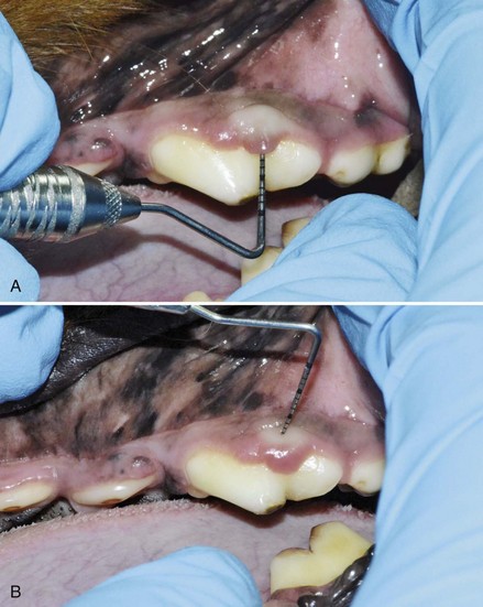
The pocket depth and contour is determined by inserting a probe to the depth of the pocket at several areas around the tooth. The corresponding depth is measured on the outside of the gingiva, also with the probe. A bleeding point is made by placing the tip of the probe perpendicular to the gingiva and applying slight pressure to make a small hole or by using a small-gauge needle. Bleeding points are made around the contour of the pocket and are used as a guide for the gingivectomy. The gingivectomy is made at an angle apical to the bleeding point to create a beveled margin. At least 2 mm of healthy, attached gingiva must be present apical to the base of the incision. A multifluted bur, laser, scalpel blade, or electrosurgery blade is used to excise the gingiva by cutting below the bleeding points, with the blade held at approximately a 45-degree angle and the tip of the blade toward the crown. The ends of the excision should be tapered into the surrounding gingiva to create the normal scalloped contour, particularly if several adjacent teeth are treated. Gingival tags can be removed with the blade or a sharp curette. The exposed tooth and root surface can now be scaled and planed smooth. Hemorrhage is controlled by applying pressure with wet gauze pads or hemostatic agents. If electrosurgery is being performed, caution must be exercised because the collateral damage may extend past the desired surgical line. In Figure 13-1, a periodontal probe is used to measure and mark the pocket depth. Once marked, a scalpel blade is used to perform the gingivoplasty.


Endodontics and Restorations


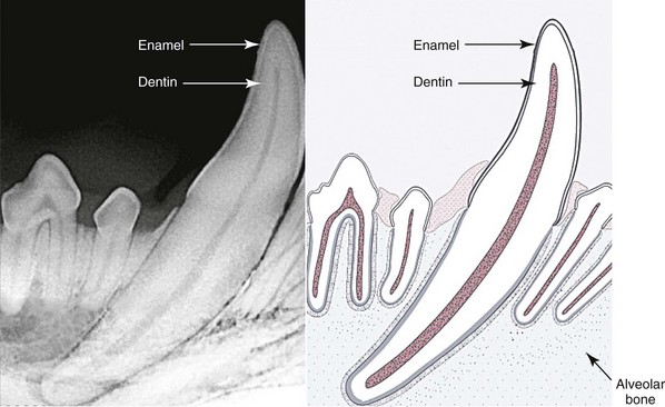
At one time, veterinarians did not treat fractured teeth. Consequently, many patients suffered silently as the tooth first died and then became abscessed. Endodontic therapy is a better option. Endodontic therapy is a general term for treatment of the dental pulp and may be used to save vital pulp, remove live or dead pulp, and prevent or treat infection (Figure 13-2).


Pulp tissue consists of blood vessels, nerves, and connective tissues that support the odontoblastic cells lining the pulp chamber and root canal. Throughout life the odontoblasts produce dentin that fills in the canal. As a result, the dentin layer thickens with age.


Bacteria usually gain entry to the pulp chamber via a fractured tooth. The pulpal tissue becomes inflamed and edematous and dies. Then bacteria move into the apical region of the tooth. From this area, they spread through the canals in the apical delta of the tooth, which formerly served as tunnels for the nerves and blood vessels. Once the bacteria enter the apical bone, an abscess starts. The periapical abscess may eventually (i.e., years later) become walled off, cause inflammatory resorption of the root, spread along the periodontal ligament, and cause ankylosis (fusion) of the tooth root with the surrounding bone.


Discolored teeth, especially those that are pink and purple, indicate pulpal hemorrhage and necessitate root canal therapy. Caution should be exercised in young animals because the tooth may be discolored as a result of trauma yet retain sufficient blood supply to survive and continue to develop. If a root canal is performed on this type of tooth and the tooth walls are not completely developed, the tooth may fracture. In young patients, evaluation through radiography is advised.


Indications for Endodontic Therapy


The most common indication for root canal therapy is fractured teeth. If the tip of the crown appears black, the patient is a candidate for a workup and evaluation of the tooth. Cats are particularly susceptible to canine tooth abscess. The pulp chamber extends close to the tip, and any exposure of dentine allows bacteria into the pulp chamber. Chronic abscess of the canine teeth is extremely common in cats; all fractured teeth necessitate root canal therapy, extraction, or close monitoring.


A worn tooth with a brown covering in the area where the pulp chamber used to be indicates that the wear has occurred slowly enough that secondary dentin was deposited by the odontoblasts lining the root canal and pulp chamber. In this case, the tooth should be evaluated radiographically. Occasionally, teeth that are completely normal in appearance may have apical disease that can be diagnosed only radiographically.


The rationale for endodontic treatment is to maintain optimal health. A tooth with endodontic disease produces various signs, including pain and irritability. Many patients with endodontic disease exhibit fluctuations in appetite. Clients frequently report halitosis, or bad breath, in pets with undiagnosed endodontic disease.


Once pulpal death occurs, most dogs and cats do not show pain. However, if the tooth is alive and recently traumatized, many patients flinch when the tooth is percussed with a probe or other instrument. Some animals chew food only on the side of the mouth opposite the traumatized tooth, or they drool and produce increased calculus on the injured side. Hunting dogs may refuse their training dummies, utility dogs may refuse their dumbbells, and attack dogs may either hesitate or bite and release repetitively because of the pain (this is referred to as typewriting). Dogs that are used for tracking scents may be less effective than usual because of the odor from the oral infection, which overwhelms the scent.


When pulp exposure is secondary to a coronal fracture or a carious erosion, pathogenic bacteria soon descend into the pulp canal and cause an abscess either within the canal itself or periapically by extension of the infection. Infection that has penetrated the apical end of the root canal can cause osteomyelitis and the subsequent loss of surrounding bone. Periapical infection can spread, contributing to pathologic fractures of the lower jaw, or the infection can extend through weakened necrotic bone and develop into an oronasal fistula from any of the maxillary teeth.


Stay updated, free articles. Join our Telegram channel

Oct 9, 2016 | Posted by in GENERAL | Comments Off on Advanced Veterinary Dental Procedures: Periodontal Surgery, Endodontics, Restorations, and Orthodontics

Full access? Get Clinical Tree

Get Clinical Tree app for offline access