Rebecca S. McConnico
Acute Colitis in Horses
Acute colitis is a common cause of rapid debilitation and death in horses. More than 90% of untreated horses with this condition die or are euthanized, but horses that are treated appropriately usually respond and gradually recover over a 7- to 14-day period. Colitis-associated diarrhea is sporadic in occurrence and is characterized by intraluminal sequestration of fluid, moderate to severe colic, and profuse watery diarrhea, with resultant endotoxemia, leukopenia, and hypovolemia. The condition can affect adult horses of all ages but usually affects horses 2 to 10 years of age. Disease onset is sudden, and progression is rapid. The condition is often preceded by a stressful event. A definitive diagnosis is made in only about 20% of cases. Most antemortem and postmortem tests to determine etiology do not yield a definitive diagnosis.
Treatment of colitis can be extremely costly because of the substantial volumes of intravenous replacement fluids required. At present, there is no curative treatment, and treatment strategies are directed at rehydration, electrolyte and plasma protein replacement, prevention or amelioration of the effects of endotoxemia, provision of nutritional support, and administration of antimicrobials when indicated.
Pathophysiology and Clinical Signs
Diarrhea associated with acute colitis is a result of abnormal fluid and ion transport by cecal and colonic mucosa, with fluid loss resulting from a combination of both malabsorption and hypersecretory processes. Under normal conditions, water and electrolytes are secreted by epithelial cells in the intestinal crypts, and most of this fluid is reabsorbed by the surface epithelial cells. Abnormal rates of secretion and absorption result in massive secretion and malabsorption by large intestinal mucosal epithelial cells, leading to severe dehydration and death.
Acute colitis is a general term referring to inflammation of the cecum (typhlitis), colon (colitis), or both (typhlocolitis), with subsequent rapid onset of diarrhea in the adult horse. In contrast to other domestic animals and humans, horses have sudden, massive fluid loss and severe electrolyte imbalances that can result in death in hours. This distinctive clinical presentation in horses may result from several unique features of the large bowel of Equidae. Some of these include the large population of gram-negative endotoxin-bearing bacteria that reside in the large intestine and the markedly high mucosal prostaglandin concentrations, manifested by a marked chloride secretory response, compared with other species. Another possible reason for the distinctive clinical signs is the intense inflammation that results from activation of resident intestinal mucosal and submucosal phagocytic granulocytes by intestinal bacterial products after mucosal barrier disruption.
The causes of acute colitis are reviewed in the fourth edition of Current Therapy in Equine Medicine, (pp 197 to 203). There is some agreement that certain distinctive clinical, pathologic, or diagnostic characteristics may help in differentiating between specific acute colitis–associated conditions (Table 68-1). Regardless of the initiating cause, common clinical and pathologic features suggest a common pathophysiologic pathway. Typical hematologic findings include hypovolemia, dehydration, metabolic acidemia, electrolyte derangements, leukopenia with a left shift, toxic neutrophils, lymphopenia, and azotemia. Clinical features include depression, inappetence, fever, tachycardia, dry mucous membranes, skin tenting, prolonged capillary refill time, colic, and watery, often fetid, diarrhea.
TABLE 68-1
Characteristics Helpful in Differentiating Among Infectious and Noninfectious Conditions That Cause Colitis in Horses
| Etiology | Differentiating Characteristics |
| Salmonella spp infection | Identification by bacterial culture or PCR analysis |
| Clostridium spp (C cadaveris, C difficile) infection | >103 colony-forming units/g feces or intestinal contents; demonstration of enterotoxin or cytotoxin A or B (for C difficile) |
| Neorickettsia risticii infection | Seasonal (July–October) incidence; geographic location (California, Minnesota, mid-Atlantic states, New York, Ohio, Canada, and Europe) near freshwater rivers or ponds; biphasic fever often associated with laminitis; significant rise or fall in serum antibody titer; positive PCR results for organism in feces or blood |
| Cyathostome and strongyle infestation | Seasonal (late winter–early spring) disease; associated with anthelmintic therapy, inadequate deworming programs, parasite resistance |
| Nonsteroidal antiinflammatory drug toxicity | Slower onset, oral cavity ulcers; early ventral edema associated with hypoproteinemia; gastric or intestinal ulceration |
| Antimicrobial administration (tetracyclines, macrolides, cephalosporins, clindamycin, lincomycin, florphenicol, potentiated sulfas, other antimicrobials) | History of antimicrobial use |
| Arsenic poisoning | Marked tenesmus; muscle tremors; extreme toxemia; hemorrhagic diarrhea; extremely short clinical course |
| Cantharidin toxicosis | Blister beetles in hay (usually alfalfa); skin acantholysis; oral erosions; painful urination, blood in urine; synchronous diaphragmatic flutter, hypocalcemia, hypomagnesemia; cantharidin in urine or stomach contents |
| Colitis X/necrotizing enterocolitis | Necropsy findings: massive hemorrhage and necrosis of cecum and large colon |
| Sand ingestion | History of sand in housing or pasture or stable area; detection of sand in feces; abdominal borborygmal sounds compatible with sand friction |
PCR, Polymerase chain reaction.
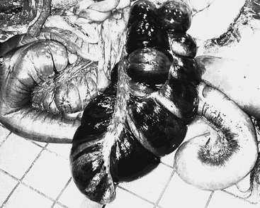
Gross necropsy findings usually reveal edematous, sometimes hemorrhagic, typhlitis-colitis with intraluminal sequestration of fluid ingesta (Figure 68-1). Common microscopic abnormalities include superficial mucosal injury affecting the distal portion of the ileum and the cecum and large colon. Injury is characterized by mucosal epithelial ulceration and erosion, mucosal and submucosal edema, and various degrees of mucosal inflammation. These lesions may enhance net fluid movement into the intestinal lumen by decreasing net solute absorption, increasing mucosal permeability, and stimulating prostaglandin-mediated ion secretion. Disrupted epithelium allows transmural migration of endotoxin.
Clinical Evaluation
In horses with impending colitis, lethargy, inappetence, and colic are frequently noticed several hours before the appearance of liquid feces. Physical examination during this early period may reveal high respiratory and heart rates secondary to abdominal discomfort from intraluminal sequestration of fluid or gas, or secondary to inflammatory mediator activity. Rectal temperature may also be high as a result of the inflammatory response to toxin absorption through a disrupted intestinal mucosal barrier. Signs of abdominal discomfort can range from mild, such as recumbency or inappetence, to severe, with the horse rolling and thrashing. Abdominal distension is often evident. These cases may be confused with other large bowel disorders, such as large colon torsion.
Acute equine colitis should be considered a potentially life-threatening emergency, and early evaluation and treatment by a veterinarian are critical. Horses with sudden onset of colitis will sequester a large volume of fluid intraluminally and begin to pass the liquid material within several hours. The volume of fluid lost from the intestinal tract can equal the horse’s entire extracellular fluid volume; hence, signs of dehydration and hypovolemia may be severe. Mucous membranes may be brick colored and sticky, capillary refill time is prolonged, and skin turgor is reduced. Progressively severe hypovolemia and subsequent circulatory shock lead to purple mucous membranes and a weak peripheral pulse. Horses with acute colitis are prone to laminitis and may develop signs of this additionally life-threatening complication (e.g., lameness, bounding digital pulses, high hoof temperature) any time during the course of disease.
Laboratory Tests
Assessment of data from the horse’s blood tests is important for determining the degree of systemic illness and plasma volume replacement needs. Packed cell volume (PCV) and total plasma protein (TPP) values are often high initially and indicate the severity of dehydration. Total protein values that are in or below reference range in a clinically dehydrated horse with a high PCV indicate overall protein loss. Daily PCV and TPP assessment is useful for monitoring daily fluid and protein needs.
Total and differential white blood cell (WBC) counts usually reveal neutropenia with a left shift, and granulocytes often have toxic morphologic changes, including cytoplasmic vacuolation, basophilia, “toxic” granule formation, and Döhle bodies. Signs of overall improvement usually correlate with a decrease in abnormal WBC features, including morphologic changes. Horses in the later stages of acute colitis may have high fibrinogen concentration and neutrophilic leukocytosis, indicating a generalized inflammatory response. Horses with colitis usually have metabolic acidemia; electrolyte derangements including hyponatremia, hypochloremia, hypocapnia, hypokalemia, and hypocalcemia; and azotemia.


