14 Aberrant conjunctival overgrowth in rabbits INITIAL PRESENTATION Colour change – red PRESENTING SIGNS Owners present the rabbit due to change in appearance of the affected eye(s) – it appears red. Normally they have noticed this for a few weeks and it is worsening. The eye is open and comfortable with no significant ocular discharge. Adult rabbits are presented and normally one, but occasionally both, eyes are affected. Dwarf rabbits and their crossbreds are possibly more commonly affected. No visual problems are reported and the rabbit is eating normally such that the owner has no other concerns. CASE HISTORY The history is minimal. Usually the rabbit has had no previous ocular problems and is in good general health. Occasionally the owner will have thought that the current ocular problem is conjunctivitis and tried treating with antibiotic drops with no improvement – this is particularly the case in multi-rabbit households where the owner has some medication for another pet. CLINICAL EXAMINATION 
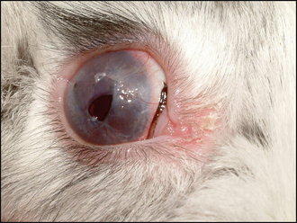
General clinical examination is unremarkable – the rabbit is normally in good body condition with no dental problems. On ophthalmic examination the rabbit is normally visual (although menace responses are not always present in rabbits). No ocular discharge is present and the eye is open and comfortable. A pink, fleshy, vascular membrane is present over the cornea through a full 360 degrees extending centrally. It can extend only a couple of millimetres, but might cover virtually the whole of the cornea such that only a small central hole remains (see Figure 14.2). Intraocular examination, if possible, is normal. Rabbits have a merangiotic retina – a horizontally oval optic disc with vessels radiating from either end (laterally and medially) – and this is most easily examined by looking up at the rabbit from below, while its head is held over the end of the consulting table. No corneal ulceration is present. If a drop of topical anaesthetic is placed in the affected eye, the pink membrane can be lightly touched with a cotton bud and can be gently moved over the corneal surface – it is not attached centrally. Figure 14.2 Right eye of dwarf-cross rabbit showing conjunctival membrane across cornea. DIFFERENTIAL DIAGNOSES • Aberrant conjunctival overgrowth • Conjunctivitis • Keratitis • Vascularizing corneal ulcer • Intraocular haemorrhage • Prolapsed nictitans membrane CASE WORK-UP The clinical appearance is pathognomonic – once seen this condition is not forgotten and, as such, no further work-up is normally necessary. NURSING ASPECTS Apart from the postoperative application of topical medication, and the usual precautions necessary for hospitalization and anaesthesia of rabbits, there are no specific nursing issues. EPIDEMIOLOGY Aberrant conjunctival overgrowth, circumferential conjunctival hyperplasia and placation, epicorneal conjunctival membranes and pseudopterygium are all terms used to describe this abnormality. The condition is poorly understood and appears to be unique to the rabbit. The pink tissue is a fold of conjunctiva which grows centri-petally from the bulbar conjunctiva at the limbus to obscure the cornea. It remains attached at the limbus but the central fold of tissue is freely mobile over the corneal surface. Vision is maintained until the central circular ‘hole’ is occluded by the progression of the membrane. Histologically the lesion consists of normal conjunctiva lining both the inner and outer surfaces of the membrane and is thought to result from a focal excess of conjunctival collagen. Any breed of rabbit can be affected, but dwarf and dwarf-crosses might be predisposed. A similar condition is seen in humans – pterygium – where a fold of conjunctiva grows across the cornea from the nasal limbus. It does become attached to the cornea, unlike in rabbits, and is more common in warm, dry, sunny climates and in certain ethnic groups, especially black people. Exposure to UV light, wind, dust and trauma predispose people to developing pterygium but none of these factors appears applicable to rabbits. It is postulated that local immunity factors are involved but no research has been undertaken to date to prove this. CLINICAL TIPS • Once seen never forgotten (see Figure 14.2) Only gold members can continue reading. Log In or Register to continue You may also needRabbit conjunctivitisEpiscleritisRabbit uveitisBlepharitisCanine intraocular neoplasiaChronic gingivostomatitis associated with FeLV and FIVExophthalmosDeep corneal ulcer Share this:Click to share on Twitter (Opens in new window)Click to share on Facebook (Opens in new window)Click to share on Google+ (Opens in new window) Related Tags: Saunders Solutions in Veterinary Practice Small Animal Ophthalmo Sep 10, 2016 | Posted by admin in SMALL ANIMAL | Comments Off on Aberrant conjunctival overgrowth in rabbits Full access? Get Clinical Tree