When faced with a veterinary patient that may have cancer it is important to evaluate the patient in its entirety. The patient is very much more than a mast cell (MCT) or bone tumour and this should always be borne in mind when approaching the diagnostic work-up. What is appropriate will vary depending on the cancer that has been detected or the clinical signs exhibited by the patient. We all know the diagnosis of cancer is often made after the patient has presented with non-specific signs such as vomiting.
Physical examination
The value of good physical examination cannot be overemphasized. Early detection of neoplasia can, in many cases, give a good outcome. Thorough examination of lumps and bumps via fine needle aspirates (FNAs) can lead to the early detection of tumours such as MCT. Such tumours are much easier to remove when small and therefore will have potentially a better outcome for the individual and certainly reduce the requirement for extensive surgery or surgery and adjuvant therapy, or no possibility of surgery (Figure 3.1). If a lump is non-diagnostic on an initial FNA, do not discount it but alert the client to watch for any change and to come back if the lump grows or changes. Unfortunately, we see many veterinary patients where the initial evaluation was inconclusive and the lump had been left to grow into what may become an inoperable tumour, often because the original FNA was inconclusive and no follow-up was discussed with the client.
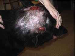 |
| Figure 3.1 |
A complete physical examination should include routine rectal examinations for all dogs over 5 years of age; the early detection of tumours such as anal sac adenocarcinomas would dramatically affect the outcome with surgery, as too often the client goes to the veterinary surgeon because they have noticed a lump protruding close to the anus. By the time the tumour is so large as to be visible to the client it cannot be removed with adequate margins to guarantee complete excision and in some cases cannot be removed at all (Figure 3.2). The key to improving prognosis for many veterinary patients is both early detection and immediate action. Abnormalities on physical examination should always be followed up.
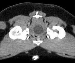 |
| Figure 3.2 |
If an external lump is found on examination always palpate the draining lymph node for signs of enlargement; this is the beginning of the important process of staging.
Establishing a minimum database
The majority of animals with cancer are middle-aged to older and therefore it is just good medicine to establish a minimum database consisting of haematology, biochemistry and urinalysis. Other problems that may influence your treatment plan may be present, including use of cytotoxic drugs in patients with renal or liver disease. In some cases it is the abnormalities on blood tests that draw the clinician towards a diagnostic plan aimed at identifying neoplasia. For example, hyperglobulinaemia may be an indication of lymphoma or multiple myeloma. A leucocytosis may be indicative of a leukaemia. An elevated packed cell volume (PCV) may occur with renal carcinoma. Hypoglycaemia can occur due to an insulinoma. Anaemia, thrombocytopenia and granulocytopenia may indicate bone marrow disease. Increased alkaline phosphatase (ALKP) may occur with an osteosarcoma. Hypercalcaemia is a paraneoplastic syndrome that can occur with many cancers, but is seen most frequently with anal sac adenocarcinoma, lymphoma or multiple myeloma. However, it is important to remember that none of the above tests is diagnostic in itself for neoplasia and other medical reasons may underlie each clinical abnormality.
Urinalysis is also important – for example, renal function may be compromised with hypercalcaemia of malignancy. A low urine specific gravity (SG) may indicate polyuria/polydipsia (PU/PD), which can occur with many cancers. Haematuria may occur with urinary bladder neoplasia, a large number of patients with cancer have signs of proteinuria, and so the list goes on.
Clotting profiles are indicated in cancer patients prior to major surgery, particularly patients with suspected haemangiosarcoma, large mast cell tumours, haemolymphatic tumours, etc. Most patients with advanced neoplasia are in a state of chronic disseminated intravascular coagulation (DIC) but this is not always clinically relevant.
Cytology or biopsy
Ultimately, the nature of a tumour must be determined either by cytology or histology, and this is discussed in more detail in Chapter 4. Evaluation of draining lymph nodes by cytology, if appropriate, should be carried out at the same time. In many instances it is necessary to remove the sentinel node for histopathological evaluation.
Stay updated, free articles. Join our Telegram channel

Full access? Get Clinical Tree


