The Vertebral Column
 . Approach to Cervical Vertebrae 1 and 2 Through a Ventral Incision
 . Approach to Cervical Vertebrae 1 and 2 Through a Dorsal Incision
 . Approach to Cervical Vertebrae and Intervertebral Disks 2-7 Through a Ventral Incision
 . Approach to the Midcervical Vertebrae Through a Dorsal Incision
 . Approach to Cervical Vertebrae 3-6 Through a Lateral Incision
 . Approach to the Caudal Cervical and Cranial Thoracic Vertebrae Through a Dorsal Incision
 . Approach to the Thoracolumbar Vertebrae Through a Dorsal Incision
 . Approach to the Thoracolumbar Intervertebral Disks Through a Dorsolateral Incision
 . Approach to the Thoracolumbar Intervertebral Disks Through a Lateral Incision
 . Approach to Lumbar Vertebra 7 and the Sacrum Through a Dorsal Incision
 . Approach to the Lumbosacral Intervertebral Disk and Foramen Through a Lateral Transilial Osteotomy
 . Approach to Lumbar Vertebrae 6 and 7 and the Sacrum Through a Ventral Abdominal Incision
 . Approach to the Caudal Vertebrae Through a Dorsal Incision
Approach to Cervical Vertebrae 1 and 2 Through a Ventral Incision
Based on a Procedure of Sorjonen and Shires47
Patient Positioning
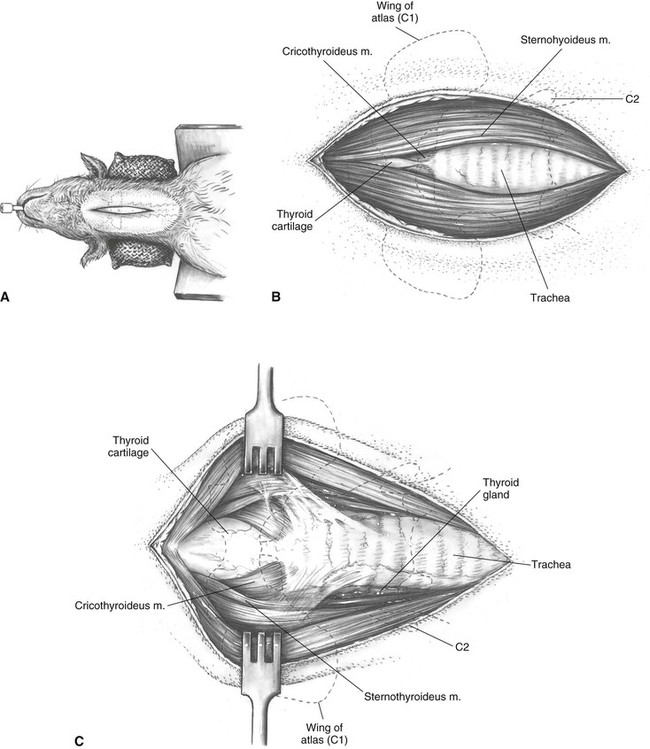
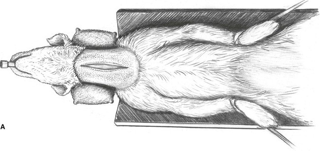
The animal is placed in the supine position with a sandbag under the neck to cause marked extension of the cranial cervical vertebrae. Placing the animal in a V-shaped trough will elevate the shoulder region and increase the extension of the cervical spine (see Plate 12A).
Description of the Procedure
A The skin incision begins on the midline between the angles of the mandible and ends in the midcervical region.
B The incision is deepened through the subcutis and between the paired bellies of the sternohyoideus muscles to expose the trachea.
C Retraction of the sternohyoideus muscles exposes the larynx and its muscles. The right sternothyroideus muscle is isolated and detached from its insertion on the thyroid process of the larynx. The thyroid gland should be protected during this dissection.
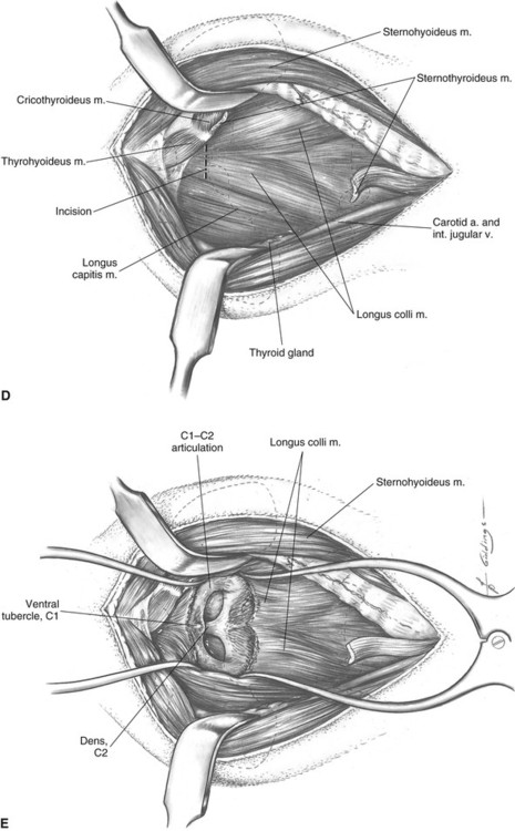
D Retraction of the larynx and trachea to the left side (medially) is preceded by ligation or cautery of several small vessels running between the carotid artery/internal jugular vein and the thyroid gland or trachea. The recurrent laryngeal nerve must be protected during this dissection and retraction. Muscle retraction to the right side (laterally) also includes the carotid artery, the internal jugular vein, and the vagosympathetic trunk. The longus colli muscles are now exposed. The ventral tubercle of C1 is located by palpation, and the longus colli muscle fibers are transected close to the tubercle.
E Elevation of muscle fibers from the ventral arch of C1 and the body of C2 proceeds laterally until the articulations are exposed.
Additional Exposure
This approach can be extended caudally (see Plate 14) to obtain ventral exposure of the entire cervical spine.
Approach to Cervical Vertebrae 1 and 2 Through a Dorsal Incision
Based on a Procedure of Funkquist15
Patient Positioning
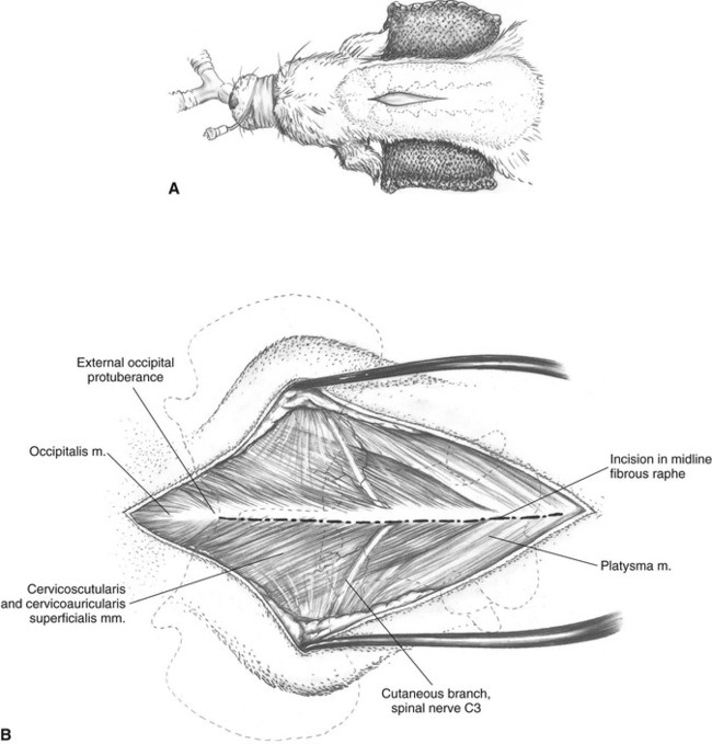
Dorsal recumbency with the neck slightly flexed and supported by a sandbag bolster or folded towel (see Plate 13A).
Description of the Procedure
A The skin incision is made on the dorsal midline starting at the level of the occipital protuberance, extending caudally to the level of the third or fourth cervical vertebra.
B Skin is undermined and retracted and subcutaneous fascia incised on the midline to expose the occipitalis, cervicoscutularis, and cervicoauricularis superficialis muscles. Caudal and lateral to these muscles are the thin fibers of the platysma muscle. These muscles are incised on the midline fibrous raphe to allow elevation and lateral retraction of these muscles.
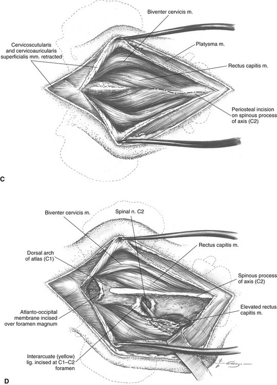
C Deepening the midline incision will allow separation of the paired bellies of the biventer cervicis superficially and the deeper rectus capitis attached to the dorsal spine of C2. The insertion of the rectus capitis muscle is incised along the lateral border of the spine of C2 to allow its elevation from the bone by combined sharp and blunt dissection.
D As the dissection is carried deeper onto the lamina of C2, care should be taken to avoid the vertebral artery, which courses through the muscles slightly ventrolateral to the articular processes. The interarcuate (yellow) ligament covering the foramina between C1 and C2 is carefully incised to expose the spinal cord and the root of spinal nerve C1. The dorsal atlanto-occipital membrane of the foramen magnum may also be incised to expose the cranial rim of the dorsal arch of C1.
Additional Exposure
This approach can be extended caudally (see Plates 15 and 17) to obtain dorsal exposure of the entire cervical spine.
Approach to Cervical Vertebrae and Intervertebral Disks 2-7 Through a Ventral Incision
Based on a Procedure of Olsson33
Indications
1. Fenestration and curettage of intervertebral disks C2-C7.
2. Decompression of cervical spinal cord by ventral slot of intervertebral disks C2-C7.
3. Distraction and fusion of intervertebral disks C5-C6 and C6-C7 for caudal cervical spondylomyelopathy.
4. Open reduction and ventral stabilization of fractures and luxations of C2-C7.
Patient Positioning
The animal, with tracheal catheter in place, is secured in the supine position. Passage of an esophageal stethoscope will facilitate subsequent identification of the esophagus during the approach. A sandbag is placed under the neck to cause definite extension of the cervical vertebral column. It is often useful to elevate the body by positioning the animal’s trunk in a V-shaped trough to gain more extension of the cervical spine (see Plate 14A). However, extreme cervical hyperextension should be avoided because it can exacerbate intervertebral disk protrusion, causing spinal cord compression and injury.
Description of the Procedure
A The skin incision extends from the manubrium to the larynx.
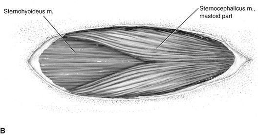
B Continuing the skin incision through the subcutaneous tissues, small transverse bundles of the sphincter colli superficialis muscle are identified and transected in the ventral midline. Retraction of the subcutaneous tissues exposes the mastoid part of the sternocephalicus muscles arising from the manubrium. Underlying the sternocephalicus muscles are the sternohyoideus muscles.
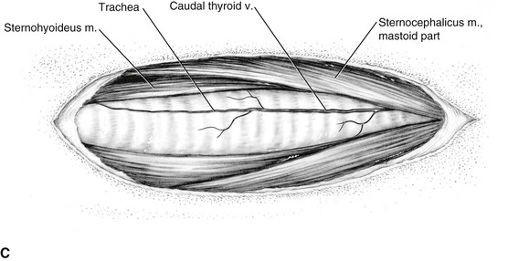
C The incision is deepened by midline separation of the paired bellies of the mastoid part of the sternocephalicus muscles and the underlying sternohyoideus muscles. With separation of the median raphe between the paired sternohyoideus muscles, the caudal thyroid vein is found in the fascia overlying the trachea. The caudal thyroid vein should be preserved, and lateral branches of the vein arising from the adjacent right sternohyoideus muscle are divided and cauterized as necessary.
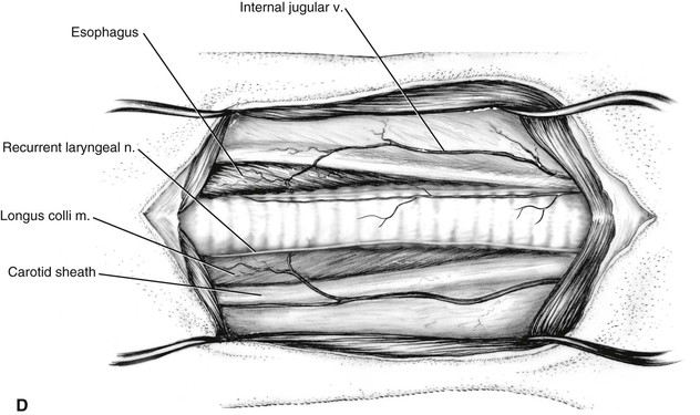
D Lateral retraction of these muscles exposes the trachea, esophagus, deep cervical fascia, carotid sheath, and internal jugular vein. At this stage of the approach, the location of the esophagus is readily determined by palpation of the esophageal stethoscope that is within the esophageal lumen.
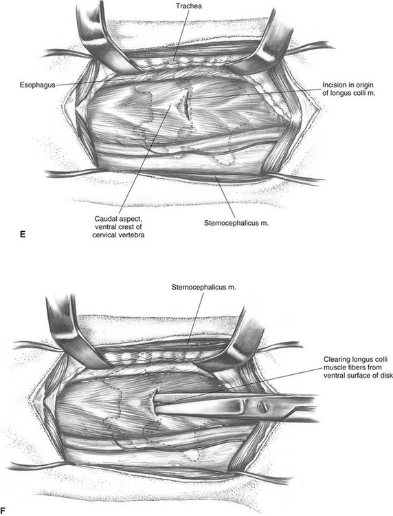
E Left lateral retraction of the trachea and esophagus using nontoothed retractors allows blunt dissection close to the trachea through the deep cervical fascia to the longus colli muscle, which covers the ventral surfaces of the cervical vertebrae. Care should be taken not to injure the recurrent laryngeal nerve or the esophagus during this dissection. The right carotid sheath containing the right carotid artery, the vagosympathetic nerve trunk, and the internal jugular vein is usually retracted to the right side of midline, but alternatively it can be moved to the left, along with the trachea. The midline ventral crest of the vertebrae can be palpated through the longus colli muscle. A short transverse incision is made through the longus colli tendon of insertion just caudal to the crest.
F Separation of longus colli muscle fibers overlying each ventral crest exposes the disk.
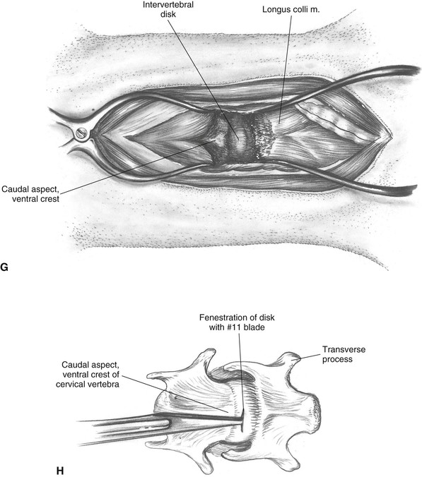
G By working caudally from the prominence, the tendon is gently scraped from the bone until the ventral longitudinal ligament is exposed. The exact location of the intervertebral space can be identified by exploration with a 22-gauge needle, which is walked off the crest caudally until it penetrates the ventral longitudinal ligament and the annulus fibrosus of the disk.
H Fenestration is accomplished by a stab incision through the ventral longitudinal ligament and the annulus fibrosus. This opening into the disk may have to be enlarged for disk curettage.
Additional Exposure
This approach can be extended cranially (see Plate 12) to obtain ventral exposure of the entire cervical spine.
Comments
Care must be used in the retraction of tissues to avoid damage to the carotid sheath; the esophagus and trachea; and the right recurrent laryngeal nerve, which lies on the right dorsolateral aspect of the trachea. The location of a specific intervertebral space is determined by first identifying the caudal borders of the wings of the atlas by palpation. The ventral midline crest that lies on a line directly between the wings is the ventral tubercle of the atlas (C1). Other vertebrae can then be numbered by counting caudally from C1. Alternatively, the large transverse processes of C6 are easily palpated. The C5-C6 disk is between and slightly cranial to the cranial edges of the processes.
Approach to the Midcervical Vertebrae Through a Dorsal Incision
Based on a Procedure of Funkquist15
Patient Positioning
The animal is positioned in sternal recumbency, with a sandbag placed under the neck to elevate it and to cause flexion of the cervical spine. A tracheal catheter is imperative to maintain a patent airway in this position (see Plate 15A).
Description of the Procedure
A The midline skin incision extends from the external occipital protuberance to the first thoracic vertebra.
B As the subcutaneous fascia is incised and the skin margins retracted, the almost transparent fibrous aponeurosis of the platysma muscle comes into view.
C The dorsolateral cervical muscles separated by this incision are retracted laterally to expose the nuchal ligament. The spinous processes can now be palpated under the ligament.
D Elevation with a periosteal elevator and retraction of the muscles from the vertebrae are done first on the side that was incised. The insertion of the nuchal ligament is now elevated from the spinous process of the axis, and the ligament is retracted with the muscles on the side opposite the incision. The ligament remains firmly attached to the muscles of one side and cranially to the axis.
Stay updated, free articles. Join our Telegram channel

Full access? Get Clinical Tree


