CHAPTER 3. Clinical Pathology: Hematology
Patricia A. Schenck
COMPLETE BLOOD COUNT (REQUIRES SAMPLE COLLECTED INTO AN EDTA TUBE)
I. Packed cell volume (PCV)
A. Percentage of whole blood composed of red blood cells (RBCs)
B. Collect in microhematocrit tube and centrifuge in a microhematocrit centrifuge. Three layers will be formed:
1. Plasma column at the top
2. Erythrocytes at the bottom
3. Buffy-coat layer in between the plasma and erythrocytes. The buffy coat layer is a small white band containing leukocytes and platelets. It may be red if many nucleated RBCs are present
II. RBC count is performed by instruments designed for particle counting. It generally parallels the PCV and hemoglobin concentration
III. Plasma protein concentration is determined by refractometry typically. Hyperlipemia can falsely increase the plasma protein concentration by 2 g/dL
IV. Total leukocyte concentration
A. Done by either Unopette dilution or by instruments designed for particle counting
B. Both methods detect all nuclei in solution; thus nucleated RBCs will be included in this count
V. Hemoglobin concentration is an index of the RBC mass per unit volume of blood
A. Provides information similar to that of PCV
B. In most species (other than the camel family), hemoglobin concentration is about a third of the PCV
VI. Mean cell volume (MCV) reflects RBC size
A. Macrocytic suggests increased red cell turnover. Some toy poodles, miniature poodles, and greyhounds normally have macrocytic RBCs
B. Microcytic suggests defective red cell growth. Akita and Shiba Inu dogs normally have microcytic RBCs
C. Normocytic means red cell size is unchanged
D. Comparing most species, dogs have the highest MCV values (largest RBCs), whereas sheep, llamas, and goats have the lowest MCV values (smallest RBCs)
VII. Mean corpuscular hemoglobin (MCH) and mean corpuscular hemoglobin concentration (MCHC) help classify anemia
VIII. Red cell distribution width (RDW) describes the relative width of the size distribution curve of the RBCs
IX. Platelet concentration
A. In most species, platelets are much smaller than RBCs
B. In cats, platelet volume is about twice that of most other species. Macroplatelets are also common in cats with hematology disorders and may be counted as RBCs with particle-size analyzers
X. Blood smear
A. The counting area is the small area between the feathered edge and the thick portion of the smear. The feathered edge should be observed for platelet clumps, large cells, and microfilaria
B. Reticulocyte count evaluates regeneration. A reticulocyte count should be performed if the PCV is below 30% in dogs or below 20% in cats
1. Reticulocytes are evaluated with methylene blue staining
2. A corrected percentage for a reticulocyte value greater than 1% or a count of greater than 60,000 cells/μL indicates RBC regeneration. Regeneration takes at least 3 days before reticulocytes appear in the circulation
3. Horses do not release reticulocytes
C. Morphology of RBCs
1. Changes in size
a. Anisocytosis is variation in RBC size
b. Microcytic RBCs are smaller than normal RBCs, with a decreased MCV
c. Macrocytic RBCs are larger than normal RBCs, with an increased MCV
2. Changes in shape ( poikilocytosis)
a. Poikilocytes are abnormally shaped RBCs
b. Schistocytes are RBC fragments usually caused by intravascular trauma (i.e., DIC). When two or more spicules are present, the cells are called keratocytes
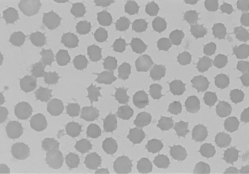
c. Acanthocytes (spur cells) are irregular, spiculated RBCs with unevenly distributed surface projections (Figure 3-1)
 |
| Figure 3-1 Acanthocytes demonstrating irregularly sized spicules in a blood smear from a dog with cholestatic liver disease. Wright’s stain, original magnification 132×. (From Cowell RL et al. Diagnostic Cytology and Hematology of the Dog and Cat, 3rd ed. St Louis, 2007, Mosby.) |
(1) May result from changes in cholesterol or phospholipid concentrations in the RBC membrane
(2) Acanthocytes are commonly seen in cats with hepatic lipidosis and dogs with hemangiosarcoma
(1) May be artifactual from slow drying of blood smear
(2) Have been observed in renal disease, lymphoma, rattlesnake envenomation, and chemotherapy. Also observed after exercise in horses
e. Spherocytes are dark-staining RBCs that lack central pallor. They are easiest to detect in the dog because dog RBCs have the most central pallor normally. Their presence suggests immune mediated hemolytic anemia
f. Eccentrocytes are characterized by a shifting of the hemoglobin concentration to one side, resulting in a loss of central pallor with a clear eccentric zone. They are associated with oxidative damage and may occur in conjunction with Heinz bodies
g. Leptocytes are RBCs in which there is excess membrane relative to internal contents. These may occur in vitro when cells contact excess EDTA. Membrane folding causes target cell formation (codocytes)
h. Codocytes are thin and bowl-shaped with a dense central area of hemoglobin (the appearance of a target). They may be seen in animals with increased serum cholesterol concentrations but have little significance
i. Stomatocytes are RBCs with a mouthlike clear area in the center of the cell. Found in dogs with hereditary stomatocytosis
3. Changes in color
a. Polychromasia indicates the presence of young erythrocytes, polychromatophilic cells characterized by being larger and slightly bluer than mature RBCs. The degree of polychromasia correlates to the reticulocyte response
b. Hypochromic RBCs are pale and have a decreased hemoglobin concentration, usually from iron deficiency
4. Structures in or on RBCs
a. Heinz bodies are caused by oxidant damage to RBCs, with denaturation of hemoglobin. Heinz bodies appear as small, pale structures near the margin of the RBCs, which may protrude. With methylene blue staining, these appear blue
b. Basophilic stippling is caused by aggregation of ribosomes into small granules. It is associated with immature RBCs in ruminants. Lead poisoning often causes basophilic stippling
c. Nucleated RBCs are RBCs in the peripheral circulation that have retained their nucleus. They are an indication of regenerative anemia, a nonfunctioning spleen, or steroids (endogenous or exogenous)
d. Howell-Jolly bodies are nuclear remnants in RBCs that appear as dark staining, round inclusions. They are associated with regenerative anemia or suppressed splenic function
e. Siderotic granules are visible iron granules in RBCs (siderocytes). They are associated with chloramphenicol, myelodysplasia, and impaired heme synthesis
f. Viral inclusions are rarely seen but may be seen in canine distemper. They are most commonly found in polychromatic RBCs
g. Parasites (see later)
5. Rouleaux formation is the stacking of RBCs. This is normal in horses and is enhanced when plasma protein concentration is increased (Figure 3-2)
 |
| Figure 3-2 The pattern of erythrocyte adhesion that occurs with rouleau is compared with the pattern that occurs with agglutination. (From Meyer D, Harvey JW. Veterinary Laboratory Medicine: Interpretation and Diagnosis, 3rd ed. St Louis, 2004, Saunders.) |
6. Agglutination results in clumps of RBCs and is associated with immune-mediated hemolytic anemia (see Figure 3-2)
D. Leukocytes
1. Neutrophils
a. Neutrophils have small granules in the cytoplasm that stain differently, depending on species. In cows, these granules stain faintly pink, giving the cytoplasm a pink tint
b. Neutrophils are important in an inflammatory response with chemoattraction to the site of inflammation and phagocytosis of organisms or foreign material
d. Band cells may be present normally in small numbers. They have a characteristic horseshoe-shaped nucleus
e. Segmented (mature) neutrophils normally predominate in peripheral blood
2. Lymphocytes
a. Responsible for humoral immunity, cell-mediated immunity, and cytokine responses
b. Round to oval nucleus with minimal clear cytoplasm
c. Normal lymphocytes have smaller diameter than neutrophils. In ruminants, lymphocytes are more irregular in size and may be the same size as neutrophils
d. Reactive lymphocytes are probably B lymphocytes producing immunoglobulin. They have a basophilic cytoplasm with irregular nuclear shape. Nucleus may be indented, giving it a bean-appearance
e. Granular lymphocytes may be natural killer or T cells and contain a small number of pink-purple granules. More prominent in ruminant blood
3. Monocytes
a. Participate in the inflammatory response. Monocytes migrate into tissues and develop into macrophages
b. Commonly misidentified on a blood smear
c. Nucleus may be oval, bean-shaped, or segmented
d. Larger diameter and grayer coloration to the cytoplasm than neutrophils. Cytoplasm may contain fine light purple granules
4. Eosinophils
a. Function not well understood. Contain proteins that bind to parasite membranes and are also involved in allergic responses
b. Vary in morphology among species. All have prominent red to orange cytoplasmic granules. Granules are rod- or barrel-shaped in cat eosinophils. Granules may wash out during staining, leaving empty vacuoles; this is most commonly seen in greyhounds
5. Basophils
a. Function is unknown. Basophils contain histamine and heparin, and their membrane has bound IgE
b. Normally not found on a blood smear
c. Larger than neutrophils, with a segmented nucleus and dark-violet granules in the cytoplasm. Cat basophils have large, faint gray cytoplasmic granules
CLASSIFICATION OF ANEMIA
I. Erythrocyte volume and hemoglobin concentration
Stay updated, free articles. Join our Telegram channel

Full access? Get Clinical Tree


