CHAPTER 35. Endocrine Disorders
Rebecca S. McConnico
EQUINE CUSHING-LIKE SYNDROME (ECD) OR PARS INTERMEDIA DYSFUNCTION (PID)
I. Previously known as
A. Equine hyperadrenocorticism
B. Equine diabetes
C. Equine Cushing disease
D. Equine cushinoid-like syndrome
II. Role of the hypothalamus
A. Translator between central nervous system (CNS) and pituitary gland
B. Equine pars intermedia hyperplasia may be related to hypothalamic dysfunction
III. Pituitary anatomy
A. Pituitary gland weighs about 1 to 3 g; 2 × 1 cm
B. Pars intermedia
1. Secretory control: Tonic inhibition is via dopamine
2. Other modulators
a. Serotonin
b. Adrenergic stimulation
c. α-Amino butyric acid
d. γ-Aminobutyric acid (GABA)-ergic
IV. Pathophysiology
A. Functional adenoma (hypertrophy) or adenomatous hyperplasia of the pituitary pars intermedia associated with a clinical syndrome of hirsutism
1. Increased β-endorphin
2. Increased α-melanotropin
3. Increased endogenous adrenocorticotropic hormone (ACTH) (small amount)
a. Similar to human and canine pituitary-dependent hyperadrenocorticism (increased ACTH) though not identical
b. Initial loss of neurotransmitter—dopamine (dopamine is inhibitory)
B. Gross pathology
1. Pars intermedia is grossly enlarged
2. Compression of pars nervosa and the rest of the adenohypophysis as a result of pars intermedia expansion
3. Adrenal gland hyperplasia (bilateral)
C. Histopathology
1. “Abnormal” cells are polyhedral, spindal-shaped, or cylindrical. Usually arranged in columns or rosettes around capillary beds and connective tissue septae
2. Mitotic index is low
3. Controversial as to whether this is neoplastic or true hyperplasia
V. Signalment (ECD or PID)
A. Average age = 19 years (range = 7 to 40 years)
B. No sex predilection
C. No breed predilection
D. Ponies >> horses (not scientifically proven)
VI. Clinical signs of ECD or PID
A. Chronic or recurrent laminitis
1. May be secondary to increased cortisol levels (or vice versa)
2. Sole abscesses—chronic
3. Chronic increased cortisone levels
4. May be associated with equine metabolic syndrome (insulin resistance and glucose intolerance)
B. Polyuria (PU), polydipsia (PD)
1. Result from destruction of pars nervosa by enlarging pars intermedia leading to decreased antidiuretic hormone (ADH) secretion
2. Chronic hyperglycemia → osmotic diuresis
3. Cortisol → increases glomerular filtration rate
C. Hyperhidrosis: Sweat glands are under β-adrenergic control, catecholamine response
D. Muscle wasting
1. Disease aging
2. Corticosteroid myopathy
3. Weight loss: Parasitism, chronic infection
E. Hirsutism (85% of cases) (Figure 35-1)
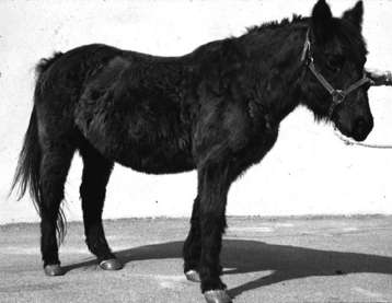 |
| Figure 35-1 Horse with pars intermedia dysfunction demonstrating the classic sign of hirsutism: a long, shaggy hair coat that fails to shed in the spring. (From Bertone J. Equine Geriatric Medicine and Surgery. St Louis, 2006, Saunders.) |
1. Most common clinical sign
2. Thick, long, curly coat; not shed
3. Heavier than normal winter coat
4. Slow shedding
5. Patchy alopecia (usually not a hallmark), shed abnormally
6. Development of “guard” hairs (chin and jugular furrow)
7. Differential diagnosis = curly-coated fox-trotter
F. Additional clinical abnormalities
1. Narcolepsy
2. Seizures, ataxia, other neurologic signs
3. Weight loss
4. Supraorbital fat pads (redistribution of fat)
5. Decreased responsiveness to painful stimuli
6. Increased appetite
7. Recurrent infections
8. Sinusitis
9. Pneumonia
10. Skin infections
VII. Differential diagnosis
A. PU/PD
1. Renal disease
2. Primary diabetes insipidus (DI)
3. Psychogenic DI
4. Hyperglycemia secondary to pancreatic destruction
5. Equine Cushing disease (hyperadrenocorticism)
B. Hyperhidrosis
1. Functional pheochromocytosis
2. Adrenal tumor
C. Additional concerns (ECD/PID)
1. Chronic dental problems (no teeth); feed pellets
2. Severe endoparasitism
a. Check fecal egg counts for intestinal parasites
b. Some veterinarians recommend deworming every 8 weeks
3. Supraorbital swellings
4. Abnormal fat distribution
5. Elevated respiratory rate: Chronic infections and opportunistic infections
6. Nocardia, Coccidioides, fungal → cell-mediated immunity compromised
VIII. Diagnosis
A. Minimum database
B. History and clinical signs
C. Complete blood cell count
1. May have slight stress leukogram
2. Neutrophilia
3. Lymphopenia
D. Serum chemistry profile: Usually normal
1. Alkaline phosphatase is sometimes elevated
2. Hyperlipemia (fat ponies)
3. Hyperglycemia
a. Incidence is variable and occurs in approximately 26% to 85% of patients
b. Hyperglycemia is most likely due to insulin resistance (see equine metabolic syndrome)
E. Resting cortisol: Usually normal
1. Diurnal variation
a. Higher in the morning (normal horse). The highest level occurs between 8:00 am and noon; the lowest level occurs in late evening
b. ECD/PID lose the diurnal variation in cortisol concentration
c. Fall
2. Increased
a. Stress
b. Exercise
c. Hypoglycemia
3. Resting tri-iodothyronine (T 3) and thyroxine (T 4) levels: Normal (not affected by hormonal influence)
F. Endocrine function testing
1. Dexamethasone suppression test: Test of choice (40 μg/kg or 2 mg/100): ∼20 mg
a. Overnight test
(1) Draw a pretest for serum cortisol (heparinized tube)
(2) Administer dexamethasone intramuscularly (IM)
(3) Draw blood at noon the next day (19-21 hours post-dosing)
(4) Excellent screening test
(5) Interpretation: Normal horse less than 1 μg/dL (or less than 30 nmol/L) cortisol 19 hours after administration
b. Standard test
(1) Draw presample at midnight
(2) Administer dexamethasone at midnight
(3) Draw blood at 8:00 am, 12:00 noon, 4:00 pm, and midnight
c. Limitations of the dexamethasone suppression test
(1) Normal cortisol level is much higher in the fall; important to interpretation!
(2) Age
(3) Gender
(4) Gestation
2. Thyroid-releasing hormone (TRH) stimulation test
a. Poor specificity, although gaining in popularity
b. May be useful to use in horses with laminitis
c. 1 mg administered intravenously (IV) after baseline insulin and cortisol is taken
d. In horses with pituitary adenomas → cortisol and insulin should rise in 15 minutes (cortisol) and 1 hour (insulin) and stay elevated for 90 minutes
e. TRH may be difficult of obtain
f. Mechanism is unclear but possibly a paradoxical response of diseased pituitary tissue resulting from alterations in the receptor-adenylate cyclase system
3. ACTH stimulation test
a. Dose is 1 unit/kg ACTH gel administered IM or 100 international units synthetic ACTH (cosyntropin) IV
c. Useful test for adrenal exhaustion syndrome
d. Normal response = 2-3 × increase in 4 to 8 hours
e. 4 × increase is extremely variable!
4. Insulin tolerance test: Useful only in hyperglycemic Cushing horses (pancreatic disease)
5. Combined dexamethasone suppression test and ACTH response test: Not useful because does not allow distinguishing normal from Cushing horses
Stay updated, free articles. Join our Telegram channel

Full access? Get Clinical Tree


