CHAPTER 40. Oncology
Yvonne A. Elce
BASIC PRINCIPLES
I. Neoplasia can occur in horses of any age, breed, or sex. The geriatric population of horses is increasing with improvements in management and veterinary care, so the equine veterinarian must attempt to keep up with the rapidly advancing field of equine oncology through continuing education
II. Definitive diagnosis of tumors is through histopathology and is highly recommended in all instances of neoplasia as the treatment and prognosis vary widely depending on the type of tumor
III. Most common tumors seen by veterinarians in horses are skin tumors (sarcoids, melanomas, squamous cell carcinomas), tumors of the sinuses and nasal passages, and lymphosarcoma
IV. Health management and regular examinations will enable early detection, which is crucial to improving the prognosis
V. Treatment should be undertaken only after discussions with the owner involving the prognosis, quality of life, and economic issues surrounding the decision
TUMORS OF THE SKIN AND UNDERLYING TISSUES
I. Most common form of tumor in the horse
II. Not confined to geriatric horses
III. Tumor types
A. Most common are sarcoids, squamous cell carcinoma, and melanomas
B. Less common are lymphosarcoma, mast cell tumors, basal cell tumors, and hemangiomas
IV. Common differentials for skin tumors include fungal lesions, cutaneous habronemiasis, pythiosis, phycomycosis, exuberant granulation tissue, and varying types of tumors.
V. Sarcoids
A. Causes (Figure 40-1)
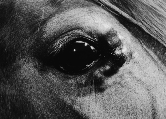 |
| Figure 40-1 Equine sarcoid. Multiple nodular lesions in medial canthal area. (From Scott DW, Miller WH. Equine Dermatology. St Louis, 2003, Saunders.) |
1. Related to interaction between genetics and infection with bovine papillomavirus
2. Found on limbs, ears, eyelids, head, and trunk but can occur anywhere
3. Often found in areas of repetitive trauma or injury
4. Four main types: Occult (flat, thickened areas of skin lacking hair), verrucous (warty and small), nodular (under-the-skin small nodules), and fibroblastic (aggressive, resemble exuberant granulation tissue)
5. Fibroblastic types are locally invasive and grow rapidly; no type is metastatic
B. Signalment
1. Breed predilections in quarter horses, Appaloosas, and Arabians; not commonly found in standardbreds
2. Generally in younger horses 1 to 6+ years of age
3. No sex predilection
C. Clinical signs
1. Depends on type of sarcoid; noticed usually as small area of abnormal skin
2. Fibroblastic types found in areas of previous trauma or wounds
3. Do not interfere with function unless grow large and impinge on adjacent structures such as the eye, joints, or tendons
D. Diagnosis
1. It is not recommended to biopsy occult, small verrucous and nodular sarcoids as trauma may transform them into fibroblastic types
2. However, definitive diagnosis is based on histopathology
E. Treatment
1. Many different treatments available
2. Surgical excision as the sole treatment has a high rate of recurrence (50%)
4. Many commercial topical preparations available usually containing caustic substances to cause sloughing of the sarcoid
5. Injections of EqStim or bacille Calmette-Guérin (BCG) can be given to stimulate the immune system to reject the sarcoid. There have been reports of anaphylaxis after multiple BCG injections, so it is recommended to pretreat with antiinflammatory drugs.
VI. Squamous cell carcinoma
A. Causes
1. Most common tumor of the eye and external genitalia
2. Also found in the stomach, oral pharynx/larynx, and bladder
3. Light-colored breeds and nonpigmented areas of skin are predisposed because solar radiation plays a role in carcinogenesis. Also local carcinogens (smegma accumulations in the sheath) may be important
4. Locally invasive and can become metastatic
5. Can be ulcerative or proliferative lesions
B. Signalment
1. Common in older horses
2. Draft horses, Appaloosas, Arabians, paint, and pinto horses are predisposed (light- colored horses)
C. Clinical signs
1. Depends on location of tumor
2. Foul smell common with all locations (oral, genital)
3. Ocular tumors usually start as small white lesions on the eye or adnexal structures with blood-stained tears
4. Common with genital tumors to have discharge or bleeding, dysuria, preputial edema
D. Diagnosis is through histopathology or cytology (biopsy, fine-needle aspirate, impression smear)
E. Treatment
1. Many treatments available
2. Surgical excision is best combined with adjunctive therapy (radiation or local chemotherapy)
3. Local chemotherapy can be performed with intralesional 5-fluorouracil (5-FU) or cisplatin
4. In addition, 5-FU cream can be applied every 1 to 2 weeks as a thin coating until resolution
5. Other treatments include cryotherapy, hyperthermia, radiation therapy
6. New reports of successful resolution with administration of systemic nonsteroidal antiinflammatory drugs (NSAIDs), particularly piroxicam at 0.2 mg/kg once daily orally. Squamous cell carcinomas produce cyclo-oxygenase-2 (COX-2), which aids in tumor growth and metastasis; treatment with COX inhibitors can cause tumor regression. Need to monitor for NSAID side effects (renal damage, ulcers in the stomach or colon)
F. Prognosis
1. Good if tumors are diagnosed and treated when they are small and before local spread or metastasis
2. Improved with use of ancillary therapies such as radiation and chemotherapy
3. Large invasive tumors often involve local lymph nodes and should provoke examinations for metastases, which decrease the prognosis significantly
4. Regular examinations of older horses (sheath cleanings and inspection of external genitalia) are recommended to aid in early detection
5. Recommend reducing exposure to solar radiation of light-colored horses with areas of nonpigmented skin through nighttime turnout routines or solar radiation–blocking blankets
VII. Melanomas
A. Cause
1. Unknown
2. May be due to disturbed melanin metabolism or production of new melanoblasts
3. Single nodules or coalescing nodules in or under the skin
4. Commonly slow growing and invade locally but may become fast-growing, malignant, and metastasize to internal organs (lungs, GI tract, spleen)
5. Most tumors produce melanin but particularly in non-gray horses may have amelanotic melanomas. Amelanotic melanomas in colored horses are usually aggressive and metastasize
B. Signalment
1. Older (15 years+) gray horses are most at risk (70% to 80% of older gray horses develop melanomas)
2. Melanomas can develop in horses of any color but are more often malignant in non-gray horses
3. No sex predilection
C. Clinical signs
1. Formation of masses under the skin or as dark black masses when the skin has ulcerated
2. Common areas include under the tail, perineal region, and external genitalia
3. Other areas include the neck, the parotid salivary gland, the head, limbs, and guttural pouches
4. Can develop difficulty defecating, urinating, or breathing, depending on the location of the tumor as the result of a local mass effect but often not clinically significant early
5. Signs of metastasis relate to organs affected (e.g., weight loss, respiratory signs)
Stay updated, free articles. Join our Telegram channel

Full access? Get Clinical Tree


