16. Tumours of the hepatobiliary system and exocrine pancreas
Hepatic tumours are rare, accounting for about 1% of canine and 2% of feline neoplasms. Average age is 10 years (Liptak 2007). There is no known breed or sex predisposition. Aetiology is unknown, although exposure to aflatoxins (Hammer & Sikkema 1995), radiation and various chemical carcinogens (diethylnitrosamine, dichlorobenzidine, aramite, 3-acetylaminofluorene and others) has been shown to induce them (Thamm 2001). Metastatic liver tumours are much more common than primary tumours (perhaps due to dual afferent blood supply from the portal vein and hepatic artery) (Hammer & Sikkema 1995). These most commonly arise from the gastrointestinal tract, splenic haemangiosarcoma, pancreatic tumours, mammary adenocarcinomas and anal sac adenocarcinomas.
Canine hepatic tumours
There are three morphological types of hepatobiliary tumour: massive, nodular and diffuse.
1. Massive: Solitary, large. Metastatic rate is variably reported but generally low (Liptak et al 2004, Patnaik et al 1980). Confined to one liver lobe (especially left) (Patnaik et al 1980). Most commonly hepatocellular carcinomas, but can also be biliary carcinomas or sarcomas (Liptak 2007).
2. Nodular: Multifocal disease usually involving multiple lobes, typically sarcomas, carcinomas, or carcinoids (Liptak 2007). Nodular hyperplasia is a benign condition seen in older dogs and is a major differential for nodular hepatic disease. Focal hepatic lesions in dogs using ultrasound were diagnosed as nodular hyperplasia in 25% and 36% of cases (Cuccovillo & Lamb 2002, Vörös et al 1991).
3. Diffuse: May represent a late stage of tumour development when coalescing nodules cause effacement of hepatic lobe parenchyma. This is primarily seen with carcinomas and carcinoids (Liptak 2007).
Feline hepatic tumours
The primary hepatobiliary tumours seen in cats are bile duct adenomas and carcinomas, hepatocellular adenomas and carcinomas, hepatoblastomas and myelolipomas. The most common secondary liver tumour is lymphoma (Neiger 2003a). Pancreatic, intestinal and renal tumours not uncommonly metastasize to the liver in cats.
Gall bladder tumours are <5% of biliary carcinomas in cats and dogs (Hayes et al 1983, Patnaik 1992, Patnaik et al 1981a). Biliary carcinomas may be intra- or extrahepatic, with no clear predominance of either location (Lawrence et al 1994, Patnaik 1992). Cats with malignant liver tumours have a poor prognosis, with 86% of the cats dying or being euthanized during hospitalization (Lawrence et al 1994).
Paraneoplastic alopecia has been reported in one cat with hepatocellular carcinoma (Marconato et al 2007). A hepatic abscess was found secondary to hepatocellular carcinoma in another cat (Singh et al 2005). Benign liver tumours in cats have a good prognosis with partial or complete resection; survival times are prolonged and extend to several years (Trout 1997, Trout et al 1995). Complete resection is recommended as neoplastic changes have been seen in some feline adenomas.
Myelolipomas present as single or multifocal masses of well-differentiated adipose tissue with normal haematopoietic elements. They have an excellent prognosis when treated with liver lobectomy (McCaw et al 1990).
Hepatocellular tumours
Hepatocellular carcinomas (HCC)
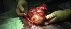
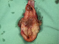
HCC is the most common primary hepatobiliary tumour in dogs (>50%), with a reported metastatic rate ranging from 22 to 61% (Patnaik et al, 1980 and Patnaik et al, 1981b, Trigo et al 1982). The massive form is most common, and usually affects a single lobe (Figure 16.1). Left lobes are more often affected (Patnaik et al 1980). Distant metastatic rate is high, depending on type: 100% for diffuse form, 93% for nodular form and 36% for massive type (Patnaik et al 1980). HCCs usually metastasize to regional lymph node, lung and peritoneum.
Benign tumours are more common than carcinomas. They are usually incidental but have been reported to cause clinical signs of vomiting, diarrhoea, lethargy and inappetence (Eves 2004). Usually they are single masses but may be multiple and pedunculated.
Hepatoblastoma
There is only one reported case (Shiga et al 1997) of hepatoblastoma in a dog.
A hepatic carcinoid is an APUDoma. These arise from neuroectodermal tissue of the amine precursor uptake and decarboxylation cells (APUD) of biliary epithelium. They may secrete vasoactive peptides and amines. Hepatic carcinoids comprise a small percentage of primary intrahepatic tumours in dogs and cats (also primary gall bladder in one dog) (Willard et al 1988). They are known to behave very aggressively, with diffuse liver involvement and early metastasis (metastatic rate 93%), most commonly to the peritoneal wall and adjoining lymph nodes (Patnaik et al 1981c).
Sarcomas
Sarcomas comprise a small percentage of hepatic tumours. Most are locally aggressive and diffuse with early metastasis (metastatic rate 86%), commonly to spleen (Patnaik et al 1980). Leiomyosarcoma is the most common, then haemangiosarcoma, haemangioma, fibrosarcoma, osteosarcoma, chondrosarcoma, malignant mesenchymoma, botryoid rhabdomyosarcoma and liposarcoma.
History and clinical signs
More than 50% of cats have no clinical signs (most of these tumours were benign) (Lawrence et al 1994). When clinical signs do occur in dogs and cats, they are generally vague, non-specific signs such as anorexia, weight loss, vomiting, lethargy, pyrexia, polyuria/polydipsia (PU/PD), abdominal distension and hepatomegaly. Between 50 and 75% of dogs and cats have a palpable cranial abdominal mass (Kosovsky et al 1989, Lawrence et al 1994, Post & Patnaik 1992, Trigo et al 1982). Icterus is usually not a feature. Hepatoencephalopathy can be seen if advanced.
Haematological changes include non-regenerative anaemia (common) and leucocytosis as non-specific findings (Kosovsky et al 1989, Patnaik et al 1980). Decreased clotting factors may cause coagulopathies, but clinical problems with coagulation are uncommon except for hepatic haemangiosarcoma (Thamm 2001). Platelet count is also important; thrombocytosis is seen in about 50% of dogs with the massive form of HCC (Liptak et al 2004).
Staging
Radiography
Thoracic radiographs rarely show metastatic disease at diagnosis (Evans 1987).
Ultrasound
Abdominal ultrasound is important for staging and biopsy. Ultrasound-guided biopsies may be obtained if the clotting profile is normal. Ultrasound appearance does not correlate well to histological type but focal masses are more likely to be HCC.
Ultrasound-guided fine needle aspirate (FNA) may be beneficial as a preliminary screen. There is overall agreement between histopathological diagnosis and cytological diagnosis in about 30% of canine and 50% of feline cases (Wang et al 2004). Ultrasound-guided tru-cut biopsy of various abdominal structures (not just liver) gave a correct diagnosis in 93.5% of cases with 5.6% minor complications and 1.6% major complications (Léveillé et al 1993).
Only gold members can continue reading. Log In or Register to continue