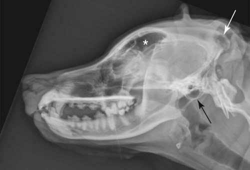
The skull encompasses the brain and houses the sense organs for hearing, equilibrium, sight, smell, and taste. The skull provides attachment sites for teeth, tongue, larynx, and muscles.1 There is pronounced variation in the shape of the skull in the dog. Three terms are used to designate the different shapes. Dolichocephalic breeds, such as collie and Russian wolfhound, have long, narrow heads with an extensive nasal cavity from rostral to caudal. Mesaticephalic breeds, such as German shepherd and beagle, have heads of medium proportion (Fig. 8-1). Brachycephalic breeds, such as Boston terrier and Pekingese, have short, wide heads. Cats are more uniform in their skull conformation. However, Siamese tend to have longer heads as compared with Himalayan and Persian breeds. The calvaria comprises the bones of the brain case, with the occipital bone forming the base of the skull. The occipital crest is the most dorsocaudal aspect of the skull (see Fig. 8-1), and the occipital condyles are caudoventral as seen on lateral radiographs. The foramen magnum, centered between the occipital condyles, forms an orifice for passage of the spinal cord. The nasal passage extends caudally from the external nares to the cribriform plate and nasopharynx. The cribriform plate is a sievelike partition between the olfactory bulb and nasal passage. The nasal passage is divided in half by the nasal septum and is filled with thinly scrolled conchae. Caudally, the nasal septum is osseous and fuses with the cribriform plate; it becomes cartilaginous as it extends rostrally.1 The vomer bone is unpaired and forms the caudoventral bony part of the nasal septum; it is visible radiographically.2 The cartilaginous nasal septum cannot be seen in radiographs, although it can be distinguished in computed tomography (CT) and magnetic resonance (MR) images. Both dogs and cats have frontal sinuses (see Fig. 8-1), lateral maxillary recesses, and small sphenoidal sinuses. These are named for the bones in which they are located. The tympanic bullae (see Fig. 8-1) form the ventral part of the temporal bone. These air-filled cavities of the middle ear communicate with the nasopharynx via the auditory tube. The temporal bone consists of the petrosal, tympanic, and squamous sections that are fused in the adult. The petrosal portion is medial and dorsal to the tympanic bulla and is composed of dense bone in the mature animal. The squamous portion of the temporal bone extends rostrally and laterally to form the zygomatic arch. The teeth are anchored in alveoli within the mandible and maxilla. The dental formulas for the dog and cat are provided in Box 8-1. Components of the tooth include the root (embedded in bone) and the crown (within the oral cavity); the bone between teeth is referred to as the alveolar crest. The dentin, enamel, and lamina dura of the tooth are radiopaque. The pulp cavity and periodontal membrane are of soft-tissue opacity (Fig. 8-2). The size of the pulp cavity becomes smaller with age.3 Specifics on radiographic technique and positioning for tooth evaluation can be found elsewhere.4–7 Cross-sectional imaging techniques, CT, and MR imaging are being used more commonly for imaging of the head. CT and MR technology provides images without superimposition of structures and better soft tissue delineation compared with radiography (Fig. 8-3).8–12 There are several references describing the normal CT and MR image anatomy of the dog and cat head.9,13–22 Hydrocephalus is excessive accumulation of cerebrospinal fluid within the ventricular system of the brain. Congenital hydrocephalus occurs secondary to structural defects that obstruct cerebrospinal fluid outflow or impede its absorption.23,24 Canine breeds affected with congenital hydrocephalus include the Maltese, Yorkshire terrier, English bulldog, Chihuahua, Lhasa apso, Chinese pug, toy poodle, Pomeranian, Pekingese, Cairn terrier, and Boston terrier.23 Hydrocephalus is less common in cats.23,25,25 Radiographic signs associated with hydrocephalus include doming of the calvaria and cortical thinning, persistent fontanelles, and a homogeneous appearance to the brain, resulting from the loss of normal convolutional skull markings (Fig. 8-4). Radiographs are very insensitive for detection and characterization of hydrocephalus, and that information is now acquired using CT or MR imaging.23 With persistent fontanelles, ultrasound can also be used to assess ventricular size,27–34 and normal ventricular appearance and size have been quantified in the dog.27,28,35,36 The advantage of CT and MR imaging for evaluating ventricular size is the ability to assess the entire brain for causes of hydrocephalus (Fig. 8-5). Asymmetry in ventricular size is often normal in dogs, and correlation between ventricular size and clinical signs is poor.27,28,30,33,34 Occipital dysplasia is the dorsal extension of the foramen magnum, secondary to a developmental defect in the occipital bone;37 it has been related to clinical signs of neurologic disease and is usually found in miniature and toy breeds.38–40 Foramen magnum size and shape can be evaluated in a rostrodorsal-caudoventral skull radiograph,41 but this projection is generally not used now that CT and MR imaging are more available. The characteristics of the foramen magnum can be assessed more accurately using CT rather than radiographs (Fig. 8-6). Occipital dysplasia may be a normal morphologic variation in brachycephalic dogs.42–44 Occipital bone malformation may result in overcrowding of the caudal fossa, leading to obstruction of cerebral spinal fluid (CSF) flow, hydrocephalus, and secondary syringomyelia. This hereditary defect, termed Chiari-like malformation, is common in the Cavalier King Charles spaniel45–49 but is also found in other brachycephalic breeds. CSF flow is obstructed by the malformation, and the cerebellum may be herniated through the foramen magnum with dorsal deviation of the brainstem.47 Clinical signs vary in severity and usually are seen between the age of 6 months and 2 years; however, neurologic signs may not appear until later in life.47 Neurologic signs are consistent with a central spinal cord lesion, and dogs with clinical signs generally have a significantly larger syrinx than asymptomatic dogs.49 Clinically, dogs often present with persistent scratching of the shoulder region, with no dermatologic cause and thought to be a paraesthesia secondary to syringomyelia.45 Radiographs are not useful for diagnosis of Chiari-like malformation. Definitive diagnosis is made with MR imaging,47 whereby the crowding of the cerebellum in the caudal fossa can be detected. There may be herniation of a portion of the vermis of the cerebellum (see Fig. 12-46. A positive correlation has been found between foramen magnum size and length of cerebellar herniation.49 Open-mouth jaw locking can be caused by temporomandibular joint (TMJ) dysplasia. This congenital condition is uncommon; it is reported most frequently in the basset hound, but has also been seen in Irish setters.50 The open-mouth jaw locking occurs after hyperextension of the jaw, excessive lateral movement of the condyloid process, and subsequent entrapment lateral to the zygomatic arch. Physical entrapment usually occurs on the side opposite from the joint with the most severe dysplastic changes. Yawning often precipitates jaw locking when it results in extreme opening of the mouth.50 In spaniels, Pekingese, and dachshunds, TMJ dysplasia is an asymptomatic anatomic anomaly.20,51,51 Open-mouth jaw locking has also been seen in cats,53,54 and CT is routinely used to diagnose TMJ dysplasia, with three-dimensional reconstructions aiding in surgical planning.55 Mucopolysaccharidoses are a group of hereditary disorders of lysosomal storage, which occur in humans, dogs, cattle, and cats.56 Mucopolysaccharidosis VI (MPS-VI) is an autosomal recessive lysosomal storage disease recognized in Siamese cats.57–60 Radiographic skeletal changes in cats with MPS-VI include epiphyseal dysplasia, generalized osteoporosis, pectus excavatum, and vertebral and skull changes.61 Specific skull changes seen on radiographs include shortened nasal conchae, aplasia and hypoplasia of the frontal and sphenoid sinuses, and shortened dimensions to the incisive and maxillary bones.61 Another form of mucopolysaccharidosis, MPS-I, has been documented in the domestic shorthaired cat62 with radiographic skeletal changes that are similar to those in MPS-VI; however, the facial dysmorphia may not be as pronounced as it is in the Siamese.59,63 Mucopolysaccharidosis in animals has clinical and pathologic manifestations similar to people and therefore represents an excellent model for studying approaches to therapy and care.58,60,64,65 Primary or secondary hyperparathyroidism can result in overall decreased bone opacity, often noted easily in the skull. A solitary parathyroid adenoma or carcinoma, or adenomatous hyperplasia of one or both parathyroid glands, causes primary hyperparathyroidism. This results in excessive synthesis and secretion of parathyroid hormone, which leads to hypercalcemia and subsequent bone resorption.66–68 Secondary hyperparathyroidism, which includes renal and nutritional secondary hyperparathyroidism, is subsequent to nonendocrine alterations in calcium and phosphorus homeostasis that lead to increased levels of parathyroid hormone and ultimate bone resorption.68 An early radiographic sign of hyperparathyroidism (primary and secondary) is loss of the lamina dura (Fig. 8-7). This will be followed by overall demineralization of the skull bones as the disease progresses (Fig. 8-8). In fact, it is uncommon to see loss of the lamina dura without some concurrent generalized skeletal demineralization. The level of cortical thinning and degree of overall osteolysis and osteomalacia depend on duration and severity of the hyperparathyroidism. Also, because young animals are growing and have rapid skeletal turnover, they are affected more severely than older animals. In extreme hyperparathyroidism, demineralization is followed by fibrous tissue hyperplasia, termed fibrous osteodystrophy. This uncommon development leads to skull thickening caused by fibrous tissue proliferation. Ultrasound evaluation of the cervical region can be used to evaluate dogs with hypercalcemia to search for a parathyroid mass.69,70 In 210 dogs with primary hyperparathyroidism, a parathyroid mass with a range of 3 to 23 mm in diameter was identified in 129 of 130 dogs that were imaged sonographically.67 Thirty-one percent of the dogs had cystic calculi, and all were either calcium phosphate or calcium oxalate (radiopaque).67 Tumors of the nasal cavity in dogs and cats account for approximately 1% to 2% of all neoplasms.71–73 These tumors occur in older dogs and cats; approximately two thirds of nasal tumors are epithelial (adenocarcinoma, squamous cell carcinoma, undifferentiated carcinoma), and the other one third are mesenchymal (fibrosarcoma, chondrosarcoma, osteosarcoma, undifferentiated sarcoma).74–77 Intranasal lymphoma can also occur, with a higher prevalence in cats.75,76,78–80 Tumors of the nasal cavity are locally invasive but have a relatively low metastatic potential. External beam radiotherapy is the current treatment of choice,79,81–84 with many centers using advanced radiotherapy techniques such as intensity-modulated radiation therapy and image guidance.85–87 Unfortunately, diagnosis of these tumors often occurs late in the course of disease, resulting in a poor prognosis for outcome in many patients. Nasal cavity tumors have an aggressive radiographic appearance, with bony invasion and loss of conchal detail being common radiographic features.12,80,88–90 Tumors may be unilateral or bilateral and cause increased soft-tissue opacity in the nasal cavity with underlying conchal destruction. Destruction of bones adjacent to the nasal cavity is also common in advanced tumors. Nasal tumors may result in increased opacity within the frontal sinus;80,89–91 it is usually impossible to determine on radiographs whether frontal sinus opacification is caused by tumor extension or by occlusion of the nasofrontal communication with subsequent mucus accumulation in the sinus. Making this distinction can be important for treatment planning. MR imaging, which is based on the chemical composition of the tissue rather than the electron density, is helpful in distinguishing tumor from mucus in the frontal sinus (Fig. 8-9), but contrast-enhanced CT provides similar information.92 The most useful radiographic views for evaluating nasal disease include the intraoral dorsoventral and/or the open-mouth ventrodorsal view for detailed evaluation of the nasal cavity without superimposition of the mandible (Fig. 8-10). The open-mouth ventrodorsal view is better for cribriform plate assessment because radiographic film cannot be positioned physically to include the cribriform plate in the intraoral view. The cribriform plate is represented by a V-shaped to C-shaped bony opacity on radiographs, varying according to skull shape (dolichocephalic vs. mesaticephalic and brachycephalic).93 Evaluation of the cribriform plate is important because nasal tumors often originate from the ethmoid conchae and cribriform plate,74 and bony lysis detected on radiographs indicates potential tumor extension into the cranial cavity (see Fig. 8-10), which signifies a poorer prognosis. The rostrocaudal frontal sinus projection is necessary for evaluation of individual frontal sinuses (Fig. 8-11) and is useful especially if CT or MR imaging are not available.94 General anesthesia is an absolute requirement for achieving accurate radiographic positioning, and it facilitates evaluation and comparison of the complex nasal passages. Techniques for obtaining radiographic views of the nasal cavity and paranasal sinuses can be found elsewhere95 and were discussed briefly in Chapter 7. Aggressive nasal tumors and those with a prolonged duration are more destructive and less confined radiographically, often exhibiting an external soft-tissue mass that represents tumor extension through overlying bone. Conchae destruction and deviation and destruction of the bony nasal septum are apparent on radiographs. Radiographic evidence of bony destruction is an important prognostic sign because it is associated with a poor outcome.75,90,91,96 Less aggressive tumors and ones that are detected early are difficult to differentiate from rhinitis on radiographs.90 Radiographic detection of bony lysis of the cribriform plate and naso-orbital wall is difficult radiographically,93 and better suited to CT or MR imaging. CT of the nasal passage is superior to routine radiography for accurate tumor staging (Figs. 8-12 and 8-13)97 and is useful for attempting to differentiate infection from neoplasia.8,10,12,98–103 It is impossible to determine the stage of a nasal mass adequately from radiographs, and CT is the preferred screening modality for nasal disease. The presence of a mass effect (increased soft tissue in the nasal cavity) along with bone destruction is a hallmark sign of nasal neoplasia (see Fig. 8-13). A destructive pattern without a marked mass effect is more typical of aspergillus infection (discussed later), whereas a mass effect without turbinate destruction is also more typical of infection, although usually not aspergillus. CT images of patients with nasal cancer are also used in computerized radiation therapy planning systems. Use of this anatomic information allows optimization of dose distribution across the tumor volume104 and probable improved survival and decreased normal tissue side effects.82,87 MR imaging of the nasal passage provides excellent, true three-dimensional images of nasal tumors. Differences in MR signal intensity between sarcomas and carcinomas have been found.105 Tumors of the oral cavity account for approximately 6% of all canine cancers and 3% of feline cancers.106,107 Squamous cell carcinoma commonly affects the mandible or maxilla in the dog and cat. Fibrosarcoma, malignant melanoma, and tumors of the periodontal ligament (epulis) are common in the dog but occur rarely in cats.108,109 In the dog, the rostral mandible is a common site for oral squamous cell carcinoma. This tumor has variable bony lysis, and regional or distant metastasis is rare.110 Oral fibrosarcoma in dogs can affect the maxilla or mandible with a predilection for the palate.110 In dogs, 82% of squamous cell carcinomas and 78% of fibrosarcomas were characterized radiographically by bone involvement.96 Often, oral fibrosarcoma appears benign histologically, but biologic activity is aggressive. These tumors, often found in the maxilla and mandible of large-breed dogs and commonly in golden retrievers, are histologically low-grade yet biologically high-grade aggressive tumors. Bone lysis is a common feature.111 Dogs with oral fibrosarcoma have a lower median survival as compared with those with soft-tissue sarcomas at other sites112 (Fig. 8-14). In contrast, malignant melanoma tends to occur in small-breed dogs, commonly metastasizes to regional lymph nodes and lungs, and has variable bony lysis radiographically.113 Squamous cell carcinoma in cats affects the mandible or maxilla, causing sclerotic and/or lytic changes to bone (Fig. 8-15); common CT features include sublingual and maxillary locations with marked heterogeneous contrast enhancement (Fig. 8-16).114 Flea control products and diet may play a role in development of squamous cell carcinoma in cats.115 Unlike squamous cell carcinoma in the dog, these tumors have a poor prognosis and are less responsive to radiotherapy in cats.110,116 The epulides of periodontal origin have been divided into three categories: fibromatous epulis, ossifying epulis, and acanthomatous epulis.117 Fibromatous and ossifying epulides are similar benign growths cured by surgical excision; the distinctive feature of ossifying epulis is the histologically large segments of osteoid matrix.108 The predominant feature of acanthomatous epulis, now termed acanthomatous ameloblastoma,118,119 is the sheets of acanthomatous epithelial tissue noted histologically108 and the local invasion, which often causes bony destruction on radiographs. Although rare, multiple epulides in cats have been reported, tend to recur after surgical excision, yet do not exhibit metastatic behavior.109 Canine epulides are radiosensitive120,121 with few complications.122 Tumors originating from dental laminar epithelium in dogs and cats include ameloblastoma, odontoma, and inductive fibroameloblastoma. Although rare, ameloblastoma is the most common tumor of dental origin in the dog and presents as a slowly growing, expansile mass.108 Inductive fibroameloblastoma (feline inductive odontogenic tumor) is a rare tumor of the rostral maxilla found in young cats.108,123,123 It is impossible to determine the histologic type of an oral tumor from radiographs. Radiographic changes are not dependent on tumor type; some tumors will be lytic, some osteoproductive, and some characterized by a combination of these changes. A sense of biologic aggressiveness can be obtained based on the radiographic changes, but a biopsy is necessary for definitive diagnosis. It is also impossible to determine the extent of normal tissue involvement of a tumor from radiographs. If therapy is being considered, either CT or MR imaging should be considered to more accurately determine the extent of tumor involvement.125 Treatment options for oral tumors consist of surgical excision alone, radiotherapy alone, a combination of surgery and radiotherapy,111,120,121,126–128 photodynamic therapy,129
The Cranial and Nasal Cavities
Canine and Feline
Normal Anatomy
Calvaria and Associated Structures
Nasal Passages and Paranasal Sinuses
Tympanic Bullae and Temporomandibular Joint
Teeth
Cross-Sectional Imaging
Congenital Anomalies
Hydrocephalus
Occipital Dysplasia
Occipital Bone Malformation and Syringomyelia (Chiari-Like Malformation)
Temporomandibular Joint Dysplasia
Mucopolysaccharidosis
Metabolic Anomalies
Neoplastic Abnormalities
Nasal Tumors
Mandibular and Maxillary Tumors
![]()
Stay updated, free articles. Join our Telegram channel

Full access? Get Clinical Tree


The Cranial and Nasal Cavities: Canine and Feline


