Cardiovascular Diseases

Chapter 3 Cardiovascular Diseases


EXAMINATION OF THE CARDIOVASCULAR SYSTEM


The cardiovascular system is assessed by observation of the animal’s general state, mucous membrane appearance, and presence of venous distention or pulsation, as well as by examination of arterial pulse quality and rate and auscultation of the heart rate and rhythm.


Inspection of the patient may raise suspicion of cardiac disease if edema is observed in the submandibular space, brisket, ventral abdomen, udder, or lower limbs, or if abdominal contours suggest the presence of ascites. Obviously this requires differentiation from hypoproteinemic states, vasculitis, thrombophlebitis, lymphadenitis, or other less common diseases. Dyspnea, tachypnea, and grossly distended jugular or mammary veins are possible signs of cardiac disease that may be observed during general inspection of the patient. Weakness and exercise intolerance are other signs that require consideration of cardiac disease. In calves, overt abnormalities such as microphthalmos, wry tail, or absence of a tail signal the possibility of an accompanying ventricular septal defect, and ectopia cordis is grossly apparent by inspection of the thoracic inlet or caudal cervical area. However, many cases of congenital heart malformations occur in the absence of other defects.


During physical examination, mucous membranes should be evaluated for pallor, injection, or cyanosis. The visual appearance of the oral mucous membranes can vary with normal pigmentation patterns specific to the breed (e.g., Brown Swiss and Channel Islands cattle) and often appear pale to the inexperienced examiner in variably pigmented breeds such as Holsteins. In general, inspection of conjunctival and vulval mucous membrane appearance and refill time is preferable. Cyanosis is rare in dairy cattle with the exception of animals that are dying of severe pulmonary disease. However, cattle having advanced heart failure, right to left congenital shunts, and combined cardiopulmonary disease may have cyanotic mucous membranes. Capillary refill time often is prolonged in cattle with advanced cardiac disease.


Close inspection of the jugular and mammary veins for relative distention and presence of abnormal pulsation is a very important part of every physical examination. Proficiency and practice at palpation of major veins is essential before an examiner can differentiate an abnormal finding from the normal range of variation found in cattle of various ages and stages of lactation. Normally mammary veins are more sensitive indicators of increased venous pressure than jugular veins and therefore should be palpated routinely during the physical examination. Jugular veins should be observed during the general inspection and again during thoracic auscultation. Jugular veins should not be palpated until the end of the physical examination because many cattle become apprehensive when the neck region is palpated; this apprehension and subsequent excitement could affect baseline parameters or data being collected during the physical examination. This evaluation of the jugular veins, if deemed necessary, should be done at the end of the physical examination during examination of the head.


Mammary veins should be palpated by applying fingertip pressure. First the vein is palpated gently to detect pulsations suggestive of right heart failure; then the vein is compressed against the abdominal wall by gentle fingertip pressure. The amount of pressure necessary to compress the vein against the abdominal wall normally is minimal. When the vein is difficult to compress or, more commonly, seems to roll away from the fingertips, increased venous pressure from right heart failure may be suspected. These evaluations of the mammary veins obviously are subjective techniques but can be helpful adjuncts to other physical examination findings when practiced during every physical examination. Although pulsations in the mammary veins are considered abnormal findings suggestive of right heart failure, an occasional healthy older cow with a large udder and rich mammary vein branching may have slight mammary vein pulsation and distention.


Evaluation of the jugular veins for pulsation and distention requires differentiation of the “notorious” false-jugular pulsation commonly observed in thin-necked dairy cattle from pathologic true jugular pulsation and distention. False or normal jugular pulsation is a product of reverse blood flow from atrial contraction at the end of diastole and expansion of the right atrioventricular (AV) valve during systole. Passive jugular filling during systole also may contribute, as does a “kick,” or referred carotid artery pulsation. False jugular pulsation arises as a wave that winds its way from the thoracic inlet to the mandible when the cow has her head and neck parallel to the ground. When the head and neck are raised, the false jugular pulse may only ascend a portion of the cervical area or may disappear. A true jugular pulse fills the whole jugular vein rapidly when the head and neck are parallel to the ground or slightly raised. This rapid filling is similar to filling a garden hose with the end held off when water to the hose is turned on full force. In addition, distention of the jugular veins is more obvious with true jugular distention as found in right heart failure (Figure 3-1). When confusion exists, the jugular vein may be held off near the ramus of the mandible, blood forced distally toward the thoracic inlet, and the vein observed. Emptying the vein in this fashion will eliminate a false jugular pulse, but a true jugular pulse will refill the emptied vein quickly and indicates right heart failure, increased central venous pressure, or right AV valve insufficiency. Some examiners suggest applying light pressure that partially occludes the jugular vein at the thoracic inlet, thereby mildly distending the jugular vein. This is thought to eliminate false (or normal) jugular venous pulsations from a referred carotid arterial impact. In general, the degree of gross distention of the jugular veins in cattle having right heart failure is more impressive than the degree of pulsation (see video clip 1).
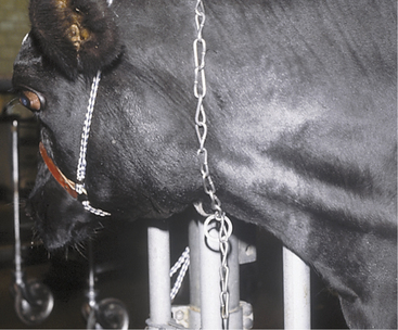


Taking the arterial pulse may be helpful in the assessment of cardiac disease. The middle coccygeal artery is the first artery palpated for pulsation during the physical examination. The facial artery is utilized when treating recumbent (hypocalcemic) cattle, and the median artery is the most convenient to palpate when performing simultaneous cardiac auscultation and pulse monitoring. Pulse rate, rhythm, and quality should be assessed. Pulse quality implies considerations of the size, strength, and duration of the pulse wave and distention of the artery. Most cattle with heart failure have decreased pulse strength, unevenness of the pulse, increased pulse rate, or a pulse rate that is different than the heart rate. Abnormalities in pulse rate or rhythm should alert the examiner to the possibility of cardiac arrhythmias.


Proficiency at auscultation of the heart requires some basic knowledge, willingness to auscult both sides of the thorax carefully during every physical examination, and patience. Many cattle object to stethoscope placement over the sites on the chest wall necessary for cardiac auscultation and will adduct the forelimb tightly against the thorax. This is noticed especially on examining the right side, where cardiac auscultation in cattle requires the stethoscope to be placed very cranial in the axillary area around the third intercostal space. Dairy bulls and large or fat cows have thick chest walls that reduce the intensity of heart sounds. Heart sounds are easier to hear on the left side of normal cattle. The pulmonic valve region is best heard in the left third intercostal space at a level between the shoulder and elbow. The aortic valve region near the heart base is best heard in the left fourth intercostal space at approximately shoulder level. The mitral (left AV) valve region coincides with the cardiac apex and is best heard at the left fifth intercostal space just above the elbow. The right AV (tricuspid) valve is heard far forward in the right third intercostal space at a level halfway between elbow and shoulder.


Although clinicians generally discuss two heart sounds in normal cattle, it is possible to hear four heart sounds in some cattle as it is with horses. Although the potential for four heart sounds is somewhat confusing and may be impossible to differentiate in most clinical patients, examiners should be aware of these facts. The first heart sound (S1) heralds the beginning of systole, is associated with the final halting of AV valve motion after closing and is best heard at the apex regions coinciding with AV valves in the cow. A slight splitting of S1 into separate mitral and tricuspid valve components is possible but is rarely audible in normal cattle. S1 tends to be of lower frequency and longer duration than S2.


S2 usually is not as loud as S1 and coincides with aortic and pulmonic valve closure. Current theory suggests that valve closing sounds associated with the generation of S2 result from the sudden halt in valve motion when it closes. Asynchronous closure of the aortic and pulmonic valves results in audible splitting of S2 in many normal cattle, especially during the inspiratory phase of the respiratory cycle.


Although S1 and S2 comprise the major heart sounds for cattle, S3 and S4 have been described. Ventricular vibrations at the end of rapid filling in early diastole are thought to cause S3, a low frequency sound seldom heard in cattle. S4 sometimes is heard late in diastole and is related to atrial contraction. In cattle with tachycardia, it has been suggested that S4 may in fact closely precede S1 and be mistaken for a split S1. The tripling or quadrupling of heart sounds that resembles a horse’s cantering gait is commonly referred to as a gallop rhythm and occurs in the higher range of normal heart rates or when tachycardia exists in some cows. Gallops are diastolic sounds related to atrial contraction (S4 gallop), to ventricular filling (S3), or to both (summation gallop). A prominent and persistent gallop rhythm in a cow with tachycardia may be the first indication of heart disease.


The heart rate of normal cattle is 60 to 84 beats/min. Neonatal calves may have normal heart rates as high as 110 to 120 beats/min, but frequently heart rates this high are brought about by the excitement of being handled or in anticipation of being fed. Not everyone agrees on the aforementioned range of normal heart rates for cattle, and several points should be addressed regarding this topic. Oxen and fat, persistently dry cows used only for embryo transfer may have a slower metabolism than lactating dairy cattle. Therefore somewhat like draft horses, these cattle may have heart rates at the low end of the normal range or even less than 60 beats/min. Conversely healthy but excited, nervous, or aggressive cattle may have heart and pulse rates more than 84 beats/min when approached by any examiner. Therefore the range of 60 to 84 beats/min really is an average and must be interpreted in light of the patient, its surroundings, and its intended use. Following the work of McGuirk et al with fasted cattle, a low normal range of 48 beats/min has been proposed. However, fasted healthy cattle seldom are encountered in the world outside of academic settings, and veterinarians are not frequently asked to examine healthy fasted cattle. An exception may be a cow off feed secondary to the classic broken water cup syndrome because she will become anorectic secondary to water deprivation. Sick cattle seldom have a heart rate less than 60 beats/min only because they are anorectic. Sick cattle that do have heart rates less than 60 beats/min usually have a vagal nerve-mediated bradycardia. Therefore 60 to 84 beats/min is still our preferred normal range for heart rate in adult dairy cattle.


Excited or nervous cattle may have an increased intensity or loudness of the heart sounds in addition to an increased heart rate. Other conditions that increase the intensity of heart sounds may be relative or pathologic. Relative factors include thin body condition, younger animals with thin chest walls, and excitement. Pathologic factors include anemia, the “pounding” heart rate sometimes heard in cattle with endocarditis, and displacement of the heart to a position closer to the thoracic wall by a diaphragmatic hernia or an abscess or tumor in the contralateral hemithorax. “Muffling,” or decreased intensity of heart sounds, may occur for relative reasons such as the increased thickness or fat on the chest wall of adult bulls or heavily conditioned cattle. Muffling also results from pathologic conditions such as pericarditis, pneumomediastinum, diaphragmatic hernia, and displacement of the heart toward the opposite hemithorax by an abscess or tumor in the hemithorax being ausculted. Cattle in shock may have either decreased or increased intensity of heart sounds, depending on the duration and severity of the condition. “Shocky” cows that are weak but still ambulatory tend to have increased intensity of heart sounds, whereas those that are recumbent or moribund have decreased intensity.


Auscultation combined with percussion provides the best subjective means to estimate the position and size of the heart. Heart sounds may radiate over a wider area than normal when transmitted by consolidated lung lobes or pleural fluid or when there is cardiac enlargement.


In calves and thin adult cattle, palpation of the apex beat is possible around the left fourth or fifth intercostal spaces at a level halfway between the elbow and shoulder. Palpation of an apex beat on the right side of adult cattle seldom is possible unless profound cardiac disease or displacement of the heart to the right by space-occupying masses has occurred. Deep palpation with the fingertips over the intercostal regions overlying the heart may elicit a painful response in conditions such as endocarditis, pleuritis, traumatic reticuloperitonitis, and rib fractures.


As in other species, bovine heart murmurs are classified based on intensity and timing. Intensity may be ranked subjectively on a 1 to 6 basis, with 1 of 6 being a faint, barely detectable murmur; 2 of 6 as soft but easily discernable; 3 of 6 as low to moderate intensity; 4 of 6 moderate but lacking a thrill; 5 of 6 loud with palpable thrill; and 6 of 6 so loud that it can be heard with the stethoscope off the chest and evincing a palpable thrill. Classification relative to timing of the cardiac cycle further defines murmurs as systolic, diastolic, or continuous. Further division is provided by terms such as “early systolic” or “holosystolic.” In general, systolic murmurs in cattle reflect AV valve insufficiency or, much less commonly, aortic or pulmonic stenosis, whereas diastolic murmurs reflect aortic or pulmonic valve insufficiency or rarely AV valve abnormalities. Benign systolic murmurs occasionally are heard in excited, tachycardiac calves or cows with anemia, hypoproteinemia, or in those being given rapid intravenous (IV) infusions of balanced fluids. Pathologic systolic murmurs most commonly are found in calves with congenital heart abnormalities such as ventricular septal defect or tetralogy of Fallot and in adult cows with endocarditis. Continuous murmurs are rare but may be encountered in calves having a patent ductus arteriosus or in cows with pericarditis. The point of maximal intensity for each cardiac murmur may add subjective data as to the valve involved in the cardiac abnormality.


Arrhythmias may be benign, pathologic, or secondary to metabolic disturbances in cattle. Sinus bradycardia and arrhythmia have been confirmed in cattle held off feed, in hypercalcemic adult cattle, and in hypoglycemic or hyperkalemic young calves. Sinus tachycardia may result from excitement, pain, hypocalcemia, and various systemic states such as endotoxemia and shock. Cattle with severe musculoskeletal pain often have normal heart rates while recumbent but have tachycardia when forced to rise and stand. Persistent tachycardia should be considered abnormal and may reflect cardiac disease unless other systemic conditions coexist.


Hyperkalemia may cause a variety of arrhythmias and is most commonly observed in neonatal calves that develop acute metabolic acidosis associated with secretory diarrhea caused by Escherichia coli or acute diffuse white muscle disease. Atrial standstill and other arrhythmias have been documented in diarrheic calves having metabolic acidosis and hyperkalemia. Extreme hyperkalemia (.7.0 mEq/L) may lead to cardiac arrest and should be corrected immediately, especially in calves that may require general anesthesia. Because severe hyperkalemia may be associated with pathologic bradycardias, even without confirmatory blood work, the experienced clinician should be alert to the therapeutic need for fluids that will specifically address hyperkalemia in severely dehydrated, diarrheic calves with discordantly low heart rates for their systemic state. Calves with white muscle disease also may have direct damage to the myocardium, which may be manifested by arrhythmias, murmurs, or frank cardiac arrest. Hypokalemia and hypochloremia in cattle with metabolic alkalosis may predispose to the most common arrhythmia of adult cattle—atrial fibrillation.


Hypocalcemia may be present or contribute to cattle having abdominal disorders that lead to metabolic alkalosis. Metabolic alkalosis may be a factor that triggers atrial fibrillation in cattle with normal hearts. Atrial fibrillation causes an irregularly irregular rhythm, with a rate that may be normal or increased (88 to 140 beats/min), depending on the presence of heart disease or the underlying predisposing condition. Atrial fibrillation is associated with irregular intensity of heart sounds. A pulse deficit may be present in any cow with a rapid or irregular cardiac rhythm, especially when the rate exceeds 120 beats/min. Atrial premature complexes (APC) may also occur in cows with gastrointestinal disease and electrolyte abnormalities. APC may preceed or immediately follow atrial fibrillation in some cows. Variation in intensity of the first heart sound during auscultation is characteristic of APC.


Other causes of arrhythmia in adult cattle include sporadic cases of lymphosarcoma with significant myocardial infiltration often causing atrial fibrillation, and ventricular or atrial arrhythmias associated with septic or toxic myocarditis. IV administration of calcium solutions is the major drug-related cause of arrhythmias in cattle, but intravenous administration of antibiotics or potassium-rich fluids occasionally prompts transient arrhythmias.


Sounds ausculted in pericarditis patients are variable, often confused with murmurs, and tend to change on a daily basis if affected cattle are available for daily reevaluation. Classic pericardial “friction” rubs occur at different stages of each cardiac cycle unlike murmurs, which tend to occur at a distinct phase of each cardiac cycle. Squeaky sounds, often similar to that made in compression of a wet sponge, may be heard as a result of pericardial disease. Rubs caused by contact between fibrin on the visceral and parietal pericardium also may be heard. The heart sounds tend to be muffled, and either free fluid or fluid-gas interfaces may lead to splashing or tinkling sounds or to complete muffling of all sounds. During the acute phase of traumatic reticulopericarditis, the character of the sounds tends to change each day. In those with subacute or chronic disease, muffling of the heart sounds or distinct tinkling or splashing tends to be consistently present.


Presence of an arrhythmia or murmur alerts the examiner that the heart may be abnormal. However, heart failure may or may not be present. In cattle, right heart failure is more common than left heart failure. The general signs of right heart failure include:


In addition to the general signs, specific cardiac signs such as a murmur, arrhythmia, or abnormal intensity of heart sounds usually are present and contribute to the diagnosis. Probably the most difficult set of differential diagnoses involves diseases that result in hypoproteinemia. Hypoproteinemia also causes ventral edema and may cause exercise intolerance and tachycardia. However, hypoproteinemia would not cause jugular and mammary vein distention and pulsation. Therefore venous distention and pulsation coupled with abnormal heart sounds or rhythm are the key signs when diagnosing heart failure in dairy cattle.


Left heart failure causes dyspnea, pulmonary edema, and exercise intolerance and may lead to cyanosis and collapse or syncope. Specific left heart failure seldom occurs in cattle, but left side failure combined with worsening, antecedent right heart failure may develop as the animal progresses into fulminant congestive heart failure.


Ancillary Procedures


Electrocardiography


The electrocardiogram (ECG) is essential for definitive categorization of arrhythmias in cattle. Vector analysis of ECG tracings to determine cardiac chamber enlargements and other pathology seldom is used in cattle because ventricular myocardial depolarization tends to be rapid and diffuse rather than organized, as in some other species. ECG is also indicated when cattle have variation in heart sound intensity, require monitoring for anesthesia or treatment of cardiac arrhythmias, or show signs of heart failure. Cardiac ultrasound, however, has superseded the ECG as a diagnostic tool in determining chamber enlargement and other cardiac pathology.


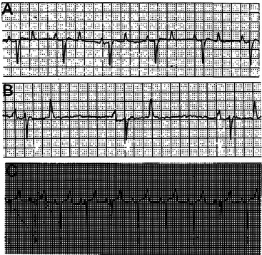
The base-apex lead system is most commonly used in cattle. The base-apex lead system results in an ECG with large wave amplitude and is sufficient for evaluating most arrhythmias. The positive electrode is placed on the skin over the left fifth intercostal space at the level of the elbow; the negative electrode is placed on the skin over the right jugular furrow roughly 30 cm from the thoracic inlet; and the ground electrode is attached to the neck or withers. The resultant ECG recorded through the base-apex lead system has a positive P wave with a single peak, a QRS complex with an initial positive deflection followed by a large negative deflection, and a variable (positive or negative) T wave (Figure 3-3, A to C).


Echocardiography


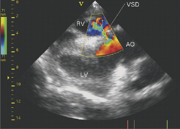
Two-dimensional echocardiography and Doppler echocardiography have greatly enhanced our ability to assess cardiac function and visualize anatomic variations and pathologic lesions in cattle. Valvular, myocardial, pericardial, congenital, and acquired lesions can be visualized in real time, measured, and monitored. Qualitative and quantitative assessment of the impact of congenital anomalies and monitoring treatment response of endocarditis, pericarditis, or other myocardial lesions are possible with the appropriate equipment and people trained to conduct and interpret a systematic cardiac examination. In short, echocardiography is now an essential component of a cardiology workup. Sector scanners utilizing a 3.5-mHz (or lower) transducer are most useful for adult cattle. A 5.0-mHz transducer may be helpful when evaluating neonatal calves suspected of having congenital anomalies or other cardiac conditions. Although some clinicians may lack the equipment or expertise with echocardiography, current graduates are being trained in the technique, and continued competition among manufacturers may allow more veterinarians to own this equipment. In any event, referral for echocardiography is indicated for valuable cattle whenever cardiac disease is apparent but a specific diagnosis is lacking. In most cattle, a systematic examination can be conducted from the right parasternal window to provide a long axis four-chamber view of the heart, a long axis view of the left ventricular outflow tract, and a short axis view of the left ventricle just ventral to the mitral valve and at the papillary muscle level. From the same window, all four heart valves can be visualized, chamber sizes can be measured, myocardial functional measurements can be made, and abnormalities of the pericardium can be seen. Congenital lesions like ventricular septal defect (VSD) (Figure 3-4) and acquired pericarditis (Figure 3-5) are easily visualized by echocardiogram.


SPECIFIC CARDIAC DISEASES IN CALVES


White Muscle Disease


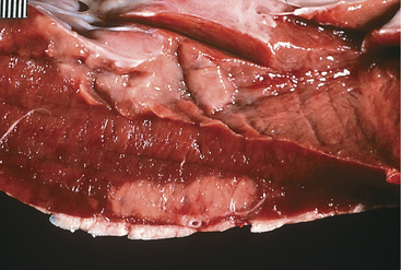
Myocardial damage from vitamin E and selenium deficiency may occur at any site in the heart and may be focal, multifocal, or diffuse (Figure 3-6). Signs may develop at any time from birth to 4 years of age but are more common in calves less than 3 months of age. Specific cardiac signs are variable and include arrhythmias, murmurs, exercise intolerance, cyanosis, dyspnea, congestive heart failure signs, and acute death. Signs may be subtle or dramatic, depending on the magnitude and locations of myocardial damage. Sudden death can occur spontaneously or following exercise or restraint. Other signs of white muscle disease such as stiffness, difficulty in prehension or swallowing, inhalation pneumonia, and myoglobinuria may or may not be present. Dyspnea may be directly related to the cardiac lesions or may be caused by Zenker’s degeneration in the diaphragm or intercostal muscles. Tachycardia (.120 beats/min) and arrhythmias are the most common specific cardiac signs, but murmurs may be present as well.


Diagnosis can be confirmed by measuring blood selenium values, urine dipstick testing to look for positive “blood” (myoglobin) and protein, and serum biochemistry to evaluate creatine kinase (CK) and aspartate aminotransferase (AST) enzymes. If the heart is the only muscle involved, serum enzymes may not be greatly elevated; however, the heart seldom is the only area involved.


Treatment should be instituted immediately with vitamin E and selenium injected at the manufacturer’s recommended dosage. Although some commercial preparations include label instructions that include IV use, it is suggested that vitamin E/selenium be given intramuscularly (IM) or subcutaneously to avoid the occasional life-threatening anaphylactic-type reaction seen with these products. The calf should be kept in a small box stall, straw bale enclosure, or hutch, so it can move about but not run freely, lest further muscle damage be precipitated. If pulmonary edema is present, furosemide (0.5 to 1.0 mg/kg) may be given once or twice daily. Concurrent aspiration pneumonia would require intense antibiotic therapy. Vitamin E and selenium injections are repeated at 72-hour intervals for three or four total treatments. Herd selenium status and preventive measures to address the problem should be discussed. Calves that survive for 3 days following diagnosis have a good prognosis.


Hyperkalemia


Cardiac arrhythmias or bradycardia associated with hyperkalemia is primarily observed in neonates having severely acute diarrhea. Enterotoxigenic E. coli causing secretory diarrhea, metabolic acidosis, low plasma bicarbonate values, and hyperkalemia appears to be the most common causative organism. Rotavirus or coronavirus also may be involved in calf diarrhea, but they seldom produce as profound a metabolic acidosis as E. coli.


Less common causes of hyperkalemia include severe diffuse white muscle disease involving heavy musculature of the limbs, ruptured bladders, renal failure, urinary obstructions, and nonspecific shock.


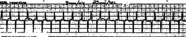
Hyperkalemia reduces the resting membrane potential, which initially makes cells more excitable, but gradually (with further elevation in potassium and further reduction in resting membrane potential) the cells become less excitable. Atrial myocytes seem more sensitive to these effects than those within the ventricles. Cardiac conduction is affected, and several characteristic ECG findings evolve in a typical sequence that correlates well with increasing K+ values: ECG changes include peaking of the T wave, shortening and widening of the P wave, prolongation of the PR interval, eventual disappearance of the P wave, widening of the QRS complex, and irregular R-R intervals (Figure 3-7). Atrial standstill characterized by bradycardia and absence of P waves may occur and has been documented in association with hyperkalemia in diarrheic calves. Further progression may lead to AV block, escape beats, ventricular fibrillation, asystole, and death.


In neonates, hypoglycemia is the major differential diagnosis when bradycardia is present. Septic myocarditis or white muscle disease also may be considered if an arrhythmia is present.


Calves less than 2 weeks of age that have developed acute diarrhea, are recumbent, dehydrated, and have bradycardia or arrhythmia should be suspected of being hyperkalemic. Obviously only an acid-base and electrolyte analysis of blood and an ECG can confirm this. However, these may not be available in the field. The consequences of underestimating the life-threatening importance of the heart and K+ relationship in these patients are severe.


Calves suspected to be hyperkalemic based on history, physical signs, and arrhythmia or bradycardia should receive alkalinizing fluids and dextrose. Being neonates, hypoglycemia may contribute to bradycardia when this sign is present. One way to treat metabolic acidosis and hyperkalemia is by IV infusions of 5% dextrose solution containing 150 mEq NaHCO3/L. Usually 1 to 3 L is necessary, depending on the magnitude of the metabolic acidosis and bicarbonate deficit. Glucose and bicarbonate help transport K+ back into cells, and the glucose also treats or prevents potential hypoglycemia. Once the acute crisis has been resolved, the calf may be safely treated with balanced electrolyte solutions containing potassium. Calves with diarrhea, despite having plasma hyperkalemia, have total body potassium deficits and require potassium supplementation. This may be true even in the acute phase of disease, but when serum K+ is 5.0 to 8.0 mEq/L there is no time to worry about a “total body potassium deficit.” We have treated hundreds of calves as suggested above, and those with a venous blood pH of 7.0 or greater have a good to excellent prognosis unless they have had failure of passive transfer of immunoglobulins and subsequent septicemia. Specific insulin therapy as an adjunct to bicarbonate and glucose to correct hyperkalemia is not necessary in calves.


Congenital Heart Disease


Virtually all types of congenital cardiac anomalies occur in cattle. Most congenital anomalies appear to be sporadic, but inheritance may play a part in some of the most common anomalies. The most common congenital anomalies in cattle appear to be VSDs (Figure 3-8), tetralogy of Fallot, atrial septal defects, and transpositions of great vessels.


Most congenital cardiac defects cause distinct murmurs. Calves affected with the most common defects such as VSDs, atrial septal defects, tetralogy of Fallot, or aortic or pulmonic stenosis usually have systolic murmurs. Patent ductus arteriosus, which is rare as a single defect in calves, can cause a systolic or continuous murmur.


Most calves with congenital cardiac defects appear normal at birth but eventually are noticed to have dyspnea, poor growth, or both. Many calves with congenital heart defects are eventually examined by a veterinarian because of persistent or recurrent respiratory signs or generalized ill thrift. The respiratory signs may be real in the form of pulmonary edema associated with heart failure and shunts or be caused by opportunistic bacterial pneumonia secondary to pulmonary edema and compromise of lower airway defense mechanisms. The owners may already have treated the calf one or more times for coughing, dyspnea, and fever, only to have the signs recur. Usually only one calf is affected, thus making enzootic pneumonia an unlikely diagnosis. Regardless of whether pulmonary edema or pneumonia plus pulmonary edema are present, veterinary examination usually detects the cardiac murmur that allows diagnosis. Venous pulsation and distention of the jugular veins may be present, but calves seldom show ventral edema as distinctly as adult cattle with heart failure.


Calves with congenital heart defects that do not develop respiratory signs usually show stunting compared with herdmates of matched age. The degree of stunting varies directly with the severity of the congenital lesions in regard to blood oxygenation but usually becomes apparent by 6 months of age and is very dramatic in calves that survive to yearlings. Some cattle with small defects survive and thrive as adults, but this is rare.


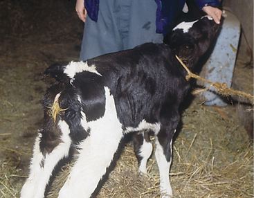
VSDs are the most common defects in dairy calves and are found in all breeds. In Guernseys and Holsteins, VSD may be linked to ocular and tail anomalies. Microphthalmos and tail defects, including absence of tail, wry tail, or short tail, frequently signal VSD (Figure 3-9). Sometimes ocular, tail, and cardiac defects all are present in the same calf, but it is more common to find tail or ocular pathology plus VSD. Depending on the size of the VSD, affected calves have a variable life span. Prognosis for most is hopeless because of eventual respiratory difficulty and stunting. However, calves do, in rare instances, survive to productive adult states. The genetics of these multiple defects (eye, tail, and heart) have not been investigated in Holsteins but have been assumed to be a simple recessive trait in Guernseys.


Tetralogy of Fallot and other multiple congenital defects that allow right to left shunting of blood provoke marked exercise intolerance, cyanosis, and dyspnea and may lead to polycythemia secondary to hypoxia. Prognosis for long-term survival is grave in these calves. Ectopia cordis in a calf creates a dramatic sight, with the heart beating under the skin in the neck, but is extremely rare.


SPECIFIC CARDIAC DISEASES IN ADULT CATTLE


Neoplasia


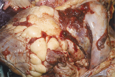
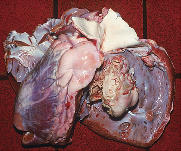
The heart is one of the common target sites of lymphosarcoma in adult dairy cattle. Many cattle with multicentric lymphosarcoma have cardiac infiltration based on gross or histologic pathology, but fewer of these cattle have clinically detectable cardiac disease. When the heart is a major target site, cardiac abnormalities are more obvious. The heart seldom is the only organ affected with lymphosarcoma. Therefore detection of cardiac abnormalities coupled with other suspicious lesions (e.g., enlarged peripheral lymph nodes, exophthalmos, melena, and paresis) simply helps to make a lymphosarcoma diagnosis more definite.


Depending on the anatomic location and magnitude of the tumors, cattle with cardiac lymphosarcoma may have arrhythmias, murmurs, jugular venous distention, jugular venous pulsations, or muffling caused by diffuse cardiac or pericardial involvement. Muffling and splashing sounds are possible if a pericardial transudate or exudate is present. The most common site of tumor involvement is the right atrium, but nodular or infiltrative tumors can be found anywhere in the myocardium or pericardium (Figure 3-10). The color and consistency of the tumors may vary. Mediastinal lymph nodes also are commonly involved. Cattle with signs of heart disease should be thoroughly examined for other lesions consistent with lymphosarcoma. When multiple lesions exist, the diagnosis is easy. However, cattle examined because of vague signs such as hypophagia and decreased milk production that are found to have tachycardia or other cardiac abnormalities can present diagnostic challenges. Although ECG and thoracic radiographs have seldom helped make a definitive diagnosis, ultrasound may be very helpful to image nodular or large masses of lymphosarcoma. Thoracocentesis and pericardiocentesis to obtain fluid for cytologic evaluation are the most helpful ancillary aids when cardiac lymphosarcoma is suspected. A complete blood count (CBC) and assessment of bovine leukemia virus (BLV) antibody status are indicated, but a positive BLV agar gel immunodiffusion (AGID) or radioimmunoassay (RIA) test does not ensure an absolute diagnosis because most positive cattle never develop tumors (see the section on Lymphosarcoma in Chapter 15). Therefore simply assuming a cow with a positive BLV test and heart abnormality detected on physical examination has lymphosarcoma may be an incorrect assumption. A double line-positive BLV-AGID may add further weight to the suspected diagnosis, as would the finding of a persistent lymphocytosis in a set of CBC results. Clinical identification of masses in other locations or cytology from thoracocentesis or pericardiocentesis provides the best means of definitive diagnosis.


Fever usually is absent in cattle with cardiac lymphosarcoma. Occasional cattle with large tumor masses in the thorax or abdomen may have fever because of tumor necrosis or nonspecific pyrogens produced by neoplasms. Secondary bacterial infections of the lungs or other body systems also may lead to fever, which confuses the diagnosis.


Prognosis is hopeless for cattle with cardiac lymphosarcoma, and most cattle with the disease die from cardiac or multisystemic disease within a few weeks to a few months. Successful attempts at chemotherapy have not been reported to our knowledge. One author has successfully prolonged life up to 6 months in a few cattle with cardiac lymphosarcoma that had significant pericardial fluid accumulations by intermittent pericardial drainage. Occasionally valuable cattle may justify such treatment to allow a pregnancy to be completed or to be superovulated. However, as with many catabolic conditions, owners should be cautioned that maintaining the dam with advanced heart disease for more than a few weeks may produce a gestationally dysmature fetus even if the pregnancy is carried to term, or may seriously affect the cow’s ability to superovulate or even produce viable oocytes for in vitro fertilization. There is also the risk of vertical transmission of BLV from dam to fetus in utero, which is greater should the dam have clinically apparent tumors. In one late pregnant cow with severe pericardial effusion caused by lymphosarcoma, we were able to maintain the cow for several weeks by surgically opening the pericardial sac into the pleural cavity, which significantly improved venous return to the heart and the overall condition of the cow. The cow was also treated with isoflupredone.


Neurofibroma, although uncommon, frequently causes arrhythmia and variable intensity of heart sound in affected cattle and bulls. Further the cardiac arrhythmia may coexist with paresis or paralysis caused by neurofibroma masses in the spinal canal. Because lymphosarcoma more commonly causes paresis coupled with cardiac disease, this combination of signs is most suggestive that lymphosarcoma is present. Although perhaps a moot point because both diseases are fatal, further medical workup of neurofibroma patients fails to provide confirmation of lymphosarcoma. To date, postmortem examination has been the only means of definitive diagnosis for cardiac neurofibroma. Examiners talented in ultrasound may be able to diagnose these lesions based on the typically gnarled, raised cords of tumor involving the cardiac nerves.


Myocardial Disease


Infections


Septic Myocarditis.


Neonatal septicemia caused by gram-negative bacterial organisms, acute infection with Histophilus somni, and chronic infections in any age of cattle resulting from Arcanobacterium pyogenes are the most common cause of septic myocardial lesions in cattle. Septicemic calves, calves suspected of having H. somni infection, or calves with chronic infections should be suspected of having septic myocarditis if an arrhythmia or other signs of abnormal cardiac function develop during their illness. White muscle disease, hyperkalemia, and hypoglycemia should be considered. Septicemic calves have a guarded prognosis, and septic myocarditis worsens it. Septic myocarditis foci in adult cattle with chronic, active infection or abscesses associated with mastitis, localized peritonitis, foot lesions, or chronic pneumonia are more commonly identified by pathologists than clinicians. Although tachycardia is likely to be present, this finding often is assumed to result from the primary illness rather than from myocarditis. As with calves, adult cattle with septic myocarditis may have paroxysmal cardiac arrhythmias that alert the clinician to the diagnosis. Definitive diagnosis has been difficult in the living patient because test for cardiac muscle enzymes may lack specificity for cardiac muscles. Increased concentration of troponin I may be used to help diagnose myocardial disease. An ECG showing atrial or ventricular premature depolarizations in a calf or cow with evidence of sepsis or a walled-off infection can be used to lend credence to the diagnosis.


Treatment of the primary disease remains the most important part of managing septic myocarditis. If the primary problem and myocardial lesion can be sterilized, the heart may return to normal function.


Septic myocardial disease of adult cattle, as in calves, usually follows septicemia or chronic infections. Septicemic spread of infectious organisms, thrombi, or mediators of inflammation may be involved in the pathophysiology of myocardial injury that occurs in septic cattle. Although relatively uncommon, development of persistent tachycardia with or without an arrhythmia in a patient with infectious disease may suggest myocarditis. Tachycardia is so nonspecific that most examiners attribute the tachycardia to the primary disease rather than secondary myocarditis. Only when the myocardial damage causes signs of heart failure does a diagnosis of myocarditis become easier. Acute death is possible. Arrhythmia, if present, must be assessed using ECG and blood electrolytes and acid-base status to rule out atrial fibrillation associated with metabolic abnormalities. Dairy cattle are most at risk for myocarditis with acute septic diseases such as severe mastitis, metritis, pneumonia, and infection caused by H. somni. Occasional cases also occur secondary to chronic localized infections such as digital abscesses that predispose to bacteremia. Depending on the size and location of the myocardial lesion, clinical signs range from subclinical to overt heart failure. ECG evidence of ventricular arrhythmias would suggest myocardial damage, but supraventricular arrhythmias are possible as well. Unfortunately definitive premortem diagnosis is impossible without advanced echocardiographic or invasive cardiac technique. Treatment must be directed at the primary disease. Minor myocardial lesions away from nodal and conduction tissue may heal or fibrose asymptomatically, whereas large or multifocal lesions may lead to heart failure, persistent tachyarrhythmia, or sudden death.


Toxins


Ionophores such as monensin and lasolocid are capable of damaging myocardial and skeletal muscle when ingested in toxic amounts. Improper mixing of ionophores into rations is the most common error that may lead to toxicity, but accidental exposure to concentrated products also is possible. Obviously this is a potential concern for calves and heifers being fed milk replacer or feeds containing ionophores. Fortunately cattle are much more resistant to the toxic effects of ionophores than are horses, but there is a narrow margin of safety, especially in young calves.


Many poisonous plants are theoretically capable of myocardial injury, but in reality few are likely because of increased confinement of heifers and adult cattle. Eupatorium rugosum (white snakeroot), Vicia villosa (hairy vetch), Cassia occidentalis (coffee senna), Phalaris sp., and others are capable of toxic myocardial damage. Gossypol also is capable of causing myocardial damage when fed in toxic amounts. This fact is of special concern given the increased incidence of feeding cottonseed to dairy cattle. Copper deficiency, especially when chronic, occasionally has been linked to acute myocardial lesions, resulting in death (“Falling disease” in Australia). Many other organic and inorganic toxins have the potential for causing myocardial damage but create more obvious pathology in other body systems and thus will not be discussed here.


No specific treatment is available for toxic myocarditis. Common sense dictates identification and removal of the toxin from the environment, alongside immediate administration of laxatives, cathartics, and/or protectants to decrease absorption and accelerate intestinal transit. Vitamin E and selenium administration and specific supportive treatment for cardiac disease should be instituted, but the prognosis for animals already with congestive heart failure is grave.


Parasitic and Protozoan Infections


Cysticerca bovis may cause myocardial lesions, but these appear rarely in dairy cattle in the northeastern United States. This is the larval form of Taenia saginata, the common human tapeworm. Contamination by human sewage of feedstuffs, pastures, or fields puts cows at risk for this disease.


Although Toxoplasma gondii is capable of infecting cattle, the clinical disease appears rare because cattle rapidly eliminate parasites from tissue. Cattle are exposed to and infected by T. gondii via ingestion of feedstuffs contaminated by cat feces.


Sarcocystis sp. is a common cause of myocardial disease in cattle. Although most infestations are asymptomatic, clinical illness characterized by hemolysis, myopathy, myocarditis, weight loss, rattail, and other signs is possible. Sarcocystis sp. requires two hosts, and carnivores or humans usually are the hosts that shed sporocysts in fecal material that subsequently contaminates cattle feed (see Chapter 15). Cattle then become the intermediate host as intermediate stages of the parasite invade endothelial cells and later stages encyst in muscle—including the myocardium. Subsequent ingestion by carnivores of beef containing cysts continues the cycle.


Histopathologic identification of sarcocystis cysts in myocardium of cattle is very common but seldom deemed significant. Certainly, however, heavy exposure to the organism could provoke significant myocardial damage.


Parasitic or protozoan myocarditis usually requires histopathology or serology for diagnosis. Treatment would be best provided by preventive measures to avoid contamination of cattle feeds by carnivore or human feces.


Inherited Myocardial Disease


A dilated cardiomyopathy has been described in Canadian Holstein-Friesians. This condition appears in Canadian black and white Holsteins and expresses itself as heart disease between 19 and 78 months of age. Although most cattle develop clinical signs within 4 years of birth, some have lived for 6 to 7 years.


Most cases are presented because of signs referable to heart failure such as ventral edema, exercise intolerance, inappetence, dyspnea, tachycardia, muffled heart sounds, and jugular and mammary vein distention and pulsation. Although tachycardia is fairly consistent, other auscultation findings such as arrhythmias, murmurs, or varying intensity of the heart sounds vary in each case. Hepatomegaly consistent with chronic passive congestion of the liver secondary to right heart failure also was present in some patients.


Echocardiography and ECG recordings are required for diagnosis. Ultrasound is the best aid to confirm dilated cardiomyopathy.


Long-term prognosis is hopeless, but affected cattle may be helped in the short term by management with cardioglycosides, and furosemide is indicated if pulmonary edema exists. McGuirk suggests digoxin at 0.86 mg/kg/hr as an IV infusion. This obviously requires diligence, IV catheterization, and hospitalization, or else a very attentive owner. Alternatively, 3.4 mg/kg IV every 4 hours may be utilized but creates greater variation in blood levels and increases the risk of digoxin toxicity. Furosemide is used at 0.5 to 1.0 mg/kg twice daily if pulmonary edema is present. Inappetent cattle may benefit from 50 to 100 g KCl orally each day to maintain potassium levels when being treated with digoxin. Ideally daily or every other day blood acid-base and electrolyte status should be assessed.


Endocarditis


Etiology


Bacterial endocarditis is the most common valvular disease or endocardial disease in adult dairy cattle. It also is one of the few treatable heart conditions of cattle. Therefore early suspicion, diagnosis, and appropriate treatment improve the prognosis.


Cattle with chronic infections such as septic musculoskeletal conditions, hardware disease, abscesses, lactic acid indigestion, chronic pneumonia, metritis or mastitis, and thrombophlebitis are at risk for bacterial endocarditis. In addition, cattle with long-term IV catheters have increased risk of endocardial infections. Bacteremia appears essential to the pathophysiology of bacterial endocarditis in cattle.


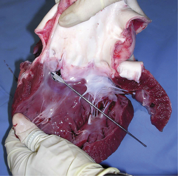
A. pyogenes is the most common organism isolated from the blood and endocardial lesions of cattle affected with endocarditis, but Streptococcus sp., Staphylococcus sp., and gram-negative organisms may also cause the disease. The right AV valve (tricuspid) is the most commonly infected valve with the left AV (mitral) being the second most common (Figure 3-11). Other valves or the endocardium adjacent to valves may also occasionally be the site of infection (Figure 3-12). Owner complaints regarding affected cattle include recurrent fever, weight loss, anorexia, poor production, and sometimes lameness.
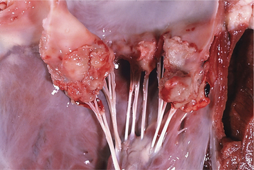


Laboratory Data


Nonregenerative anemia commonly results from chronicity of the primary infection, the endocardial infection, or both. Neutrophilia is common and was found in 24 of 31 cases in one report, whereas absolute leukocytosis was found in 14 of 31. In this same report, serum globulin values were greater than 5.0 g/dl in 19 of 23 endocarditis patients that had globulin measured. Elevated globulin was believed to be consistent with the chronicity of infection.


Blood cultures are an important diagnostic test, but echocardiography provides the definitive diagnosis. A patient suspected of having endocarditis should have a series of blood cultures submitted rather than a single time-point sample. Although blood cultures in adult cattle may be negative in as many as 50% of endocarditis patients tested, isolating the causative organism from the bloodstream provides the best opportunity for appropriate and successful treatment with a specific antibiotic. Venous blood cultures should be collected after the jugular vein has been clipped and prepared aseptically. The cow should have been held off systemic antibiotics for 24 to 48 hours before culture attempts, if possible. Although one blood culture attempt is better than none, it is preferable to obtain a series of three to four cultures when economics allow. The interval between collections of multiple samples has been debated by clinicians for decades. Some clinicians culture only during a fever spike, some at 3- to 30-minute intervals, some at 6- to 8-hour intervals, and some once daily. We prefer to obtain three cultures at 30-minute intervals in febrile patients and intervals of several hours in nonfebrile patients suspected of having endocarditis.


Diagnosis


Early signs of reduced appetite and production, fever, and tachycardia certainly are not specific for endocarditis. A pounding heart or systolic murmur should suggest the diagnosis and dictate further workup. Diagnosis may be overlooked because of more obvious primary problems such as abscesses, infected digit or other musculoskeletal infection, suspected hardware disease, or thrombophlebitis because these conditions may also cause fever and nonspecific signs of illness. Therefore heart murmurs, a pounding heart, or early signs of heart failure in addition to tachycardia merit consideration of a diagnosis of endocarditis. Lameness and stiffness may be difficult to differentiate from primary musculoskeletal disease or painful stance caused by peritonitis but can be important clinical signs that aid diagnosis. Because of fever, tachycardia, and sometimes polypnea, cattle having endocarditis often are misdiagnosed with pneumonia or traumatic reticuloperitonitis.


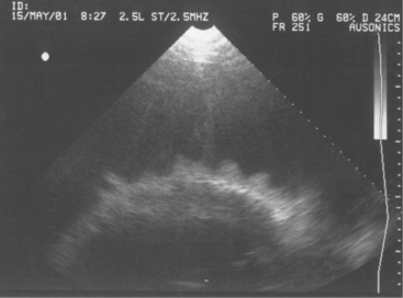
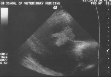
Diagnosis of endocarditis usually is based on the patient’s history and clinical signs. However, a positive blood culture and echocardiography allow definitive diagnosis. Blood cultures, as mentioned previously, may or may not be successful; however, when positive, they allow appropriate selection of antibiotics. Definitive diagnosis based on two-dimensional echocardiography has proven to be one of the most impressive uses of ultrasound since its widespread use in diagnostics began 10 years ago. Veterinarians trained in echocardiography now have a tool to confirm bacterial endocarditis in most patients (Figure 3-13).


Treatment


Long-term antibiotic therapy is required to cure bacterial endocarditis in cattle. Thus cattle selected for treatment must be deemed valuable enough to justify the cost of antibiotics and discarded milk that will be incurred. A successful blood culture allows selection of an appropriate antibiotic based on sensitivity or mean inhibitory concentration (MIC) values. Because endocarditis in cattle usually is caused by A. pyogenes or Streptococcus sp., some clinicians assume penicillin will work and do not bother to do blood cultures. This assumption would be a worthwhile gamble if economics dictate that laboratory costs be minimized.


Therefore penicillin and ampicillin are the drugs of choice for bacterial endocarditis in cattle caused by A. pyogenes and most Streptococcus sp. Although ceftiofur currently has the advantage of “no withdrawal,” it is more expensive and has been overused and abused by clinicians who hope the drug will cure all infections of dairy cattle. Penicillin (22,000 to 33,000 IU/kg twice daily) or ampicillin (10 to 20 mg/kg twice daily) is administered for a minimum of 3 weeks. If gram-negative organisms or penicillin-resistant gram-positive organisms are isolated from blood cultures, an appropriate bactericidal antibiotic should be selected based on MIC or antibiotic sensitivity testing.


Based on work by Dr. Ray Sweeney and others at the University of Pennsylvania, rifampin (rifamycin) has been shown to establish therapeutic blood levels after oral administration to ruminants. Unfortunately there is significant variability in blood levels between treated cattle, which may limit its treatment potential. Rifampin is a unique antibiotic that gains access to intracellular organisms or walled-off infections by concentrating in macrophages. Rifampin always should be used in conjunction with another antibiotic because bacterial resistance may develop quickly when the drug is used alone. The dosage is 5 mg/kg orally, twice daily for cattle. Although some maintain this dosage is too low, it has seemed effective clinically when used in conjunction with penicillin not only for chronic A. pyogenes endocarditis but also for pulmonary abscesses. Therefore if economics allow, oral rifampin has been reported to improve treatment success in cattle with bacterial endocarditis.


Occasionally cattle will become significantly anorectic while receiving rifampin (more so than was noted in association with the primary disease), but in many cases this apparent intolerance to the drug is overcome if administration is discontinued for several days and then reinstituted at the same or lesser dose.


In addition to antibiotic therapy, those cattle showing venous distention, ventral edema, or pulmonary edema require judicious dosages of furosemide. Because many endocarditis patients have reduced or poor appetites, overuse of furosemide may lead to electrolyte depletion (K+, Ca++) and dehydration. Therefore when furosemide is used, the drug should be administered on an “as-needed” basis, and 0.5 mg/kg once or twice daily usually is sufficient.


Because cattle with endocarditis often appear painful or stiff and may have either primary musculoskeletal disorders or secondary shifting lameness, aspirin is administered at 240 to 480 grains orally twice daily. Unfortunately aspirin does not appear to minimize platelet aggregation and is unlikely to prevent further enlargement of vegetative lesions. Free access to salt should be denied of cattle showing signs of congestive heart failure.


Treatment continues for a minimum of 3 weeks. Positive signs of improvement include increasing appetite and production, as well as absence of fever. The heart murmur persists and may vary as treatment progresses. Resolution of the heart murmur and tachycardia coupled with echocardiographic evidence of resolution of the endocarditis lesions are excellent prognostic signs. Many cows that survive are, however, left with persistent subtle or obvious heart murmurs caused by valvular damage. This should not be a concern as long as other signs indicate resolution of infection and heart failure is not present. Cattle with venous distention, ventral edema, or other signs of right heart failure have a worse prognosis than cattle diagnosed before signs of heart failure. However, mild to moderate signs of heart failure should not be interpreted to mean a hopeless prognosis because supportive treatment may alleviate these signs while antibiotic therapy treats the primary condition.


Prognosis for endocarditis patients is guarded. Sporadic case reports tend to highlight successfully managed individual cases, but further case series are necessary to suggest accurate recovery rates. Of 31 cattle affected with endocarditis that were admitted to our hospital between 1977 and 1982, 9 responded to long-term antibiotic (8 penicillin and 1 tetracycline) therapy. Based on these data and the experience of other clinicians, the prognosis is better when the diagnosis is made early in the course of the disease. Repeated echocardiographic examination allows for monitoring and reassessment of the valvular lesions during and after treatment. With experience and the correct software, ultrasound examination also allows for specific evaluation of cardiac function (e.g., atrial diameter, fractional shortening) that may more accurately assess the degree of cardiac dysfunction and provide valuable prognostic information.

Stay updated, free articles. Join our Telegram channel

Apr 26, 2017 | Posted by in GENERAL | Comments Off on Cardiovascular Diseases

Full access? Get Clinical Tree

Get Clinical Tree app for offline access