R. Avery Bennett, Katrin Baumgartner
Avian Deflighting Techniques
Over the years, various procedures have been developed to prevent flight in birds, either to improve their quality of life or to prevent their escape from public exhibits.33 By preventing flight, birds may often be allowed more freedom within the facilities. Birds on display in open-air exhibits may interact with conspecifics and with other species of birds as well as specimens of other taxa, thus creating a more realistic and natural display. Although housing birds in large, enclosed exhibits may also meet these goals, such arrangements tend to be very expensive and may not be an option for all institutions. It may be desirable to only temporarily render the birds unable to fly; however, in open exhibits, it may be more appropriate to take permanent measures to prevent flight. It is ideal to preserve as much as possible of the bird’s normal anatomy while taking steps to prevent flight. Unfortunately, most procedures used to deflight birds also change the appearance and function of the wing. In some countries, amputation of any body part of any animal other than for medical reasons is prohibited by law. This limits what procedures may be used to prevent flight even in birds in a zoologic setting.
The majority of the literature regarding techniques for preventing flight involves surgery, and such surgery has reportedly been performed without anesthesia or analgesia.1 Many of these articles report deaths caused by shock in some patients.
Any procedure that involves incision or transection of tissues must be considered painful and should be conducted under general anesthesia with appropriate postoperative analgesia for 2 to 5 days. In some cases, such as in chicks, a local anesthetic along with an analgesic and appropriate postoperative analgesia may be acceptable.
Most commonly, procedures are only performed on one wing to cause imbalance and thus make flight more difficult.1,16,34,36 A common comment made in many publications is that surgical deflighting procedures impair the bird’s ability to balance and that this may affect the ability of male birds to reproduce. Some reports in the literature describe that a given deflighting procedure does not affect the birds’ ability to groom and breed,1,34 and some others indicate that it does, indeed, affect the ability to breed.10,28 Interestingly, in one report, the author indicated that pinioning causes imbalance and inhibits breeding, but follicle extirpation, a procedure advocated in the study, does not.22 Intuitively, it seems that having no primary flight feathers on one wing would have the same effect on balance that pinioning would. The authors of this study were, however, unable to find scientific evidence to support these claims.
Preventing birds from flying has been discussed as a matter related to animal welfare for many years.17 Some species of birds such as waterfowl, gallinaceous birds, cranes, storks, and flamingos seem to do well even if they cannot fly, whereas other species such as hummingbirds, bee-eaters, swifts, swallows, and some raptors need flight to thrive. Small-sized aviaries expose birds to the risk of wing trauma.
Legal Issues
In zoologic collections, different deflighting techniques are used to control flight in birds. Some countries have legislation preventing any surgical procedure being performed for nonmedical reasons. Such legislative regulations are often limiting and not directed specifically toward zoo birds, so in many countries, zoo veterinarians are placed in a legal limbo. Because deflighting, including trimming wing feathers, involves removal of a body part or partial removal or destruction of tissue, the following legislations may be applied to any deflighting technique.
The German Animal Welfare Law states in paragraph 6 (1):
The complete or partial amputation of body parts or the complete or partial removal or destruction of organs or tissues of a vertebrate are prohibited.
Because this law pertains to all vertebrates, birds are not exempted from it. The amputation of parts of the body (pinioning), the complete or partial removal of tissue (tenotomy or tenectomy, follicle excision), and the destruction of tissue (laser or cryosurgical follicle ablation), and even flight feather trimming are all considered illegal, and veterinarians performing these procedures may be prosecuted. In Germany, exemptions, even for feather trimming, do not exist.
In Switzerland, legislation specifically against pinioning birds has been enacted.
The Swiss Animal Welfare Ordinance states:
Article 20: Prohibited actions in domestic poultry:
b. Trimming of the comb and wattles and the wings.
Article 24: Other prohibited actions:
b. Surgical procedures to make it easier to keep pets, such as resection of teeth, clipping of wings, or removal of secretory glands; procedures to prevent reproduction or the removal of dewclaws, are exempted.
The European Convention for the Protection of Pet Animals was opened for signature in Strasbourg on November 13, 1987, enforced on May 1, 1992. This Convention states: “Surgical operations … for other non-curative purposes shall be prohibited ….” At present, 15 of the 27 States in the European Union have ratified this Convention and have prohibited cosmetic surgical procedures. In addition, four European states have prohibited these operations, even though they did not ratify the Convention.
In the Swiss and European regulations, pet and poultry are included, but zoo birds are not. Nevertheless, some veterinary authorities in Europe consider deflighting amputation or tissue destruction and a modification of appearance; therefore, it is an illegal procedure. They cite article 10 of the European Convention to prohibit any deflighting techniques other than feather trimming. It has been suggested that these laws be changed with respect to zoo birds.27
In the United States, the Animal Welfare Act of 1996 regulates the treatment of animals in research institutions and exhibitions. Birds, rats, mice, farm-animals, and cold-blooded animals are excluded from the Animal Welfare Act.2
Anatomy and Physiology of the Wing4,7,38
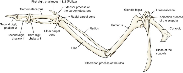
The anatomy and physiology of birds have been adapted to allow flight. The bones in the wing include the humerus, ulna, radius, radial carpal bone, ulnar carpal bone, carpometacarpus, and bones of the digits of the manus (digitus alularis, digitus major, and digitus minor) (Figure 65-1). The alula (digit II) has one or two phalanges and has an important aerodynamic function.
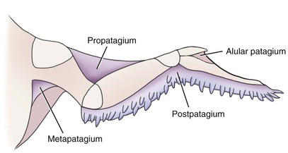
The wing has four patagia (Figure 65-2).7 The propatagium is the largest skinfold of the wing, and it fills the angle formed by the partially flexed elbow. It is composed of a network of multiple layers of collagen and elastin, which are suspended between the leading edge and the dorsal antebrachium of the wing.
The primary avian flight muscles are the pectoralis and the supracoracoideus.12,38 The pectoralis is a large muscle composed of primarily large fibers and is attached to the humerus at the deltopectoral crest. It may make up 35% of the bird’s total body weight.11 This muscle contracts during the downstroke and pronates the wing. The supracoracoideus is a smaller muscle with short fibers and originates from the sternum; its tendon of insertion passes through the triosseal canal to the dorsal surface of the humerus. This muscle elevates and supinates the wing during the upstroke. Both muscles, the pectoralis and the supracoracoideus, accelerate and decelerate the wing across the transition between downstroke and upstroke.38 The extensor carpi radialis originates from the medial epicondyle of the humerus, extends over the cranial surface of the carpal joint, and ends in the carpometacarpal extensor apophysis. This muscle extends the carpus and advances the primary flight feathers forward. The musculus pollicis brevis inserts craniodorsally on the base of the alula and abducts it or, if the musculus flexor pollicis is relaxed, raises it. The musculus pollicis longus originates on the proximal ventral surface of the ulna and the radius. The tendon of insertion attaches to the extensor process of the carpometacarpus. This muscle extends the carpus when the elbow is flexed. Two smaller muscles, the triceps brachii and the biceps brachii, control the wing’s shape by flexion and extension of the elbow.
Anatomy and Physiology of Remiges6
Feathers have different functions. The contour feathers include the feathers involved in flight (remiges) and the tail (rectrices). Remiges are divided into primary and secondary remiges. The primary remiges insert dorsally from the carpus and distally along the carpometacarpus and phalanges. In most species of birds, 10 primary remiges exist and are numbered from proximal to distal. The secondary remiges insert dorsally along the ulna and are counted from distal to proximal. They vary in number from six in hummingbirds to as many as 40 in some species. The primary and secondary remiges are not fixed rigidly in place, but they are able to twist during flight. The remiges are attached to feather follicles, which are specialized pockets of epidermal and dermal cells from which a new feather grows after the old feather is lost during molt by extrusion of the old feather from its base. When the feather is growing from the papilla of the follicle, it is called a blood feather because it has an active blood supply. As the feather matures, it loses its blood supply and becomes hollow. The follicle grips the feather at the calamus (the hollow portion of the feather shaft) by muscular contraction of the follicular muscle and friction.
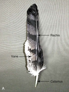
The main shaft of the feather is the scapus and is divided into the vexillum, or vaned portion, and the calamus, or unvaned portion (Figure 65-3). The vane comprises the majority of the feather, extending perpendicular to the shaft. The scapus is the central axis of a feather composed of the calamus and the rachis.7 The rachis (vaned portion of the scapus) is the solid, long, tubular portion of the shaft distal to the skin and is the continuation of the calamus, which is the hollow, short, tubular, unpigmented end of the mature feather that extends below the level of the skin.
Wing Feather Trimming
The most commonly used method to prevent flight in birds is to cut the remiges and has only a temporary effect.10,18 The advantages of this method are that it is temporary and nonpainful and therefore does not require anesthesia or analgesia, it is minimally stressful (if not performed in large groups and repeated on a regular basis), and it is inexpensive. However, the fact that it is temporary may also be a disadvantage.10,33 Feather trimming is usually done on only one wing on the basis of the concept that this will make it more difficult for the birds to fly because the birds are unbalanced. However, in some species, it may be necessary to trim the feathers on both wings to effectively prevent flight.33 Larger, heavier-bodied birds are more likely to be unable to fly having had only the feathers on one wing trimmed. Trimming the feathers on both wings may be advantageous in allowing the bird to balance better, which may be important in some situations such as during breeding.18
The duration of effectiveness depends on the rate of molt in different species. Most birds only molt once a year, and the molt occurs over a relatively brief period. Some birds such as ducks molt twice a year because they develop eclipse plumage during breeding season, and some other species such as cranes only molt every other year.36 Feather trimming should be postponed until after a molt to minimize the number of times the bird has to be caught to trim remiges.34 If trimming of flight feathers is used as a method of preventing flight, it is important to monitor the wing for feather regrowth and trim the new feathers once they have regrown into a mature, nonvital feather.
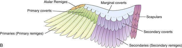
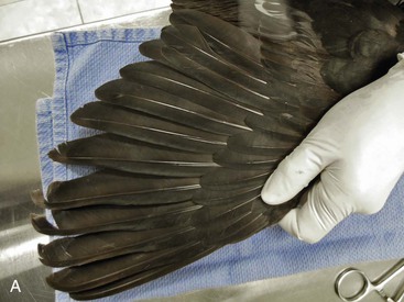
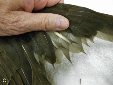
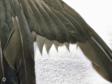
In general, all primary remiges on one wing are cut. Trimming the feathers on the same side for all specimens of one sex and the opposite side for the other sex allows for quick identification of the sex of the bird, even from a distance. When trimming remiges, blood feathers should not be trimmed.36 Generally, cutting feathers just proximal to the distal tip of the covert feathers, will prevent damage to blood feathers and is considered more cosmetically acceptable because the covert feathers cover the cut ends of the remiges. Birds may also be less likely to chew on cut feathers that are covered by covert feathers. It is considered best to cut each feather individually; cutting only the rachis results in a more cosmetically pleasing appearance because the vane then extends beyond the cut end of the rachis (Figure 65-4). Leaving the first and possibly the second primary flight feathers uncut also produces a more cosmetically acceptable result, but some birds will still be able to fly.36 In species that are heavier-bodied, it is preferable to leave these two feathers, as this allows the bird to land more gently. African gray parrots (Psittacus erithacus) are prone to developing lesions on the skin over the keel from hitting the substrate hard during attempts to fly after having their feathers trimmed.10 Some species such as cockatoos are prone to chewing on cut flight feathers, which may lead to self-mutilation.

Brailing
Brailing was first described in 193032 and involves temporarily immobilizing the carpus to prevent flight by using external coaptation. Brailing is applied around the carpus to prevent it from extending, thereby preventing flight. It is usually only applied to one wing.
Historically, brailing has been used primarily to prevent flight in juvenile pheasants prior to placing them into an enclosed flight cage. However, it has also been used to prevent birds from flying or traumatizing themselves during shipment.1,10,36 Pheasants are often reared for their feathers or for hunting, so amputation and follicle extirpation techniques are not used. Once flight feathers begin to grow in the chicks, which occurs at an early age in pheasants (10–14 days), the brail sling is applied.36 It is recommended that the sling not be left in place for more than 3 weeks or it may result in wing deformity.1,36 If it is necessary to prevent flight for a longer period, the brail sling is removed and a slightly longer one applied to the contralateral wing.36 This allows the first wing to recover and grow properly. Brailing may also cause damage to growing flight feathers.13
Radial Neurectomy
The radial nerve runs along the medial aspect of the distal humerus and has only skin covering it at this location. Excision of a portion of the radial nerve has been done in an effort to prevent flight in birds by paralyzing the muscles that flex and extend the carpus.34 Paralysis persists for months, and the wing droops after the procedure. The results are unpredictable, and most sources agree that the procedure does not permanently prevent flight.1,34,39
Tenotomy and Tenectomy Procedures
These techniques often result in short-term prevention of flight, but it is likely that even when a portion of a tendon is excised, the severed ends reunite with scar tissue resulting in return of enough function to allow birds to escape.
Tenotomy or Tenectomy of the Extensor Metacarpi Radialis Longus Muscle
In an effort to prevent flight in birds, without removing a portion of the wing for cosmetic reasons, tenotomy of the tendon of the extensor metacarpi radialis longus muscle was performed in 1940.32 It was theorized that with the cutting of this tendon, birds would no longer be able to extend the carpus enough to be able to fly. In theory, the wing should not droop because it is an extensor of the manus and the flexors remain undamaged. Although the authors reported success with this technique, they noted that it should be done bilaterally and that the birds at the San Diego Zoo were still able to “glide off (a hill) to a lower level.” Subsequently, others have recommended partial tenectomy—removing a section of the tendon, rather than just transecting it. However, this has not improved the success rate of deflighting birds.33,39 Other sources have reported that birds, in fact, do have a wing droop after the procedure.34,39
One author suggested that it is important to immobilize the wing for approximately 6 weeks following surgery to allow joint ankylosis.26 In 452 birds in his study, either tenotomy or tenectomy was performed. In 349 of the birds, the wing was immobilized for 6 weeks, and in 333 of the birds the carpi were fused; and in only 5 of the 103 birds in which the wing was not immobilized, the carpus considered fused. It is likely the inhibition in extension of the carpus resulted from joint arthrodesis rather than from transecting or excising a portion of the tendon.
Stay updated, free articles. Join our Telegram channel

Full access? Get Clinical Tree


