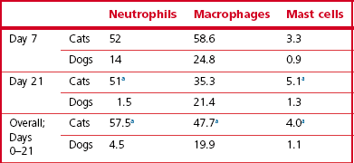
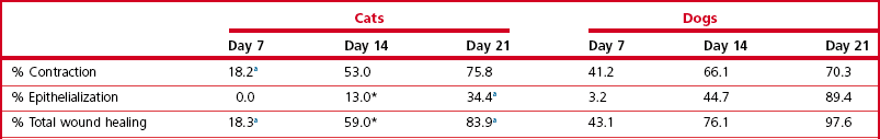
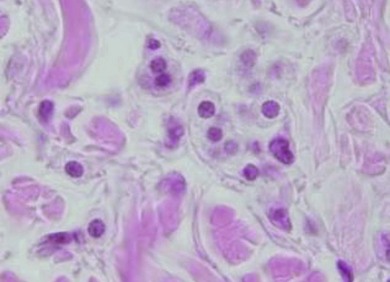
Chapter 17 Very early in the study of wound healing it was observed that wounds progress through stages: the initial phase of inflammation, followed by the phase of active proliferation (repair), and finally after the defect is closed, the phase of remodeling. Although once thought to be temporally distinct, we now understand that the phases of wound healing occur as a highly overlapping sequence of processes. We can recognize each of the phases in the healing of wounds in all vertebrates, and the phases follow the same chronological order, leading to the assumption that wound healing is more or less homogeneous across species lines, at least among mammals.1 Clinical experience reveals that there are significant differences in wound healing between mammalian species. Recently, significant differences between cutaneous healing in the cat and the dog have been described.2 These differences may cause us to reconsider the validity of long-accepted general small animal wound care recommendations when they are applied to the feline species. The normal cutaneous anatomy and histology of the cat follows the same general pattern as for other mammalian species. However, interspecies variations in feline cutaneous anatomy and histology may have significant effects on wound healing and other surgical considerations in regard to felines. As with all haired mammals, the epidermis of the cat is much thinner than that of humans, and is slightly thinner than that of the dog. The dermis comprises the majority of the thickness of the skin; it ranges from 0.4–2.0 mm thick in the cat, depending on body location. Like the epidermis, the feline dermis is somewhat thinner than the canine dermis, which ranges from 0.5–5.0 mm thick.3 The thickness of the subcutis varies greatly with body area and body condition; however, in general the subcutis of the cat is also significantly thinner than the dog. Another feature of feline skin is that the feline skin is very pliable and mobile over most of the body surface, even more so than in the dog, and certainly much more so than for ‘tight-skinned’ species such as the pig and humans. Cats also show significant differences from other mammals with respect to their cutaneous vascular supply. In one comparative study, the cutaneous angiosomes of seven mammalian species were evaluated with the purpose of validating the use of animal models for human wound healing research. Comparison of the cutaneous angiosomes of the cat with other mammals revealed that the cat has a relatively low density of tertiary and higher-order vessels, particularly on the trunk.4 This gross anatomic difference appears to translate to a functional difference based on laser Doppler perfusion studies, in which a significantly lower level of cutaneous perfusion was noted in the trunk skin of normal anesthetized cats than in dogs.2 The link between tissue perfusion and wound healing has been well established.5,6 First intention cutaneous healing (the healing of clean, sutured skin wounds) has been measured in the cat as part of an overall comparison of cutaneous healing between cats and dogs. Healing of 3 cm linear sutured wounds was evaluated via tensiometry at seven days post-wounding. Results demonstrated that the wound breaking strength in healing cat skin is significantly inferior to that of dogs. The mean wound breaking strength for sutured wounds in cats at seven days post-wounding was only one half of the wound breaking strength for dogs.2 This difference should be taken into consideration when deciding the right time for suture removal. Histologic evaluation of skin biopsies revealed that the observed difference in early first intention healing correlated with significantly lower collagen production in cat wounds compared to those in dogs.7 Interestingly, the grossly observed differences in apparent level of inflammation in open wounds do not correlate well with histopathologic evidence. Based on extensive histologic evaluation of open wound healing in cats and dogs, the picture that has emerged is one of similar levels of inflammatory cell infiltration in the first week of wound healing. However, as healing progresses, the populations of inflammatory cells, (particularly neutrophilic leukocytes and mast cells) in cat wounds appear to remain elevated for a longer period of time (Fig. 17-1 and Table 17-1).7 This observation supports other experimental and clinical evidence that, in the feline, the inflammatory phase of healing appears to follow a more chronic course compared to other species. The chronicity of inflammation in cat wound healing may in turn be at least partly responsible for delayed onset and slower progression of the proliferative phase of healing. Table 17-1 Measures of second intention healing in cats compared with dogs aIndicates a significant difference between cats and dogs for the indicated parameter on that day; (p ≤ 0.05, repeated measures ANOVA) Figure 17-1 Second intention healing in feline skin. Normal uncomplicated healing at 21 days post-wounding. Note the mast cells still present in the wound. (H&E stain, 400× magnification.) The production of granulation tissue in the wound is the first gross and histologic evidence of entry into the proliferative or repair phase of wound healing. The cat is notable for a slower rate of production of granulation tissue in open wounds, when compared to other mammals. On average, the first appearance of grossly visible granulation tissue in open wounds in cats and dogs begins at a similar time although is usually about one day later in cats than in dogs. However, once the process of granulation begins, the rate of granulation tissue production is markedly slower in cats. In an experimental model of open wound healing, cats took more than twice as long as dogs to fill the wound cavity with granulation tissue: 19 days for cats vs 7.5 days for dogs (Table 17-2).2
Wound healing
Introduction and overview of wound healing
Normal cutaneous anatomy in the cat
Wound healing in cats
First intention cutaneous healing
Second intention cutaneous healing
![]()
Stay updated, free articles. Join our Telegram channel

Full access? Get Clinical Tree


Wound healing