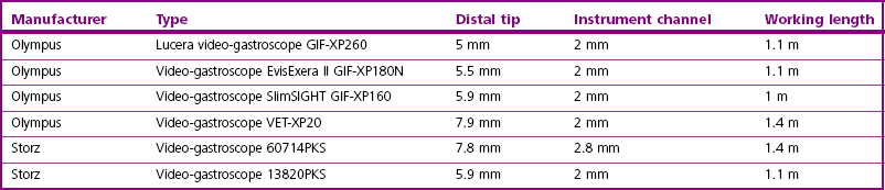
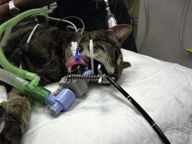
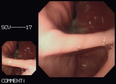
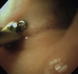
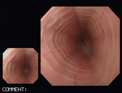
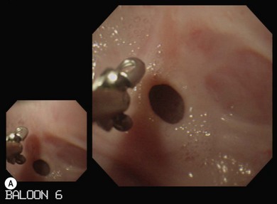
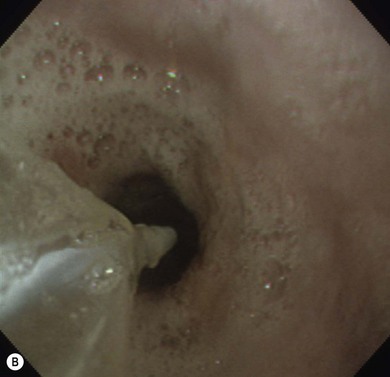
Endoscopy

Chapter 9


Endoscopy


Endoscopy can form a very useful part of the investigation of many disorders. An initial thorough assessment of any patient presenting for any form of endoscopy is of the utmost importance; the risks and benefits of endoscopy should always be carefully weighed up for the individual patient. Endoscopy is used for assessment of the upper and lower gastrointestinal tract, the respiratory tract, and the lower urinary tract. This chapter will cover diagnostic endoscopy for surgical diseases in cats.


General considerations


A thorough history and physical examination is required first, and, in most instances, endoscopy should be preceded by other investigations to assess the cat for systemic disease, and to ensure that endoscopy is the most appropriate next stage in the investigation. For the majority of cases, this assessment should include hematological and biochemical analysis of blood, other appropriate laboratory investigations depending on presenting signs (e.g., total T4, fecal analysis, FeLV/FIV testing, urinalysis), blood pressure assessment, and diagnostic imaging (e.g., radiographs, ultrasound, computed tomography). The patient should be stable for anesthesia, and any concurrent diseases or complications (e.g., dehydration) should be addressed. The endoscopic examination should always be fully documented, either by completing a report or through the use of recording equipment.


Endoscopes are expensive and delicate pieces of equipment. It is therefore essential that the surgeon is familiar with how to handle the endoscopes, and how to set up and appropriately use them, in order to avoid damaging the equipment, as well as avoiding risk to the patient through inappropriate use. It is also important to be aware of the cleaning, storage and maintenance required for endoscopes to ensure that they are kept in good working order. The reader should have basic training and be familiar with the basic technique of endoscopy, particularly how to hold the flexible endoscope and use the controls correctly.


Flexible endoscopes


Flexible endoscopy is required for the gastrointestinal tract (GIT), and feline respiratory tract. Flexible endoscopes are complex pieces of equipment with channels for suction and irrigation and passage of instruments as well as light guide fibers and optical image fibers, and guide-wires for the angulation of the tip. They are composed of the insertion tube (the part that enters the patient), the hand piece (containing deflection control knobs, suction valve, air/water valve and instrument channel port) and the umbilical cord (connects hand piece to the light source and also contains the video cable connection, suction pump connection and irrigation bottle connection).


Flexible endoscopes are either fiber optic or video. Fiber optic endoscopes are cheaper, and can be smaller in size, but the image produced is pixelated and of poorer quality than that of a videoscope. Additionally, the glass fibers are very fragile and if damaged the image quality is further reduced. Video endoscopes overcome the problem of poor image quality; however, due to the size of the distal chip sensors required, video endoscopes are not widely available under 5–6 mm diameter. Therefore, many endoscopes used in feline medicine, particularly bronchoscopes, are still fiberscopes.


Rigid endoscopes


A rigid endoscope can potentially be used for tracheoscopy and anteriograde rhinoscopy. The small size of the nose in cats makes rhinoscopy challenging but often an important part of the investigation of cats with chronic nasal discharge, in combination with advanced imaging and biopsy. Rigid endoscopes consist of a fiber optic rigid telescope and a fiber optic light cable that connects to a light source which can be xenon or halogen. Xenon provides a brighter light source and is also suitable for use with flexible video endoscopes and fiberscopes.


A 2.7 mm 18 cm long rigid telescope with a 14.5Fr 17 cm length operating sheath is useful for larger cats (Hopkins II Stortz). The 1.9 mm 19 cm endoscope (cystoscope) is more suitable for most cats. The use of a 10Fr sheath with the smaller (1.9 mm) endoscope protects the delicate scope from damage and enables flushing. Both the 2.7 mm and 1.9 mm endoscopes have a 30 degree viewing angle. These endoscopes are also suitable for cystoscopy, otoscopy (and arthroscopy).


Gastrointestinal endoscopy


Gastrointestinal endoscopy can be used for assessment of the stomach, upper small intestine, and the rectum and colon in the cat.1,2


Equipment


For gastrointestinal (GI) endoscopy, a flexible endoscope is required with four-way tip deflection (and ability to retroflex >180° in one plane), and the mechanical functions of insufflation, irrigation and suction. These functions should always be checked prior to anesthetising the cat. A single gastroscope can generally be used in all sized cats for esophagoscopy, gastroduodenoscopy and colonoscopy.


The ability to collect endoscopic biopsies is one of the major advantages and aims of performing gastroendoscopy.3 The larger the biopsy channel, the larger the biopsies that can be collected and therefore the more likely they are to be of diagnostic quality. The compromise is that the larger the distal tip size, the more difficult pyloric intubation will be. The instrument channel should be at least 2 mm in order to be able to obtain diagnostic biopsy samples. There are several scopes on the market that can be considered (Table 9-1).


Most of these endoscopes have a 2 mm instrument channel, with the largest instrument channel being 2.8 mm. A larger instrument channel has significant advantages, but there are also disadvantages to consider with the larger size of endoscope. An experienced endoscopist will be able to intubate the pylorus of most cats with a distal tip approaching 8mm in diameter, but an inexperienced endoscopist may struggle, and it may not be possible in smaller cats. A 1.4 m length insertion tube is quite long for cats, which can make maneuvering more challenging as the insertion tube outside the patient tends to loop.


The main forceps required for feline gastrointestinal endoscopy are biopsy forceps. Grasping forceps and basket forceps are also useful for removing gastric foreign bodies, although these are less commonly encountered in cats than dogs. Basket forceps are also required for placing percutaneous endoscopic gastrostomy (PEG) tubes. There are many different types of biopsy forceps, varying in shape (oval, round), edge (smooth, serrated), fenestrated or unfenestrated, those with a central spike, and swing jaw forceps. The author prefers oval fenestrated forceps without a central spike. The fenestrated cups reduce crush artefact, and the oval cups tend to collect more tissue. Central spikes can damage the biopsy tissue. The type of edge used depends on the toughness of the tissue being sampled. The author tends to mainly use smooth edged cups, but if the tissue is tough, these can slip off, in which case switching to serrated edged cups is advisable. When taking biopsy samples of the intestine, swing jaw forceps help turn the cups into the intestinal wall, but they are much more expensive forceps.


Prior to anesthetizing the patient, ancillary equipment should be prepared (Box 9-1) and endoscopy equipment checked that it is fully functional (including air pump, suction unit and valve, air/water valve, tip deflection, light source image, biopsy forceps, recording equipment).


Uses and indications


Common reasons for performing upper GI endoscopy in cats are listed in Box 9-2.


Unless a specific esophageal disorder, e.g., a stricture that may make advancing beyond this point unnecessary, is diagnosed, full esophago-gastro-duodenoscopy should always be performed.


Colonoscopy is indicated for investigation of large intestinal diarrhea, hematochezia, tenesmus, dyschezia, constipation and investigation of a palpable rectal mass or stricture. Upper GI endoscopy should always be performed whenever colonoscopy is being performed, since lesions are rarely confined to the colon even when clinical signs are predominantly large intestinal.


GI endoscopy has the advantage of allowing examination of the mucosal surface of the GIT. It is a much less invasive way of obtaining gut biopsies compared to open surgery: the patient can be discharged the same day as the procedure, there is no convalescence or time delay due to wound healing, and immediate corticosteroid treatment can be started if indicated.


Limitations and contraindications


There are relatively few contraindications for gastrointestinal endoscopy. The main one that is essential to consider is where systemic causes of clinical signs have not been excluded and there have been inadequate investigations performed prior to considering endoscopy. Other contraindications include patients that are a poor anesthetic risk, the presence of a bleeding disorder, any patient that has had recent GI surgery, and where food has not been withheld, or the stomach has been found to be full on imaging.


While being a very valuable way of assessing the GIT, there are limitations of endoscopy. Firstly, it does require appropriate and fully functioning equipment together with a competent endoscopist, with good biopsy technique, to allow adequate evaluation of the GIT and collection of diagnostic quality biopsies. It is also not possible to assess functional and motility disorders, hypersecretory disorders, the whole GIT, submucosal lesions, or intraperitoneal lesions. When endoscopic biopsies have failed to produce a diagnosis, where non-mucosal disease is suspected, or disease is known to be beyond the reach of an endoscope, or where there is evidence of intraperitoneal disease (e.g., mesenteric lymphadenopathy, ascites, hepatic/pancreatic abnormalities), then surgical biopsies are indicated.


Patient preparation


For upper GI endoscopy, patient preparation simply requires withdrawal of food for at least 12 hours prior to the procedure, to ensure that the stomach is empty. If any barium contrast studies have been performed, then GI endoscopy should not be performed for at least 24 hours. For colonoscopy, withdrawal of food for 24 hours is required, in addition to ‘cleansing’ the large intestine. There are various different laxative protocols, but the author’s preference is to administer 10–15 mL/kg of polyethylene glycol laxative (Klean-Prep), via a nasoesophageal tube (just placed for each administration and then removed in between), on two or three occasions within the period approximately four to 20 hours prior to colonoscopy, and then to administer a sodium citrate rectal enema (Micralax) two to four hours prior to the procedure.


Pre-medication prior to induction of anesthesia is routine. Atropine is not used routinely, although some advocate that it can make pyloric intubation easier, and it may reduce the risk of vagally induced bradycardia during the procedure. An intravenous catheter should be in place, and fluids administered throughout the procedure. Induction of anesthesia is routine. Nitrous oxide should not be used since insufflation of the stomach permits diffusion of nitrous oxide into it and causes gastric overdistension. Intravenous midazolam or diazepam can be useful if there is difficulty intubating the pylorus, or if the cat becomes uncomfortable (e.g., tachypneic) during this procedure, or if the GIT is particularly inflamed. For further information on anesthesia see Chapter 2.1


An endotracheal (ET) tube should be placed. Cuffed ET tubes are usually advised to prevent the risk of reflux, but caution needs to be used with cuffed ET tubes in cats.


Red rubber tubes with inflated cuffs should not be used, as these high pressure cuffs can cause tracheal necrosis; only the low pressure, high volume cuffed PVC tubes should be used. Presence of a cuff can, however, limit the diameter of the ET tube that can be passed and narrower tubes are more likely to become obstructed. The author prefers to use the largest possible uncuffed tube—in most average sized cats, a size 5 uncuffed ET tube can be passed. The tube should be secured in place, and a mouth gag must always be inserted to prevent damage to the endoscope. Pulse oximetry monitoring should be used, and appropriate warming equipment to ensure the cat does not become hypothermic during the procedure.


The cat should be positioned in left lateral recumbency for routine GI endoscopy (upper and lower), so that the gastric antrum is uppermost, allowing air to fill it and make the pylorus more visible, and that the descending colon lies ventrally, which aids intubation of the transverse and ascending colon (Fig. 9-1). For esophagoscopy where conditions such as mega-esophagus, esophageal strictures or esophageal foreign bodies are suspected, the cat should be kept in sternal recumbency with the head elevated in order to reduce the risk of aspiration. For PEG tube placement, the cat is positioned in right lateral recumbency. The endoscopist should be positioned in such a way that they have a good view of the video monitor, but also in a way that the insertion tube outside of the patient is kept in as straight a line as possible to prevent difficulty steering and advancing, which occurs if there is looping of the insertion tube outside the patient.


Endoscopic biopsy technique


An assistant is required to operate the biopsy forceps and it is important that they are familiar with how to operate them before starting. Instruments must always be passed through the biopsy channel in a closed position and never forced against resistance. Squeezing too hard to close the cups can break the wire of the forceps so must be avoided. Care must be taken when passing instruments through the deflected tip of the endoscope, as forceful passage can easily damage the inner lining of the instrument channel. The instrument channel also serves as a suction channel, so suction will be much reduced when an instrument is within the channel, especially if the instrument channel cap is open. When the scope has passed several intestinal flexures, the forceps can be difficult to open as the wire may be bent, and straightening the endoscope will help with this.


The quality of the biopsies obtained is determined mainly by the size of the forceps (dependent on the size of the scope), and the pressure exerted on the tissue by the operator. This is a big limitation in feline endoscopy as the patient size limits the size of endoscope, and therefore the size of biopsy forceps that can be used. Therefore, in order to obtain diagnostic quality biopsies, there is no room for poor operator technique. Exerting maximal pressure can be achieved by positioning the biopsy cups perpendicular to the tissue being sampled (Fig. 9-2). Being able to do this effectively and knowing how much pressure can safely be applied comes with experience. Deflating the viscus before biopsy also helps increase the size of the sample by reducing stretching of the mucosa.


Once the biopsy has been collected the forceps are removed from the biopsy channel, the cups opened and immersed in 10% formalin, releasing the tissue. The forceps must be rinsed in water before reintroducing into the endoscope. Alternatively, tissue samples can be laid on card or tissue cassettes prior to being placed in formalin.


Gastrointestinal endoscopy technique


General technique


As the endoscope is inserted into a viscus, air is instilled to obtain a clear view. The rate of air insufflation needs to be adjusted to ensure adequate inflation without over inflation. The small size of cats makes it easy to over inflate the GIT and thus this needs to be monitored carefully. Using a low setting on the air pump and ensuring the airhole is not inadvertently covered leading to continuous insufflation will help avoid over inflation. The anesthetist should also observe and palpate the abdomen regularly to ensure over inflation is not occurring. This is extremely important, as over inflation not only makes the endoscopy more difficult, particularly pyloric intubation and biopsy collection, but more importantly will lead to impairment of venous return, and can quickly result in severe cardiovascular and respiratory compromise, in addition to risking perforation of a viscus. Inability to sufficiently insufflate can be the result of a faulty air pump, leaking seal, air escaping from the viscus, or may indicate significant GI pathology.


Steering is achieved through a combination of insertion and retraction of the insertion tube, longitudinal rotation of the insertion tube (torquing), up/down and left/right tip deflection, and passive movement as the endoscope follows the wall of a viscus. Attention should be paid to keeping the insertion tube as straight as possible to assist in more accurate steering.


The lens will often become obscured by blood, mucus and GI contents, and this requires flushing by depressing the air/water button. Any fluid pooling in a viscus requires suctioning to examine the underlying mucosa. Air should always be suctioned from the viscus before withdrawing the endoscope.


Upper gastrointestinal endoscopy


Delay in intubating the pylorus, combined with insufflation of air, makes pyloric intubation more difficult. Therefore it is usual to only quickly visually inspect the esophagus and stomach on the way down, leaving more complete evaluation to the end of the procedure (except where esophagoscopy is the main purpose of the procedure). The endoscope is inserted through the upper esophageal sphincter by applying only gentle pressure and insufflating small amounts of air. Once through the sphincter the tip should be adjusted so that the esophageal lumen is in the center of the view, and the endoscope gently advanced to the lower esophageal sphincter. A quick inspection of the mucosa on the way down ensures that any pathological lesions can be distinguished from iatrogenic damage when closer inspection is performed on withdrawal. In cats, the distal esophagus has distinct circular folds and the lower esophageal sphincter may be seen as a star or slit-like opening (Fig. 9-3).


The most common pathologies observed in the feline esophagus are esophagitis, esophageal ulceration, and esophageal strictures (Fig. 9-4A). Biopsies are not routinely taken from the esophagus as the mucosa is very tough. Masses should be biopsied, but these are rare. With esophageal strictures, endoscopy can be used to guide balloon dilation, by passing a balloon catheter alongside the endoscope (Fig. 9-4B). When the stricture is very narrow it can be difficult to insert the balloon into the stricture and great care needs to be taken when directing the balloon to ensure that the tip does not cause further esophageal trauma or esophageal perforation. See Chapter 27 for more information on esophageal disease.


Once at the lower esophageal sphincter, the endoscope tip should be angled towards it with continued insufflation and gently advanced through into the stomach. As the endoscope enters the stomach the junction of the fundus and body of the stomach can be seen, with parallel rugal folds on the greater curvature running towards the pyloric antrum (Fig. 9-5). This provides an important landmark in locating the pylorus. The other most important landmark is the angularis incisura (angle of the lesser curvature) with the cardia above, and the pyloric antrum below, and this can be located by retroflexing the endoscope tip fully to first locate the cardia (Fig. 9-6) and then slightly reducing the retroflexion to bring the lesser curvature into view. Some insufflation is required to visualize these landmarks, but over inflation will make pyloric intubation difficult, and this is the most common mistake made. In cats, the angle of the lesser curvature is quite acute, and a slide-by technique can be useful for passing the endoscope into the antrum. This involves gently advancing the endoscope along the mucosal surface of the greater curvature; the endoscope tip will be impinging on the gastric mucosa and so red-out will occur, but provided this is moving and the endoscope is not advanced against any resistance, it can continue to be advanced along the greater curvature until the pyloric antrum comes into view. Ensuring that the pylorus is in the center of the screen, and suctioning as the insertion tube is advanced towards it assists with pyloric intubation.


Stay updated, free articles. Join our Telegram channel

Sep 6, 2016 | Posted by in SUGERY, ORTHOPEDICS & ANESTHESIA | Comments Off on Endoscopy

Full access? Get Clinical Tree

Get Clinical Tree app for offline access