Chapter 56 White-Nose Syndrome in Cave Bats of North America
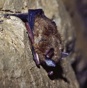
White-nose syndrome (WNS), an emerging disease of hibernating insectivorous bats, is associated with unprecedented winter mortality at cave and mine hibernacula in the northeastern and mid-Atlantic regions of the United States.2 The disease is named for the prominent white, powdery fungal growth around the muzzles of bats, although the fungus also appears on wings, ears, and tail membranes (Fig. 56-1). This fungus, which is consistently associated with WNS development and is the presumptive cause of the syndrome, is a newly discovered species named Geomyces destructans.12
The population effects of WNS have been profound and widespread. Several WNS-affected locations have reported 90% to 100% loss of the hibernating population after only two seasons of the disease.23,30 Since its first detection in New York (February 2006), WNS has spread over 950 km, seemingly advancing 200 to 700 km/year. More than one million cave bats are estimated to have died.8 The species most commonly observed to be affected is the little brown bat (Myotis lucifugus), which had been the most abundant bat species in the region prior to the emergence of WNS.11 Other hibernating species known to be susceptible to WNS include northern long-eared bats (M. septentrionalis), tricolored bats (Perimyotis subflavus), eastern small-footed bats (M. leibii), big brown bats (Eptesicus fuscus), and federally listed (endangered) Indiana bats (M. sodalis). Within the WNS-affected region, Indiana bat winter counts decreased by 25,000 bats (30%) between 2007 and 2009.29 Three additional Myotis spp. bats (M. austrorparius, M. grisescens, and M. velifer) may also be proven to be susceptible to WNS, however thus far only the genetic signature of G. destructans has been demonstrated in these species.31
The risk WNS poses to the rest of the North American bat population is unknown, but there is cause for concern. In all, 25 of 45 North American bat species hibernate in caves or mines30 and therefore may be susceptible to WNS. Some of the country’s largest and most species-rich bat hibernacula are located in the midwestern and southeastern United States,29 just beyond the current leading edge of the disease. As WNS continues to spread further south and west, extensive hibernacula in Missouri, Tennessee, and Kentucky housing more than 100,000 bats, including endangered Virginia big-eared bats (Corynorhinus townsendii virginianus) and gray bats (M. grisescens), are at risk.
Emergence and Spread
White-nose syndrome was first photodocumented in hibernating bats in a commercial cave near Albany, New York, in February 2006, although the disease went unrecognized until the following winter, when it was observed at four hibernacula within a 15-km radius near this presumptive index site. That same year (2007), the N.Y. State Department of Health received 10 times the 25-year average of bat submissions for rabies testing between January and April.2
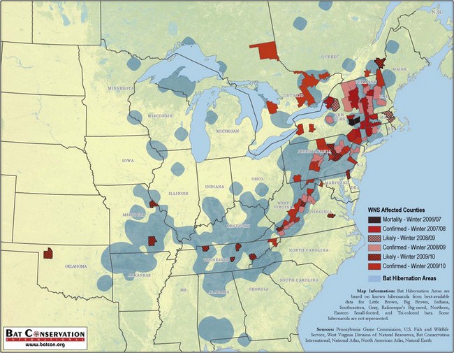
Increased surveillance during the winter of 2007 to 2008 detected WNS at 33 sites in four states (New York, Connecticut, Massachusetts, and Vermont), with approximately 25% of all surveyed bat hibernacula affected within a 210-km radius from the presumptive New York epicenter. By the winter of 2008 to 2009, WNS expanded both within its original range and spread to another 27 sites in five additional states, including New Hampshire, New Jersey, Pennsylvania, Virginia, and West Virginia (Fig. 56-2). In winter 2009 to 2010, WNS reached eastern Tennessee and north into Quebec and Ontario although G. destructans DNA was recovered from bats as far west as Missouri and Oklahoma.26 The pattern of expansion and the diversity of environmental variables among the growing number of affected sites have supported WNS as an infectious disease rather than the result of contaminant exposure.7,9
Epidemiology
Visible evidence of cutaneous fungal growth on bats during the winter months is the hallmark of WNS; however, bats may have microscopic lesions on wing, muzzle, and ear caused by G. destructans without the visible manifestation of the fungus.7,19 Even when visually present in the cave environment, the proliferative white fungal growth is easily disrupted and may disappear with handling.7,19 Field observations have described pinpoint colonies of visible fungus around the nares of bats first developing in late November, although late September marks the earliest diagnosis of WNS skin infection in bats to date,32 detected in a subclinical little brown bat collected from a hibernaculum confirmed as WNS-positive the previous winter. External signs of infection become more prominent on bats as hibernation continues.7,19 Both adult and juvenile bats (~6 months old) are susceptible to WNS and associated winter mortality. G. destructans has been recovered from bats emerging from hibernacula with moderate to severe wing damage19 and from postemergent females sampled at maternity colonies as late as May.20 It has not yet been cultured from free-ranging bats collected between June and August within the affected region, nor has WNS been associated with summer bat mortality, including that at maternity colonies,31 although samples are limited.
In addition to the physical manifestation for which WNS is named, other characteristics accompanying the disease are often readily recognized in the field during the winter. Abnormal behavior such as bats observed outside in cold temperatures during daylight hours is often an early indicator of problems in the area. Large numbers of bats roosting near the entrances of hibernacula, particularly species not known to do so, and a delayed or lack of arousal response in the presence of human disturbance are other atypical behaviors commonly reported at affected sites. The gross appearance of G. destructans on torpid bats preceded behavioral changes and mass mortality in at least one affected site in Pennsylvania.28 Most dead bats are often found at, or just outside, the hibernaculum entrance in poor body condition. Bats emerging in late winter and spring often have moderate to severe wing damage, including holes and tears, which may adversely affect their ability to forage.23 The effects of this damage on survivorship and reproductive success of postemergent bats in maternity colonies are not yet well understood.23
Presumptive Cause
The two most consistent findings from laboratory evaluations of dead bats with WNS are the presence of invasive fungi consistent with G. destructans on the glabrous skin and suboptimal body condition during late hibernation.7,19 The presence of fungal hyphae on the WNS-affected bats is in clear contrast to the absence of invasive fungi on bats examined from areas not experiencing winter mortality or displaying signs of WNS. Furthermore, no consistent internal lesions have been identified in WNS-affected bats and ancillary tests failed to reveal any other consistent pathogen, mineral, or heavy metal–based toxicities.3,9 Findings such as pneumonia and ectoparasitism and endoparasitism were interpreted as incidental because they were also found in bats from uninfected populations.2,9
G. destructans infections were prevalent on the wings where the fungal hyphae invaded into the dermis and, in severe cases, extended full thickness through the wing. On the face, fungal infections were not observed to infect mucosa beyond the anterior nares.19 Although in vivo experiments demonstrating G. destructans as the causative agent of WNS mortality are ongoing, the strong correlation between the presence of this newly described fungus and WNS development make it the prime putative agent of the syndrome.2
Classification of G. destructans
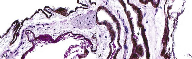
Phylogenetic analyses of two ribosomal RNA regions, the internal transcribed spacer (ITS) and small subunit (SSU), conducted on morphologically identical fungal isolates cultured from affected bats collected at different locations most closely resembled the anamorphic genus Geomyces (order Helotiales).2,12 Morphologically however, the asymmetrically curved conidia appear to make this fungus unique among Geomyces spp., which are known to be psychrophilic (cold-loving) keratinophilic fungi (Fig. 56-3).12 Other Geomyces spp. have been detected worldwide in soils and dust from cold climates as well as on the skin, hair, and feathers of animals from these areas.15 Although rarely reported as a pathogen, cutaneous infections by the closely related G. pannorum have been reported in humans.13
Culture Conditions
In the laboratory, optimum growing temperatures for G. destructans range from 4° C to 10° C (39.2° C to 50° F).2 This is similar to the seasonal temperatures reported at WNS-affected hibernacula, where year-round growth may create reservoirs for infection.2 Growth does not occur above 20° C (68° F). G. destructans grows well on antibiotic-infused Sabouraud dextrose agar16 and other standard media12,22 by placing wing or muzzle tissues directly onto culture plates. Colonies range from white to gray-green in appearance on the surface and are slow-growing, remaining small in diameter after 2 weeks of incubation.12
Other Diagnostic Criteria
In addition to isolation in fungal culture, the presence of G. destructans may be confirmed by examining fungal tape lifts from skin and fur of affected bats with light microscopy to observe the characteristic morphologic structure of conidia. Although the technique is not highly sensitive, fungal tape lifts collected from skin with grossly visible fungus (primarily the muzzle) have the advantage of being a nonlethal sampling technique. Swabbing flight membranes and muzzles of bats with a moistened sterile swab has also been used experimentally to collect G. destructans conidia for identification by light microscopy.20 If further confirmation is warranted beyond that of conidial morphology, a polymerase chain reaction (PCR) assay targeting the ITS region of G. destructans rRNA gene has been used with variable success on tape or swab samples (USGS NWHC, unpublished data). PCR when applied to bat tissues has, however, demonstrated high levels of sensitivity and specificity that are comparable to those demonstrated by histologic evaluation.16 PCR assay is a useful screening tool to detect the presence of G. destructans DNA on bat carcasses or wing biopsies collected from live animals. It does not, however, provide information about fungal viability as do fungal cultures or determine whether the fungus is associated with the cutaneous lesions that characterize WNS.
Histopathology
Full details of the histologic appearance of G. destructans infection have been described elsewhere.9,19 On hematoxylin and eosin preparations, G. destructans appears as masses of negatively stained hyphae on the surface of the skin and invading into the dermis and adnexa.19 Fungal morphology is more easily visualized by the application of a periodic acid–Schiff stain (PAS) or a Gomori methenamine silver stain (GMS). Because fungal colonies may be regional or widely spaced, special stains to highlight fungi are suggested for all skin sections being evaluated for WNS (see Fig. 56-3). PAS is favored because it highlights fungal morphology without the background staining present in the GMS.
Distinctive conidia and patterns of colonization by G. destructans hyphae are the criteria for diagnosing WNS in tissue section.19 The conidia are sickle-shaped, measuring 7.5 µm in length and 2.5 µm wide, with blunt apexes. The septate branching hyphae are more variable in appearance, ranging from 2 to 5 µm in diameter and may have parallel or irregular walls (see Fig. 56-3). When conidia are not readily apparent, the pattern of G. destructans growth and the appearance of fungal hyphae in tissue section may be used to distinguish it from incidental superficial fungal infections or postmortem invasion. In its mildest manifestation, G. destructans covers the surface of the skin and may fill adnexa. Large numbers of hyphae invade and fill the dermis in more severe infections and may breach the entire thickness of the wing membrane.19 Postmortem fungi may also invade the dermis and subcutaneous tissues and stain PAS-positive, but these do not form the cupping epidermal erosions that are densely filled with G. destructans hyphae, as is seen in WNS.19
One striking feature of G. destructans infection is the lack of inflammatory response to the fungus among torpid animals. Although some inflammation may be detected, it is not severe during the winter months.19 This lack of reaction is in contrast to the reactions noted in response to Geomyces spp. infections in other species.13 Inflammation is seen most often in bats that have recently emerged from hibernation and consists of a mixture of neutrophils and smaller numbers of lymphocytes.19 It is not clear whether this lack of a morphologically detectable inflammatory response is mediated by immune evasive features of G. destructans, or if this is a manifestation of immune suppression similar to that which has been documented in other hibernating animals.21
Stay updated, free articles. Join our Telegram channel

Full access? Get Clinical Tree


