19 Virtually edentulous, no clinical signs of root resorption
RADIOGRAPHIC FINDINGS
The series of full-mouth radiographs (eight views) identified numerous teeth in different stages of replacement resorption. See Figs 19.1, 19.2, 19.3, 19.4 and 19.5 for details of radiographic findings.

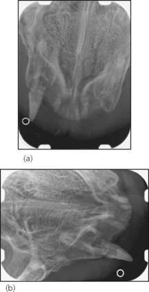
Figure 19.1 Rostrocaudal radiograph (a) and right lateral radiograph (b) of the rostral upper jaw.
(a) The upper incisors and canines are in different stages of replacement resorption. The roots of the incisors are still identifiable as ‘tooth’, but the absence of a clear periodontal ligament space indicates active external root resorption and replacement of lost tissue by bone. These roots are ankylosed, i.e. there is fusion between root and bone. The crown of 104 is still in place, but most of the root has been resorbed and replaced by bone. Tooth 204 has been almost completely resorbed and replaced by bone-like material. It is impossible to clearly differentiate between remaining root tissue and bone.
Stay updated, free articles. Join our Telegram channel

Full access? Get Clinical Tree


