Peter R. Morresey
Uroperitoneum
Uroperitoneum is a well-documented and readily diagnosed condition. Although it is reported in adult horses, the condition most commonly affects neonatal foals. Retention of urine leads to electrolyte and acid-base abnormalities. Of these, hyperkalemia is the most clinically important because it may precipitate cardiac arrhythmias and cardiac arrest. Respiratory embarrassment may also occur in advanced cases because of diminished diaphragmatic excursion caused by grossly excessive peritoneal urine accumulation. Uroperitoneum, if advanced, is therefore a medical emergency.
Pathophysiology
Loss of integrity in the urinary tract allows urine access to the peritoneal cavity where some constituents, such as electrolytes, can be rapidly resorbed while others remain entrapped. Peritonitis can also result if a devitalized septic focus was responsible for the leakage.
The most clinically important result of urine retention is disturbance in serum electrolyte values, chiefly potassium. High serum potassium concentration results in arrhythmias, especially bradycardia, and can progress to cardiac arrest in severe cases. As herbivores, adult horses have a diet high in potassium and must excrete large quantities each day. Serum concentrations of potassium can therefore rapidly rise with uroperitoneum. Similarly, in foals, a diet of milk rich in potassium and low in sodium intensifies these serum electrolyte abnormalities.
Hyponatremia and hypochloremia also develop as the volume of uroperitoneum increases. Uroperitoneum increases total body water, and sodium diffuses from the extracellular fluid (ECF) into the retained urine until equimolar concentrations are achieved. Because the total amount of body sodium is unchanged, the ECF and hence serum sodium concentration decrease (hyponatremia). Chloride diffusion follows sodium to maintain electroneutrality, resulting in hypochloremia.
Acidosis develops with uroperitoneum for two reasons. Increased intraabdominal pressure reduces cardiac output by diminishing venous return from the caudal vena cava and portal vein. Metabolic acidosis then develops as a result of lactate accumulation consequent to poor tissue perfusion. In advanced cases of uroperitoneum, respiratory acidosis may also develop because ventilation is compromised by the large volume of peritoneal urine (Figure 184-1). The acidosis further exacerbates hyperkalemia secondary to potassium release from cells in exchange for the hydrogen ion.
Azotemia is a classic finding with uroperitoneum because urea nitrogen, which is in high concentrations in the retained urine, diffuses across the peritoneum and is resorbed into the bloodstream. Creatinine, however, is a relatively large molecule that, unlike urea and sodium, cannot rapidly diffuse back across the peritoneum into the blood. This difference in diffusibility is responsible for the classic finding of peritoneal fluid creatinine concentration well in excess of serum concentrations.
Precipitating Conditions in the Neonatal Foal
Uroperitoneum is most common in the neonatal foal. Although initial investigations suggested that uroperitoneum was more common in male foals, likely a result of the longer and narrower urethra, more recent retrospective case series have not substantiated these findings.
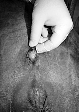
The umbilical cord contains the urachus, which connects the fetal urinary bladder to the allantoic cavity in utero. Up to four twists along the length of the umbilical cord are considered normal. A higher number of twists may cause vascular compression, intimal tearing, obstruction of blood flow, and obstruction to urine flow from the fetal urinary bladder to the allantoic cavity. Dilation of the urachus proximal to a compression site may be observed grossly. Increased pressure within the urachus and bladder may be sufficient to cause their rupture. Complete obstruction of umbilical vessel flow may result in fetal death. Increased traction on the umbilical cord from the decreased length can also compromise integrity of the urachus and the umbilical area, increasing the likelihood of urine leakage.
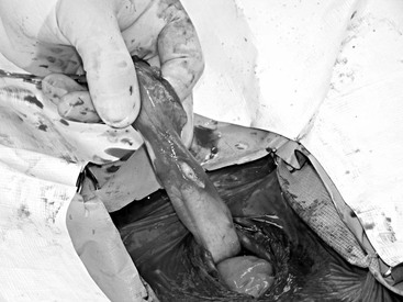
Hospitalized compromised neonates are at particular risk for developing uroperitoneum. Historical findings in such foals include dystocia, local umbilical trauma, and regional sepsis leading to omphalophlebitis or urachal abscess formation (Figure 184-2). As a consequence of systemic infection or dissemination of regional infection of the umbilicus or urachus, septic hemorrhagic foci may develop in the bladder or urachal wall and precipitate areas of tissue necrosis and urine leakage (Figure 184-3). Recent investigations revealed that 78% of foals with uroperitoneum had evidence of infection or sepsis, and 45% had positive sepsis scores. In addition, histologic lesions compatible with ischemic necrosis and sepsis are often found postmortem in foals with positive sepsis scores. The most common bacteria cultured from lesions and blood of affected foals is Escherichia coli.
Nontraumatic causes of uroperitoneum include congenital abnormalities and iatrogenic causes. Urine may leak from congenital anomalies of the distal ureters, defects in the ventral and dorsal bladder wall, and the ureterovesicular junction. Iatrogenic factors should be suspected when uroperitoneum occurs in foals that have been recumbent for prolonged periods and have received considerable volumes of intravenous fluids. Loss of conscious bladder control may enable overfilling of the urinary bladder, which markedly increases intravesicular pressure, predisposing to rupture, particularly if there are devitalized and weakened areas of the bladder wall and urachus. For this reason, hospitalized or septic neonates should be monitored for uroperitoneum. In these animals, administration of intravenous fluids can mask the serum electrolyte changes indicative of uroperitoneum.
Uroperitoneum in Adult Horses
Although it is rare in older horses, uroperitoneum may develop if any intraabdominal portion of the urinary tract (urethra, bladder, ureter, or kidney) ruptures because of internal factors or sufficient external trauma. Uroperitoneum in adults is most common in postpartum mares, in which necrosis of the bladder wall following delivery of the foal leads to perforation and urine leakage. It is theorized that the necrosis is consequent to ischemia that develops when the bladder becomes trapped between the fetus and the pelvic brim during the high intraabdominal pressures that develop with parturition. Septic peritonitis has also resulted in some cases.
Compared with other species, urolithiasis is rare in horses, especially in mares, which have a short, wide urethra. In male horses, urethral obstruction by calculi can be responsible for urethral disruption or sufficiently increased intravesicular pressure to cause bladder rupture and urine leakage into the peritoneal cavity. This occurrence is relatively rare in male horses, compared with males of other large-animal species, likely for two reasons: first, the male horse urethra is relatively uniform in diameter except for the portion at the ischial arch; and second, it has neither a sigmoid flexure (as do ruminants) nor a urethral process (as do rams).
Clinical Presentation
Physical Examination
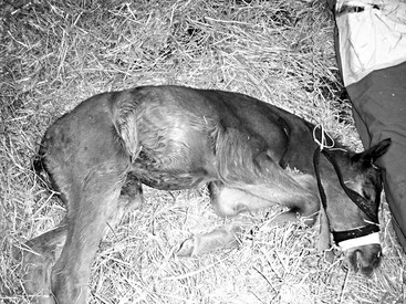
In the early stages of uroperitoneum, physical abnormalities may be inapparent. Abdominal distension only arises when the volume of urine retained is considerable and the rate of leakage from the bladder exceeds fluid absorption across the peritoneum. Especially in adult horses, considerable urine volumes are necessary to grossly enlarge the abdomen. In neonatal foals, uroperitoneum is more readily apparent, with signs of progressive abdominal distension, mild to moderate colic, generalized depression, and diminished suckling activity commonly seen.
Some horses with uroperitoneum, including those with rupture of the bladder, can still urinate normally. However, most affected horses have dysuria or pollakiuria. In neonatal foals, however, dysuria may be mistaken for straining to defecate because of meconium retention or impaction. Affected foals with meconium impaction will display kyphosis (dorsoflexion) as opposed to the lordosis (ventroflexion) that is characteristic of foals with uroperitoneum that are straining to urinate. Meconium impaction can, however, lead to urinary tract rupture and uroperitoneum. As in mares straining to give birth, the straining of foals with meconium impaction can also cause bladder rupture. In the latter case, the markedly increased intraabdominal pressure during straining and pressure of retained meconium at the pelvic inlet can cause significant trauma to the bladder wall.
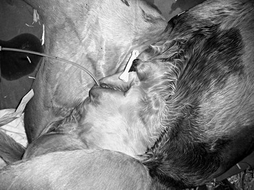
Uroperitoneum may lead to concurrent urine deposition outside the peritoneal cavity; scrotal enlargement with urine may occur in colts. To the clinical observer, subcutaneous urine accumulation appears similar to other areas of subcutaneous edema. However, on ultrasonographic examination, the urine leaking into subcutaneous tissues appears as areas of hypoechogenic fluid dispersed between hyperechogenic tissue planes. Urachal urine leakage may cause uroperitoneum and urine accumulation subcutaneously in the umbilical region (Figure 184-4). This may progress to generalized ventral edema and preputial edema in male foals. Ureteral rupture allows subperitoneal dissection of urine in addition to the other signs of uroperitoneum. Perineal swelling may develop in cases of urethral rupture. Ultrasonography of the thoracic cavity should be conducted in foals with uroperitoneum because concurrent pleural effusion consisting of urine leakage from the peritoneal cavity has been reported.
< div class='tao-gold-member'>
Stay updated, free articles. Join our Telegram channel

Full access? Get Clinical Tree


