Chapter 10 Urinary system
The parts of the urinary system are:
The functions of the urinary system are:
The kidney
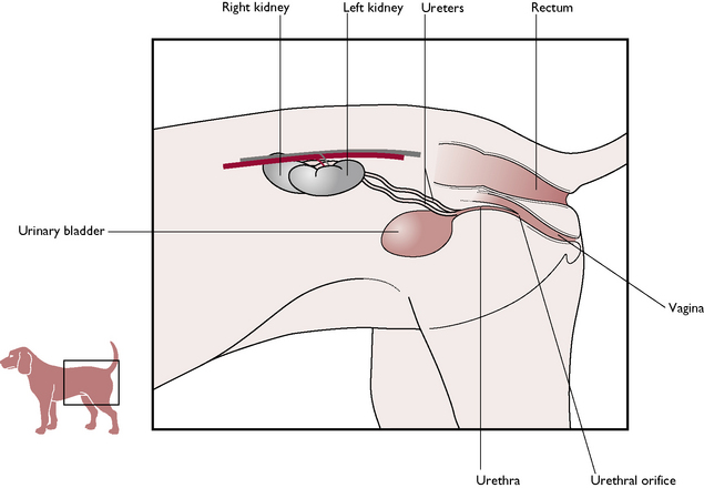
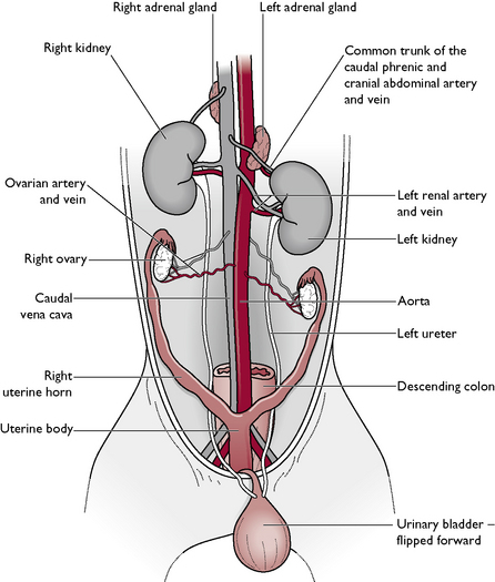
There are two kidneys lying in the cranial abdominal cavity, one on each side of the midline ventral to the lumbar hypaxial muscles (Fig. 10.1). Each kidney is closely attached to the lumbar muscles by a covering of parietal peritoneum. There is no mesenteric attachment, as seen in other abdominal organs, and the kidney is described as being retroperitoneal. The right kidney lies slightly cranial to the left because the stomach has evolved to lie on the left side of the abdomen, pushing the left kidney out of position. Lying close to the cranial pole of each kidney are the ovaries of the female and the adrenal glands (Fig. 10.2).
Macroscopic structure
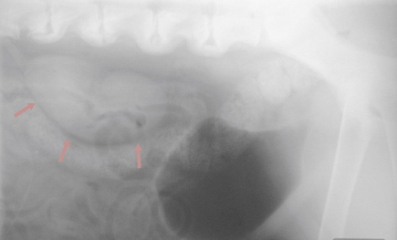
The kidneys of the cat and dog have a characteristic bean shape and the indented area is known as the hilus. This is the point at which blood vessels, nerves and the ureters enter and leave the kidney. The kidneys are normally a deep reddish-brown but the colour may be affected by any substance filtering through them. On a lateral radiograph of the abdomen, a normal kidney can be seen to be equivalent in size to approximately 2.5 vertebrae (Fig. 10.3). The outer surface may be surrounded by a layer of fat, which acts as an energy reserve and protects the kidney from external damage.
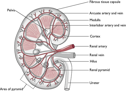
When examining the cut surface of a normal kidney cut longitudinally, it is possible to see four layers (Fig. 10.4). From the outside inwards these are:
Blood supply
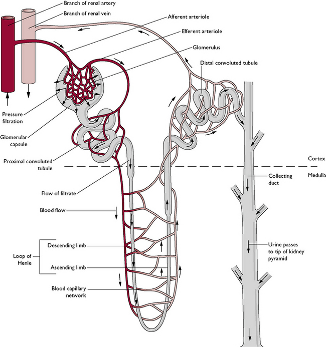
Arterial blood is carried from the aorta in a single renal artery to each kidney (Fig. 10.4). This carries 20% of cardiac output. Within the tissue of the kidney, the renal artery divides into several interlobar arteries, which pass between the renal pyramids and into the cortex. Here capillaries supply the renal tubules and also give off numerous capillary networks known as glomeruli (sing. glomerulus) (Fig. 10.5). Each glomerulus supplies an individual nephron. The capillaries then recombine to form interlobar veins, which enter the single renal vein. This carries venous blood to the caudal vena cava.
Microscopic structure
The functional unit of the kidney is the nephron (Fig. 10.5). Each kidney contains about a million nephrons, which are closely packed together. They are responsible for the filtration of blood and the production of urine. Each nephron is a long tubule divided into several parts:
Renal function – the formation of urine
The physiological processes occurring in the renal nephrons are:
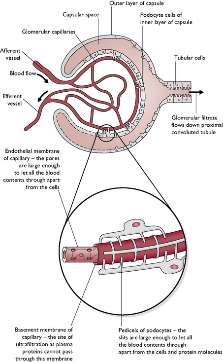
Blood enters the kidney and is carried to the capillaries forming the glomeruli.
Glomerulus
Blood pressure within each glomerulus is high because:
Stay updated, free articles. Join our Telegram channel

Full access? Get Clinical Tree


