Chapter 12 Unexplained Lameness
Lameness diagnosis is a never-ending challenge, even for an experienced clinician, because despite a logical and thorough investigation it still may prove difficult to reach a satisfactory conclusion. This chapter discusses some of the reasons why a definitive diagnosis may remain elusive. In some horses it may be possible to isolate the source of pain reasonably accurately, but it may not be possible to determine the cause of pain. In other horses the source of pain cannot be determined (see Chapter 97).
False-Negative Responses to Diagnostic Analgesia
A false-negative response to local analgesic techniques may occur for a variety of reasons, including the following:
The following are common case examples:
The horse in Figure 12-1 was admitted with suspected back pain but showed obvious bilateral forelimb lameness, with right forelimb lameness predominating. Digital pulse amplitudes were increased in the right forelimb, but there was no response to hoof testers. Nonetheless, the horse appeared clinically to have foot pain typical of laminitis. Apparent desensitization of the foot, performed to convince the owner that the horse had foot pain, produced absolutely no change in the lameness. The horse responded rapidly to treatment for laminitis.
![image](/wp-content/uploads/2016/06/B9781416060697000122_f012-001-9781416060697.jpg)
Fig. 12-1 A, Lateral and B, solar scintigraphic images of the front feet of a horse with suspected back pain but clinical signs of laminitis. There is reduced radiopharmaceutical uptake in the toe region (arrowheads). The horse showed no improvement in lameness after apparent desensitization of the lamer right front foot by palmar (abaxial sesamoid) nerve blocks, despite the firm focal pressure applied with artery forceps around the coronary band. However, the horse responded well to symptomatic treatment for laminitis.
Failure to allow sufficient time may in some circumstances result in a false-positive response and then confusion. The tibial and fibular nerves are relatively large, and it takes time for the local anesthetic solution to diffuse into them and to take effect. This time requirement, combined with the deep location of the deep fibular nerve and thus difficulty in precisely locating the site for injection, may result in a response delayed for up to an hour after injection. Testing the efficacy of these blocks through evaluation of cutaneous sensation is unreliable. If the response is deemed to be negative after 30 minutes, and intraarticular analgesia of the compartments of the stifle is then performed and the lameness improves, it may be wrongly inferred that pain originated in the stifle. However, the improvement in lameness may reflect alleviation of pain arising from the hock region. Much wasted time and money may then be spent trying to establish a cause of stifle pain.
Blocking each compartment of the stifle joint separately (e.g., the medial femorotibial joint) may not result in substantial clinical improvement in the lameness, despite the presence of stifle pain. A considerably better response is frequently seen after blocking the medial and lateral femorotibial joints and the femoropatellar joint in combination.
The importance of the clinical examination and repeated observations of a horse cannot be overemphasized. Each clinician has to learn how much to trust nerve blocks. This depends on experience and the frequency of performing blocks. An inexperienced clinician is far more likely to encounter false-negative responses. The results of nerve blocks must be compared with the clinical signs, and if the interpretation is doubtful, the block should be repeated or the area desensitized with a different technique. The clinician must develop experience in the interpretation of improvement in lameness compared with complete alleviation of pain and lameness. This contrast depends to some extent on the degree of the baseline lameness and whether the forelimbs or hindlimbs are involved.
Failure to Perform the Appropriate Nerve Blocks
Failure to perform nerve blocks in a logical and complete sequence can lead to confusion. If the response to a low six-point block in a hindlimb is negative and is followed by a positive response to tibial and fibular nerve blocks, the clinician may conclude that pain arose from the hock. Lesions of the proximal aspect of the suspensory ligament (SL) may be completely overlooked.
Blocking the Wrong Limb
Failure to appreciate that a head nod reflects hindlimb lameness and is not always a sign of forelimb lameness may result in a blocking a forelimb with negative results, when the primary source of pain is in the ipsilateral hindlimb.
Sources of Pain That Cannot Be Desensitized by Nerve Blocks
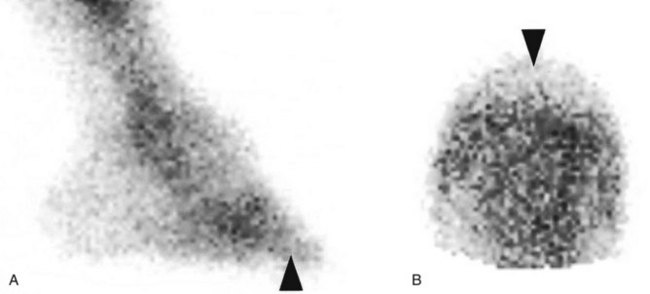
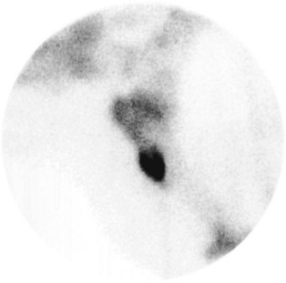
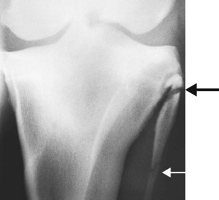
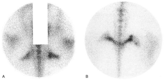
Many regions of the limbs proximal to the carpus and tarsus cannot be satisfactorily desensitized. In young horses, stress fractures are now well-recognized causes of lameness that pain from which in many circumstances cannot be blocked out. In young or older horses, fractures of the deltoid tuberosity of the humerus (Figure 12-2), the proximal aspect of the fibula (Figure 12-3), the third trochanter of the femur, and the tuber ischium (Figure 12-4) are all causes of lameness that are unaffected by nerve blocks.
![image](/wp-content/uploads/2016/06/B9781416060697000122_f012-002-9781416060697.jpg)
Fig. 12-2 Lateral scintigraphic image of the shoulder region of a 6-year-old Warmblood with acute onset of moderate left forelimb lameness. Note the marked focal increased radiopharmaceutical uptake in the region of the deltoid tuberosity of the humerus. There was a slightly displaced fracture of the deltoid tuberosity of the humerus, which healed satisfactorily with conservative management.
![image](/wp-content/uploads/2016/06/B9781416060697000122_f012-003-9781416060697.jpg)
Fig. 12-3 Caudocranial radiographic image of the left stifle of a general purpose riding horse with recent-onset, episodic, and transient severe left hindlimb lameness. There is a fracture of the proximal aspect of the fibula (large arrow). The lucent line separating the different centers of ossification further distally (small arrow) should not be confused as a fracture. There was little evidence of bony union after 6 weeks, but after 12 weeks the fracture healed satisfactorily.
![image](/wp-content/uploads/2016/06/B9781416060697000122_f012-004-9781416060697.jpg)
Fig. 12-4 A, Dorsal oblique and B, caudal scintigraphic images of the tubera ischii of an 8-year-old Grand Prix show jumper with loss of hindlimb power and a tendency to jump to the right. There is increased radiopharmaceutical uptake in the right tuber ischium and a change of contour compatible with a fracture.
Muscle injuries, such as tearing or fibrosis of brachiocephalicus or the pectoral muscles, may have no localizing signs (see page 152). Associated lameness cannot be influenced by nerve blocks. Atypical equine rhabdomyolysis can cause hindlimb lameness without any other clinical signs typical of tying up.
Periarticular lesions of the otifle such as collateral ligament injury are usually associated with detectable soft tissue swelling, but injuries of the patellar ligaments can occur with no localizing clinical signs, and associated pain is often unresponsive to intraarticular analgesia (see Chapter 46). I have also examined five horses with acute onset of severe lameness and mild swelling on the craniomedial aspect of the femoropatellar joint resulting in loss of palpable definition of the patellar ligaments. Lameness was characterized by a markedly shortened cranial phase of the stride at the walk, but less severe lameness at the trot. Ultrasonographic examination revealed the presence of a periarticular hematoma surrounding the patellar ligaments, which themselves were structurally normal.
Pain Associated with a Neuroma
A neuroma may develop secondary to external trauma to a nerve, after abnormal stretching of a nerve, or subsequent to surgery. There is usually intense focal pain on pressure applied directly over the neuroma. However, perineural analgesia of the area may fail to abolish or improve associated lameness. A much better response may be achieved by infiltration of local anesthetic solution directly around the neuroma.
Potentially Confusing Responses to Local Analgesic Techniques
Improvement without complete alleviation of lameness after perineural analgesia is not always easy to interpret. It may reflect failure to completely alleviate pain from a single source, or there may be additional sources of pain. Sometimes lameness improves with each successive block (e.g., palmar digital, palmar [abaxial sesamoid], low four-point, and subcarpal nerve blocks). However, the lameness is not associated with any detectable radiological, ultrasonographic, or scintigraphic abnormalities. Sometimes additional useful information can be obtained by performing intraarticular analgesia of the interphalangeal and metacarpophalangeal joints, but if the response is negative the diagnosis remains inconclusive.
Isolation of pain to a region but failure to define the cause is particularly frustrating. For example, intraarticular analgesia of the femorotibial joints may be positive, but no radiological or ultrasonographic abnormalities may be detectable. Nuclear scintigraphy may reveal a generalized increased uptake of the radiopharmaceutical in the distal aspect of the femur and proximal aspect of the tibia compared with the contralateral limb. Medication of the joints may result in no improvement. Exploratory arthroscopy may reveal minor findings (e.g., mild fibrillation of the cranial meniscal ligaments) of questionable relevance, but evaluation of all the joint surfaces and meniscal cartilages is impossible. The definitive diagnosis for the cause of pain remains elusive. Computed tomography (CT) and magnetic resonance imaging (MRI) may permit the identification of subchondral bone injuries and meniscal and ligamentous injuries not accessible to arthroscopic inspection.
The importance of subchondral bone pain as a cause of lameness must not be overlooked. Such pain frequently is present without associated radiological change. A comparison of the responses to intraarticular analgesia and perineural analgesia (and the response or lack thereof to intraarticular medication) may be helpful. With subchondral pain, intraarticular analgesia may have a limited effect. Nuclear scintigraphy is a sensitive indicator of increased modeling in the subchondral bone. MRI has the potential to show subtle structural changes in the subchondral bone.
Until recently, soft tissue lesions within the hoof capsule have proved elusive to definitive diagnosis. Diagnostic ultrasonography, although possible, has marked limitations. Pool-phase scintigraphic images sometimes are helpful. Examination of the navicular bursa may yield useful information about the bursa, the deep digital flexor tendon, and the distal sesamoidean impar ligament. Advanced imaging techniques such as CT and particularly MRI have the best potential to demonstrate soft tissue pathological conditions, although determining the clinical significance of lesions is not necessarily easy.
False-positive results may be obtained if the horse is only mildly lame at investigation but has a history of a more obvious lameness. The detectable mild lameness may not necessarily reflect the original cause. Lameness that is induced when a horse is lunged in small circles on a concrete surface may not reflect the primary cause of lameness. Thus eliminating this lameness by nerve blocks may be misleading. Lameness induced by flexion also may not reflect the principal cause of lameness. Blocking the flexion response does not necessarily identify the primary cause of lameness.
A pony had moderate forelimb lameness that was markedly accentuated by lower limb flexion. The response to flexion was eliminated by either regional or intraarticular analgesia of the fetlock joint. However, the baseline lameness was unchanged and did not respond to any of the nerve blocks that were repeated on several occasions. Surgical removal of a large osseous fragment from the fetlock joint did not improve the lameness.
False Positive Results of Intrasynovial Analgesia
Lameness may be abolished or substantially improved by intraarticular analgesia of the metacarpophalangeal (metatarsophalangeal) joint with no intraarticular pathology. Pain associated with proximal lesions of the cruciate, straight, or oblique sesamoidean ligaments or the insertions of the suspensory branches may be substantially improved by intraarticular analgesia of the fetlock. Therefore in the absence of both clinical signs suggestive of fetlock joint pain (e.g., synovial effusion, joint capsule thickening, pain on manipulation) and radiological abnormalities it is suggested that the distal sesamoidean ligaments and suspensory branches be evaluated ultrasonographically. Intrathecal analgesia of the digital flexor tendon sheath has the potential to remove pain from injured distal sesamoidean ligaments; it may also relieve pain from the deep digital flexor tendon within the hoof capsule. Therefore the distal sesamoidean ligaments should be included in ultrasonographic examination of the palmar soft tissues of the pastern.
Multiple Sources of Pain in a Limb and More than One Lame Limb
Problems can arise in interpretation of nerve blocks in a horse that is lame in more than one limb, especially if there is more than one source of pain in a limb. Perineural blocks usually last for up to 2 to 3 hours unless a long-acting local anesthetic agent, such as bupivacaine, is used. If a horse is lame in several limbs it is usually easiest to start with the lamest limb and block it first. Interpretation becomes difficult if there is a failure to desensitize all the lame limbs simultaneously. If the blocks in one limb are wearing off, then lameness in the least lame limb becomes less apparent. The horse’s tolerance for nerve blocks may also compromise how much can be done. It may be necessary to start again on another occasion using bupivacaine. If simultaneous lameness of the ipsilateral forelimb and hindlimb is suspected, blocking should begin in the hindlimb. In this situation a substantial amount of the head nod probably originates from the hindlimb component. Because elimination of head nod is vital to improvement after blocking, forelimb diagnostic procedures cannot be fairly evaluated.
Very-Low-Grade Lameness
Nerve blocks, especially in hindlimbs, often result in improvement rather than complete alleviation in lameness. Assessing improvement in subtle lameness is nearly impossible. If the horse has a history of more severe lameness previously, delaying further investigation often is worthwhile. The horse should be worked to accentuate the lameness and simplify interpretation of the response to diagnostic analgesic techniques.
Improvement of Lameness in Some Situations, but Unrelieved Lameness under All Situations: Which Is the Baseline Lameness?
Sometimes a horse has lameness that appears different in nature under different circumstances. Such findings may be related to more than one cause of lameness, and it is important to recognize this fact. For example, a dressage horse had left forelimb lameness that was apparent to the rider only when the horse was ridden on the right rein (to the right). Clinical examination revealed left forelimb lameness on the right rein on the lunge on a hard surface. This was alleviated by desensitization of the foot. However, desensitization did not alter the lameness that was apparent when the horse was ridden. The cause of the lameness could not be identified. It is vitally important to relate the results of the investigation to the history.
![](https://clinicalpub.com/wp-content/uploads/2023/09/256.png)
Full access? Get Clinical Tree
![](https://videdental.com/wp-content/uploads/2023/09/appstore.png)
![](https://videdental.com/wp-content/uploads/2023/09/google-play.png)