Ultrasonography and Orthopedic (Nonarticular) Disease

Chapter 18 Ultrasonography and Orthopedic (Nonarticular) Disease


The use of diagnostic ultrasonography for evaluation of tendons, ligaments, tendon sheaths, bursae, and joints is discussed in Chapters 16, 17, and 82, and horse preparation for other musculoskeletal uses is similar.


Skeletal Muscle


Normal skeletal muscle appears heterogeneous, with hypoechogenic muscle fibers laced with and surrounded by echogenic fascia, connective tissue, and fat. In transverse section a normal muscle has a marbled or speckled appearance, which is unique for each individual muscle, as is its striated appearance in longitudinal section.1,2 A non–weight-bearing muscle appears more echogenic than the same muscle when the horse is fully weight bearing.1,2 Therefore comparisons between contralateral muscles should be made when the horse is bearing weight evenly.


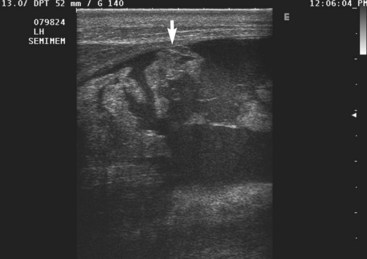
Traumatic muscle injuries, myositis, and masses infiltrating the muscles usually can be differentiated from one another by ultrasonography.1 Muscle tears in horses most frequently are seen in the hindlimb and shoulder musculature. In neonatal foals, muscle tears are most commonly seen in the hindlimb.3 The affected muscle or muscles can be identified by tracing the involved muscle from its origin to insertion. Fluid-filled anechogenic areas with hypoechogenic loculations are located within the muscle belly, associated with areas of hemorrhage and muscle fiber tearing (Figure 18-1).1,3-6 As the muscle injury becomes more severe, large areas of interfascial and subcutaneous hemorrhage are detected. The free edge of a completely disrupted muscle may be imaged floating in the anechogenic loculated fluid in the hematoma. Echogenic areas of clot are often imaged within the intramuscular, interfascial, or subcutaneous hematoma. As these echogenic clots become more organized, they may cast an acoustic shadow from the far surface. Rupture of the gastrocnemius muscle should be considered as a cause of recumbency in neonatal foals that are unable to rise and have soft tissue swelling in the hindlimb. Rupture of the gastrocnemius muscle has been reported as a complication of dystocia.3 Partial muscle tears are more difficult to diagnose because the ultrasonographic abnormalities are more subtle.1 The normal striated muscle pattern is lost with increased echogenicity of the injured muscle.1 Swelling of the affected muscle usually is present. Tears in the muscle fascia or thickening of the muscle fascia or fasciitis also may be detected ultrasonographically.1


Ultrasonography is an excellent tool for monitoring the resolution of the hematoma and the healing of the muscle.1,5 The affected horse’s exercise should be restricted until the hematoma has resolved and the muscle tear has filled in with tissue. As the muscle heals, the fluid becomes more echogenic and the area fills in. The healing muscle may become more heterogeneous. Hyperechogenic areas of muscle scarring are seen associated with fibrotic myopathy, especially in the semitendinosus (primarily) and semimembranosus muscles, but can also occur in other muscles such as the biceps femoris and gracilis (rare).1,5,7 As these areas of fibrosis become more organized, acoustic shadowing from the far side of the fibrotic area may be detected. Hyperechogenic areas casting acoustic shadows are seen in horses with an ossifying myopathy and frequently are found adjacent to areas of fibrotic myopathy.1 Areas of mineralization develop as scattered, pinpoint hyperechogenic areas that progressively become linear and result in acoustic shadowing from the near side of the areas.


Postanesthetic myopathy results in increased echogenicity of the affected muscle, with loss of the normal muscle striations.1,2 Muscle edema may result in the muscle appearing less echogenic than normal if the lesion is evaluated early in the course of the disease. Once a large influx of inflammatory cells has occurred, the affected muscle becomes more echogenic. A necrotizing myositis usually results in a more heterogeneous ultrasonographic appearance associated with the inflammatory cell infiltrate and bacterial infection.1 Cavitation of a severely affected muscle is associated with liquefactive necrosis. Hyperechoic echoes, associated with the local production of gas by bacteria, may be seen with an anaerobic necrotizing myositis.1


Muscle tumors are rare; the most common muscle tumor is hemangiosarcoma. Discrete echogenic masses in the muscle or anechogenic loculated, more heterogeneous masses may be detected.1 Central anechogenic or hypoechogenic areas representing tumor necrosis are identified in large or rapidly growing tumor masses. Primary muscle tumors such as rhabdomyosarcomas are extremely rare but can be a cause of lameness.8 Accurate identification of tumor type in muscle necessitates obtaining an ultrasound-guided biopsy and histopathological evaluation of the tissue obtained.


Jun 4, 2016 | Posted by in EQUINE MEDICINE | Comments Off on Ultrasonography and Orthopedic (Nonarticular) Disease

Full access? Get Clinical Tree

Get Clinical Tree app for offline access