Ultrasonographic Imaging of the Gastrointestinal Tract

CHAPTER 16 Ultrasonographic Imaging of the Gastrointestinal Tract


Ultrasound imaging has become widely accepted as an important diagnostic tool for imaging the gastrointestinal tract in veterinary patients. Initially it was thought that artifacts created by gas and ingested material would significantly limit or exclude the use of ultrasound for examining the gastrointestinal tract. However, experience has shown that these artifacts, although present and at times obstructive, rarely interfere significantly with a thorough ultrasonographic examination. The technology has continued to improve. Higher resolution (12 to 18 MHz and higher) transducers and more affordable equipment—along with the availability of Doppler ultrasound—have made it possible to image the gastrointestinal tract more thoroughly, enhancing the ability to evaluate and understand changes produced by diseases affecting that organ.1


Ultrasound uses sound reflected from the tissue boundaries within the body to form an image that we can recognize and interpret. At diagnostic frequencies (1 to 25 MHz) it is noninvasive and does not pose any known significant biological risk. It is particularly useful in evaluating the intestinal wall, the luminal contents, and function evidenced by peristalsis. It also can interrogate the surrounding organs such as the peritoneum, pancreas, liver, and lymph nodes. Ultrasound can guide needle placement for either an aspirate or biopsy of a detected abnormality. Intestinal ultrasound provides additional and complementary information to other diagnostic modalities (e.g., survey radiographs, computed tomography, nuclear medicine, magnetic resonance imaging, and contrast radiography).


Ultrasound imaging now has become so integral to the diagnostic evaluation of small animals with gastrointestinal signs that there has been a marked reduction in radiographic contrast procedures. Ultrasound is faster, is more cost-effective, and often provides as much or more information than these other diagnostic modalities. However, these improvements also have raised questions about the significance of some findings, and a great deal remains to be learned regarding the feline intestinal tract. One example is differentiating the various etiological causes for the appearance of the thickened muscular layer of the small intestine. It is especially important to correlate ultrasound findings with the history, clinical findings, laboratory results, and ultimately cytological and histopathological findings.


METHOD OF EVALUATION


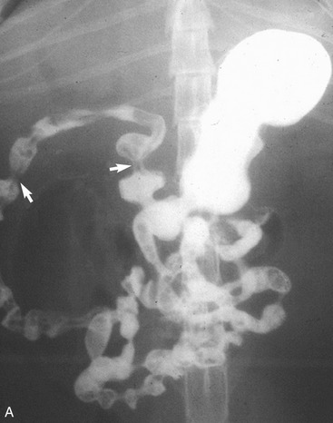
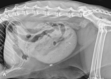
It is preferred, but not always necessary, that the patient be held off food before the ultrasonographic examination. Withholding food for 8 to 12 hours can reduce interference from gas and ingested contents. This is especially helpful when examining the patient for suspected foreign bodies within the intestinal lumen and when evaluating intestinal walls. An enema is not recommended or usually needed because this introduces gas into the colon, which may interfere with the examination. Gas within the lumen often is manifested by reverberation, comet tails, and acoustic shadowing artifacts. A comet-tail artifact is a streaklike or cone-shaped reverberation phenomenon, typically occurring deep to metallic objects. A ring-down artifact is a streaklike reverberation phenomenon, typically occurring deep to a fluid-gas interface. It has the appearance of vertical hyperechoic lines extending from the first encounter with the surface of the air interface to deep within the image. These artifacts can obscure normal or diseased tissue and also can mimic disease. Feces within the colon or barium contrast material anywhere in the intestinal tract can attenuate the sound beam (Figure 16-1). This often obscures the far wall of the intestine and limits the ability to evaluate that section of intestine. Survey radiographs can be helpful in providing an overall view of the abdomen and to confirm or identify sonographic findings.


If possible, the ultrasound examination should be performed before an upper gastrointestinal study with barium or before administration of oral medications, especially those that contain bismuth. If a diagnostic procedure requires administration of food, such as for measurement of postprandial serum bile acid concentrations, or the patient is diabetic and needs to be fed regularly, the ultrasound examination should be performed before any food is given.


Feces within the colon typically are not a problem during evaluation of the small intestine. However, if artifacts from the feces interfere with colonic evaluation or with the evaluation of an adjacent area that needs to be examined, varying the pressure applied to the transducer or imaging from a different window can be helpful. If an important region is obscured from evaluation, providing a tray with cat litter to encourage defecation or repeating the examination the day after a fast may be warranted.


To achieve the best image quality, hair should be shaved in the area that needs to be imaged using a surgical clipping blade. The shaved area usually is rectangular, extending from the epaxial muscles in the dorsocranial abdomen (usually eighth to ninth rib) to the caudal abdomen just in front of the pelvic limbs and ventrally to the midline on both sides of the cat. An attempt should be made to keep the margins of the clipped area straight because the area’s appearance is often noticed and important to the owner. Mammary papillae obscured by surrounding hair may be at risk for trauma from the clipper blades when the patient is shaved by an inexperienced individual. The area to be clipped will be more extensive when the patient is imaged from a lateral position than it will be for a patient in a dorsal recumbent position. If the animal has thin hair, it can be moistened with water and alcohol in lieu of cutting the hair. Commercially available water-soluble ultrasound gel is used as an interface between the skin and the transducer. Alcohol also may be used to clean dry, dirty, or scaly skin. This application of alcohol often reduces the amount of gel needed and also improves image quality.


The method of positioning the cat for examination often varies depending on the experience and training of the individual performing the examination. Usually either a lateral imaging plane (i.e., left or right lateral recumbency) or a dorsal recumbency imaging plane is used. Each sonologist should establish a consistent scanning protocol. A routine examination includes an attempt to identify and image each section of the intestine. If the standard viewing window does not provide an adequate image, the sonologist can change the position of the transducer as necessary to complete the evaluation. For the challenging patient, changing the position of the cat relative to the transducer can help in avoiding troublesome gas pockets. The author typically examines each patient in both recumbent lateral positions. Positional studies, such as the recumbent ventrodorsal or standing positions, can be used to afford an acoustic window not otherwise available in a specific patient.


The highest MHz transducer available should always be used. To image the layers of intestinal walls, a minimum of a 7-MHz transducer should be used. A 5-MHz transducer will allow visualization of the intestinal tract; however, the detail required to assess the intestinal wall often is not adequate. A 10- to 18-MHz transducer will allow better imaging of the intestinal wall layers. Endosonography uses a specialized intracavitary transesophageal/transcolonic transducer to image from within the lumen of the intestine. These high-resolution transducers are especially helpful in evaluating the individual wall layers of the intestine for changes secondary to neoplasia and inflammatory bowel disease. Some are equipped with endoscopic biopsy instruments. More than one transducer may be required to image the entire intestinal tract at different depths. A linear probe provides better imaging of the superficial intestinal tract. The pie-shaped image afforded by a sector transducer provides a limited view of a segment of the intestinal tract in the near field. Because each machine type is different, it is often necessary to experiment with one’s own machine for the optimal imaging transducer. Both transverse and longitudinal views of gastrointestinal segments are needed to assess both the intestinal wall and lumen. Measurements of the wall can be made from longitudinal views, but reports indicate that measurements are most accurate when made from the transverse image to avoid oblique sections through the wall. The luminal axis of the intestinal segment evaluated, and not the plane through the patient’s body, determines the plane of the image seen on the screen (i.e., longitudinal, transverse, or oblique). Color Doppler and/or pulse wave Doppler are useful aids to assess blood flow to certain segments of the intestinal tract.


NORMAL ANATOMY


Ultrasonographic study of the abdomen includes interrogation of each region of the gastrointestinal tract (i.e., esophagus, stomach, duodenum, jejunum, ileum, and colon), noting the wall thickness, distinction of the wall layers, luminal contents, motility, and vascular integrity.


In the cat abdominal visceral detail usually is good for both ultrasonographic and radiographic imaging. A significant amount of heterogeneous and mildly hyperechoic omental and falciform fat often is present, but usually does not affect image quality negatively.


Some appearances and features of the intestinal tract are found in both cats and dogs. However, there also are unique features of the feline intestinal tract. In the cat only the cervical part of the esophagus and a small segment of abdominal esophagus (between the cardia of the stomach and the diaphragm) are accessible for sonographic evaluation. The cervical portion of the esophagus can be imaged lateral to the trachea, from the larynx to the thoracic inlet. The abdominal esophagus is best seen from a right dorsal imaging window. The esophagus is seen ventral to the aorta and dorsal to the caudal vena cava as it traverses the diaphragm and courses to the cardia of the stomach. The majority of the esophagus can be imaged best with radiography and a radiographic contrast study (i.e., esophagogram).


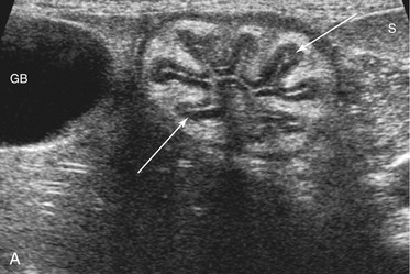
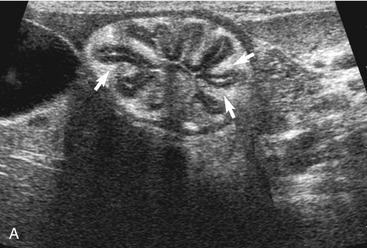
The stomach (the fundus to the pylorus), duodenum, jejunum, ileum, and colon all can be identified separately in the cat. The stomach is located in the left cranial abdomen just caudal to the liver, craniomedial to the head of the spleen, and cranial to the left kidney (Figure 16-2). The wall may be mildly thickened and contain a hyperechoic submucosal layer from adipose tissue, especially in overweight cats (Figure 16-3). The pylorus is located on the midline, as opposed to the right of midline as in dogs. During an abdominal ultrasound examination the stomach usually is empty and typically has a rosette or wagon-wheel appearance. It shows a characteristic pattern with its size, shape, and prominent rugal folds.


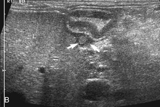
The duodenum courses in a straight superficial path from the pylorus along the right body wall for approximately two-thirds the length of the abdomen. From there it curves medially at the caudal flexure and then courses craniomedially. The sphincter of Oddi, the entrance of the conjoined bile duct and pancreatic duct, is located in the proximal flexure of the duodenum just distal to the pylorus. The pancreatic duct and bile duct join before entering the duodenum at the cistern of Vater. Approximately 90 per cent of cats have only the primary pancreatic papilla; the rest also have a minor duodenal papilla. The Peyer’s patches (lymphoid aggregates), which can be seen on the antimesenteric surface of the duodenum in dogs, are usually not seen in cats. The duodenum, and sometimes parts of the jejunum, can show segmental contractions resulting in a “string of pearls” appearance as opposed to the stripping peristaltic activity seen in dogs (Figure 16-4). The jejunum is identified as a long intestinal segment between the duodenum and the shorter ileum. The ileum is a small section of intestine that can be identified by its characteristic appearance (often contracted, with a “wagon wheel” appearance) and its position at the entrance into the colon. The ileum has a prominent hyperechoic submucosal layer and a corresponding undulating mucosal layer.


The ileum and/or the cecum can be seen connecting to the larger section of intestine, the colon. The cecum is a cul de sac. It enters the colon adjacent to the ileocolic junction, which is best identified from the right side in the midabdomen. The wall of the tip of the cecum often is thickened and hypoechoic with a reduction in wall layering. Left colic lymph nodes usually are seen in cats near the ileocolic junction, in addition to the mesenteric (jejunal) lymph nodes located at the root of the mesentery. In a recent paper, at least two colic lymph nodes were identified in all of 31 normal cats evaluated.2 The diameter of these lymph nodes ranged from 1.9 to 5.2 mm. In this study, at least one, and usually both, normal medial iliac lymph nodes were found. In interrogations of the intestinal tract, the pericolic and jejunal lymph nodes are especially useful to image and measure because they drain the intestinal tract and can be used as a sentinel reflecting an abnormality within it.


In contrast to the small intestine, the colon is recognized by its larger size, thinner wall, location (extending dorsal to the bladder through the pelvic inlet), and appearance (with sound attenuation and shadowing caused by luminal contents [feces] or reverberations by a gas-distended lumen) (Figure 16-5). The ascending, transverse, and descending colon can be followed. It is easiest to trace the short ascending segment from the right side and the descending segment from the left side of the abdomen. If it is unclear whether a loop of intestine is an abnormal segment of small intestine or colon, it is helpful to follow the loop to see if it courses through the pelvic canal or to trace a segment of bowel known to be colon to the area in question. Small intestine has a thicker wall and does not have the dilated lumen with shadowing contents seen in the colon.


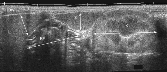
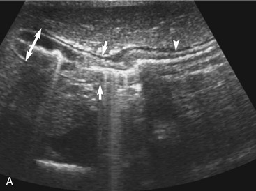
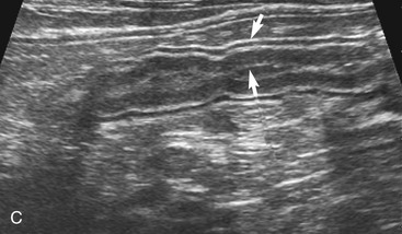
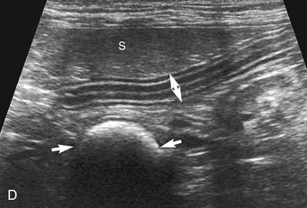
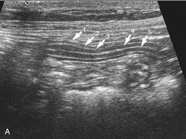
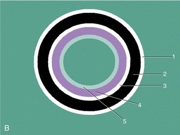
The wall layers of the intestinal tract are similar to the appearance described in human beings and dogs.3 Depending on the section of intestine examined, the wall layer thickness and appearance varies. The wall layers identified sonographically are formed by the interface of specific histological components of the layers of the intestine, which generate different acoustic impedance values between the layers, and by internal reflecting margins within tissue layers. The ultrasound measurements of the apparent layers correspond with the histological measurements but are affected by the axial resolution of the transducer used and the speed of sound in the different tissue layers. The higher the resolution of the transducer, the better the corresponding morphological correlation. The mucosa layer is most apparent in the duodenum and jejunum and less apparent in other segments of the intestine (i.e., colon and ileum), likely because of the change in function and role in the absorptive process. The wall of the intestine includes five layers with an alternating hyperechoic and hypoechoic appearance. The three hyperechoic layers (the S layers) are serosa, submucosa, and the surface of the mucosa; the two hypoechoic layers (the M layers) are the muscularis and the mucosal layers (Figure 16-6).


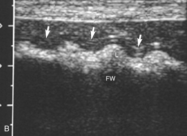
The wall thickness in part is related to the degree of distension of the loop of intestine and gets mildly thinner the more distended it becomes (Figure 16-7). Contraction during normal peristaltic activity does affect the width of the intestinal wall although the width of specific layers can change momentarily with contractions. Because the intestinal tract is dynamic, contractions shorten the loop of intestine in the contracted area and increase the width of the muscular and mucosal layers compared to adjacent dilated segments. It is important to be consistent in placing the cursors for wall measurements. The stomach wall thickness varies from 1.7 to 2.8 mm. It is important to avoid measurement of rugal folds or oblique cuts to prevent erroneously thickened measurements. It is easiest and most accurate to measure the wall when a small amount of fluid is present within the gastric lumen. This may require administering water orally if there is any question about the wall thickness. The thickness of the duodenum and jejunum usually is between 2.3 and 2.8 mm. It is more difficult to measure the wall thickness of the ileum accurately due to its “wagon wheel” appearance. The ileum has a prominent hyperechoic submucosa due to fat and lymphatics, and a hypoechoic muscularis layer. Ileal wall thickness may measure between 2.4 and 2.8 mm. The colonic wall is thinner (usually 1.4 to 2.3 mm), and the wall layers are less distinct.4,5 The far wall of the colon often is obscured by shadowing artifacts due to absorption of sound by fecal material. In general, intestinal wall thickness in cats is fairly consistent at 2.3 to 2.8 mm; it is regarded as abnormally thick when wall thickness is greater than 3 mm or the appearance of individual wall layers changes.


The appearance of luminal contents varies depending on the ingested material. Gas, fluid, mucus, food, and feces are normal contents observed. Ring down, comet tail, and sometimes attenuation and shadowing artifacts are observed frequently due to the sound interacting with the luminal contents. The luminal contents often contain mucus mixed with tiny gas bubbles, fluid, and/or ingesta. In fact, fluid (with minimal gas bubbles) may be given by mouth to accentuate imaging of the wall and the lumen of the proximal gastrointestinal tract.


Normal motility can be observed with real-time ultrasound. Four to five contractions per minute is normal in the stomach and duodenum; one to three is normal in the remainder of the small intestine. It is not common to see contractions in the colon.


The entire intestinal tract, including each segment of intestine, should be identified. Different methods for locating the intestinal segments can be employed. The abdomen can be divided into four quadrants. Each quadrant is scanned individually, with specific segments of intestine identified within that quadrant. No attempt is made to follow each bowel segment. Another method is to look for, and follow, specific segments such as the stomach, duodenum, or ileum separately, using their normal location in the abdomen and characteristic appearance as guides, and then to scan the remainder of the gastrointestinal tract systematically by imaging the quadrants without following every section of the intestinal tract. If an abnormality is detected, a known segment of bowel can be followed proximal and distal to the abnormality. Another method is to follow the entire gastrointestinal tube from proximal (i.e., stomach) to distal (i.e., colon). However, this approach is more time consuming and difficult due to patient movement and skill needed.


IMAGING OF SPECIFIC GASTROINTESTINAL DISORDERS


Disorders involving the gastrointestinal tract may be divided into those involving the lumen or the wall, or those arising outside the intestinal wall. A final diagnosis or differential diagnosis involves correlating the abnormal sonographic findings to history, clinical presentation, and laboratory results. The final diagnosis also may require an aspirate or biopsy for cytological and/or histopathological confirmation.


LUMINAL CHANGES


The normal ultrasonographic size of the lumen and the appearance of the contents vary with the segment of intestine and the type of material ingested. The stomach and the small intestine often are empty or contain only a small amount of gas, fluid, or food material. The colon often is partially distended with formed shadowing feces. Luminal abnormalities seen typically are an increased volume of contents with distension of the bowel proximal to the area of an obstruction, and the obstructing material itself (e.g., an intraluminal foreign body, an intussusception, or a mural mass). This distension of the intestine also can be uniform, consistent with a mechanical obstruction downstream (aborad) from the point of imaging. In other instances the dilation can be combined with specific wall patterns or mural changes that indicate gathering from a linear foreign body or an infiltrated area or mass secondary to neoplasia or fungal disease. A dilated intestine is not always the result of a mechanical obstruction: it may be associated with diminished motility secondary to effects of drugs or metabolic, neurological, or inflammatory disorders. The luminal contents obviously should remain within the lumen. Gas within the intestinal wall or peritoneal cavity or contents (i.e., gas, fluid, or ingesta) outside the lumen are surgical emergencies.


Obstruction


Ultrasound is a useful diagnostic modality to determine if a gastrointestinal obstruction is caused by an intraluminal foreign body, stricture, mural mass, or an extramural mass. A gas-distended gastrointestinal tract on a radiographic study does not preclude the use of abdominal ultrasound in identifying the cause for the abnormality. A distended stomach may be due to a recent meal, a motility problem, or a gastric outflow obstruction. To determine the significance of a distended gastric lumen or the small intestinal lumen, the clinician should ask the owner when the last meal was ingested or whether gastrointestinal signs are present. If the patient has eaten recently or if a question remains regarding the clinical significance of the gastric distension, food should be withheld from the patient for 12 hours and the study repeated to ascertain whether the material is still present. In a vomiting patient or a fasted patient with a distended stomach, a careful evaluation of the wall and lumen of the antrum, pylorus, and the proximal duodenum is warranted.


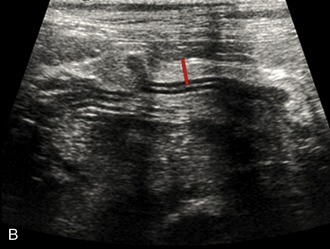
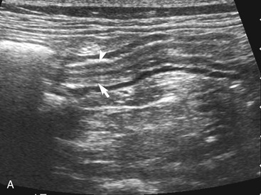
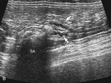
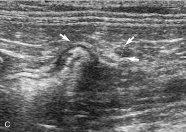
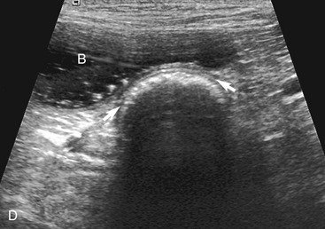
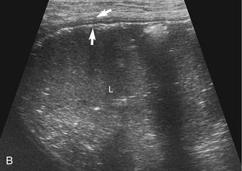
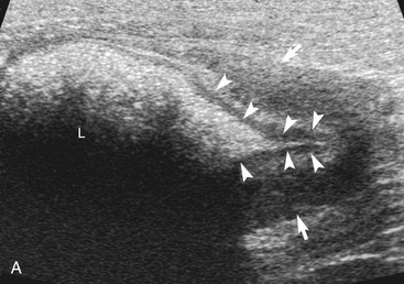
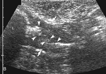
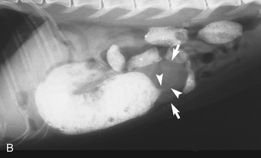
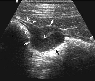
The cause of the obstruction can be intraluminal, mural, or (less often) extramural. In an intraluminal obstruction, the intestinal tract often will be distended with motile, hypoechoic fluid, and possibly gas or food, orad to the point of the obstruction. The fluid may be propelled ineffectively aborad by peristaltic waves and may move back and forth. In a patient with an acute obstruction peristalsis often is increased, but intestinal smooth muscle also may be hypomotile if the obstruction is chronic. If the high obstruction is very proximal within the duodenum, vomiting and thus periodic removal of some of the contents may decrease distension of the lumen. Distension of the intestine due to an obstruction of the distal small intestine may vary depending on the obstruction’s duration and scope (i.e., partial or complete). Segments of small intestine distal to the obstruction usually are of normal size. When a dilated segment of intestine is observed, it is useful to determine the section of intestine involved. To accomplish this, the dilated segment is followed in each direction to the point of the obstruction. By following the segment in each direction, the orad and aborad course of the intestine can be determined. The lumen and wall at the point of the change in diameter of the intestine is examined critically to determine if there is any evidence of a luminal or mural obstruction (Figures 16-8 and 16-9). The lumen is evaluated critically for a focal change in appearance of the contents. This may be manifested by a change in echogenicity, amount of sound attenuated by the contents, or by the shape, size, or margin definition observed. The wall is interrogated for a change in thickness (increased or focally decreased), for a change in echogenicity and visibility of separate wall layers, for gas in the wall, and for plication (Figure 16-10). The adjacent peritoneal cavity also is evaluated for fluid, free air, and hyperechoic omentum, which may be adhered to the serosal wall, consistent with inflammation and possibly intestinal leakage.


As discussed previously, a stricture may cause a distended intestinal lumen orad to the site of obstruction with a normal luminal size distal to the obstruction, and with minimal to no apparent change in the wall thickness at the site of the obstruction. This narrowed area should be monitored for several minutes to rule out the possibility of focal temporary narrowing due to a peristaltic wave. If an obstruction occurs within the ileum, the entire small intestine may be distended. Locating the ileum in the right midabdomen as it enters the colon is useful to make sure that the luminal distension is due to an obstructive process and not due to functional ileus. With exocrine pancreatic insufficiency, malabsorption, feline dysautonomia, or some inflammatory diseases, the intestine may be distended markedly throughout its length without any normal-sized small intestine detected. If the entire small intestine and the colon are dilated and if there is no mass or intussusception at the ileocolic junction, it is unlikely that the luminal dilation was caused by surgery or a mechanical obstruction. In this case a medical cause should be investigated. An exception would be the presence of a volvulus/torsion of the colon and the small intestine at the root of the mesentery. In this situation, which often is accompanied by markedly gas-distended intestinal loops, the patient usually is in critical condition with marked vascular compromise of the intestine.

Stay updated, free articles. Join our Telegram channel

Aug 6, 2016 | Posted by in INTERNAL MEDICINE | Comments Off on Ultrasonographic Imaging of the Gastrointestinal Tract

Full access? Get Clinical Tree

Get Clinical Tree app for offline access