CHAPTER 1 Pyothorax
EPIDEMIOLOGY
Pyothorax is predominantly a disease of young cats (mean age 4 to 6 years), although cats of any age may be affected.1–4 An increased risk for developing pyothorax has been demonstrated for cats from multiple-cat households.3 No breed or sex predisposition has been identified. Currently, there are no data on the incidence of pyothorax. Some studies suggest a seasonal trend with more cases presenting in late summer or autumn.3,5
ETIOPATHOGENESIS
An understanding of the etiopathogenesis underpins recommendations for investigation, treatment, and prophylaxis. Information on mechanisms of infection of the pleural space has remained elusive, because any inciting cause is often no longer evident by the time clinical signs are observed. Pyothorax in cats most often is caused by obligate and facultative anaerobes of oropharyngeal origin (Box 1-1).6 Oropharyngeal flora can gain access to the pleural space by aspiration, direct penetration from a bite wound, or by hematogenous spread from a distant wound. Available evidence suggests that aspiration of oropharyngeal flora, subsequent colonization of the lower respiratory tract, and direct extension is the most common route of infection of the pleural space in cats.7 In a retrospective study, aspiration of oral flora was the most likely mechanism of pleural space infection in 78 per cent of cats in whom probable mechanisms were identified.4 At necropsy, about 50 per cent of cats with pyothorax have pneumonia or pulmonary abscessation, supporting a pathomechanism of parapneumonic spread.7 Aspiration of oropharyngeal flora precedes most cases of human anaerobic pyothorax and equine pleuropneumonia.8,9 Viral upper respiratory tract (URT) infection can impair the mucociliary escalator in cats, human beings, and horses, predisposing them to pleuropneumonia.10–12 Antecedent URT infection has been recognized as a predisposing event in up to 26 per cent of cases of feline pyothorax.7 The fourfold increase in risk of pyothorax in cats from multiple-cat households compared with control cats may be related to the greater risk of developing viral URT infections in this environment.3,13
Box 1-1 Organisms Isolated from Feline Pyothorax
Obligate and facultative anaerobic bacteria, similar in composition to the normal feline oropharyngeal flora, are isolated most commonly. Less than 20 per cent of isolates involve nonoropharyngeal flora.7
OBLIGATE AND FACULTATIVE ANAEROBIC BACTERIA
Bacteroidaceae (Bacteroides spp., Porphyromonas spp., Prevotella spp.) Fusobacterium spp.
Direct inoculation of oral flora into the thorax from a bite wound is likely to be the initiating event in some cases of pyothorax. The importance of this route may have been overemphasized, or it may have become less common, perhaps due to increased neutering and confinement.7 Thoracic puncture wounds were identified in 4 per cent and 16 per cent of cases in recent studies.3,4 If cat fights were a common cause of pyothorax, then free-roaming males likely would be overrepresented, whereas neither outdoor access nor gender have been identified as risk factors for the development of pyothorax. It also might be expected that the prevalence of feline immunodeficiency virus (FIV), a virus spread predominantly by biting, would be similar in cats with pyothorax to that in cats with fight wounds. Combined data on the FIV status of cats with pyothorax indicate that 5.8 per cent of 51 cats tested were seropositive.2–414 The prevalence of FIV worldwide ranges from 1 to 14 per cent in asymptomatic cats, up to 44 per cent in sick cats, and recently has been shown to be 19 per cent in cats with bite wounds.15,16 Although the data are limited, they do not support biting as a major mechanism of infection of the pleural space.
In less than 20 per cent of cases, particularly in kittens, pyothorax is caused by infectious agents other than oropharyngeal flora, emphasizing the need for pleural fluid cytology and culture in all cases. These organisms include Staphylococcus spp., Rhodococcus equi, Nocardia spp., enteric gram-negative organisms (Escherichia coli, Salmonella spp., Klebsiella spp., Proteus spp.), nonenteric gram-negative organisms (Pseudomonas spp.), and protozoa (Toxoplasma gondii).1,2,4,17–20 Fungal causes of pyothorax are rare and include Cryptococcus spp., Candida albicans, and Blastomyces dermatitidis.4,21,22 Mechanisms of infection of the pleural space with nonoropharyngeal flora include penetrating thoracic trauma not associated with a cat bite. However, if environmental contamination of thoracic wounds were a common mechanism, a higher isolation rate of saprophytic bacteria such as Nocardia spp. , Pseudomonas spp., and Mycobacteria spp. would be expected. In contrast to the situation in canine cases, Nocardia spp. are not commonly isolated from feline septic pleural effusions. Other routes of infection with nonoropharyngeal flora include hematogenous spread from a septic focus; perforation of the esophagus, trachea, or bronchi; or migrating plant material and parasitic migration.* Pyothorax and/or pneumonia caused by Salmonella spp. has been documented in cats with concurrent Aelurostrongylus abstrusus infestation.19,28 Migrating lungworm or ascarid larvae can act as carriers for intestinal bacteria. Nonoropharyngeal pathogens are more likely to be isolated from kittens, perhaps because of concurrent age-related infectious or parasitic conditions.4
HISTORY AND CLINICAL SIGNS
Historical and physical examination findings can be attributed to the presence of a pleural effusion and/or to systemic illness. Dyspnea, inappetence, and lethargy are reported most commonly. Pleural effusion and pulmonary atelectasis cause a restrictive pattern of respiration, characterized by an increase in respiratory rate and inspiratory effort and shallow respiratory excursions. Cats typically adopt a crouched, sternally recumbent posture with elbows abducted. Poor body condition, dehydration, and abnormal lung sounds or muffled heart sounds also are common. A fluid line may be appreciated on thoracic percussion. Coughing is reported in 14 to 30 per cent of cases, reflecting pleuritis and/or concurrent pneumonia.2,4,5,22 Pyrexia has been reported in 28 to 50 per cent of cases, although some cats in these series had received prior antibiotic treatment.2,4 Therefore pyrexia at initial presentation may be more common than these figures suggest. Hypothermia, present in 15 per cent of cats, should alert the clinician to the possibility of severe sepsis, particularly when accompanied by bradycardia.3,29 In the largest retrospective study of 80 cats with pyothorax, bradycardia was significantly more common in cats who were hypothermic.3
The duration of clinical signs prior to diagnosis typically is 1 to 2 weeks, but it may be months.2,4,5,23 Because cats are prone to compensate for gradual onset respiratory compromise by reducing activity, signs may be noted only at the acute stage or not at all by the owner. Coupled with the nonspecific nature of many of the presenting signs, many cats are presented late in the course of the disease. By the time clinical signs of respiratory compromise become obvious in feline patients, minimal respiratory reserve remains. Pyothorax should be considered as a cause of sudden death.
DIFFERENTIAL DIAGNOSIS
Pyothorax can be differentiated from other causes of pleural effusion by examination of the fluid obtained at thoracocentesis. Complete diagnostic investigation, including fluid analysis, cytological examination, and culture, should be carried out in all suspected cases of pyothorax, even when the gross characteristics are highly suggestive. This will confirm the diagnosis; identify unusual infections, nonseptic processes, or intercurrent problems (e.g., lungworm infection); and direct appropriate antimicrobial treatment.7
DIAGNOSIS
STABILIZATION DURING DIAGNOSTIC INVESTIGATION
The first 48 hours from presentation is a critical time for pyothorax patients. Most nonsurvivors (60 to 100 per cent) die or are euthanized during this period. Careful handling, early stabilization, and minimally invasive diagnostic procedures are essential for any patient with pleural space disease. The additional stress imposed by transport to the hospital and examination can precipitate respiratory failure. Sepsis or the systemic inflammatory response syndrome also can contribute to these deaths.29,30
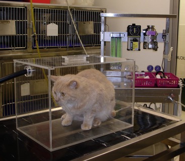
Assessment for, and treatment of, reduced oxygen saturation using supplemental oxygen and therapeutic thoracocentesis is of paramount importance. Oxygen should be administered immediately to cats in severe respiratory distress using a mask or chamber (Figure 1-1). Pulse oximetry can be used to monitor hemoglobin saturation noninvasively. Hemoglobin saturations of less than 90 per cent in patients breathing room air indicate severe hypoxemia and the need for oxygen supplementation. Other considerations for initial patient stabilization include identification and correction of hypothermia, hypotension, hypoglycemia, and fluid and electrolyte imbalances.
DIAGNOSTIC IMAGING
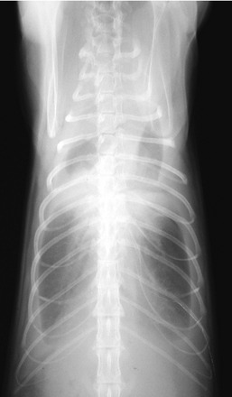
Thoracic ultrasonography is an expedient, noninvasive technique for confirmation of a moderate to large-volume pleural effusion in the dyspneic patient. In contrast to transudates, which are anechoic, the exudate in pyothorax is hypoechoic or complex echoic. The effusion often is septate due to fibrinous or fibrous tags extending between the parietal and visceral pleura. Pulmonary abscesses and restrictive pleuritis also can be identified ultrasonographically. Effusions are bilateral in 70 to 90 per cent of cases.2–417 When sonography is not available, one dorsoventral radiographic view will confirm the presence of a pleural effusion while requiring minimal restraint (Figure 1-2). Severe hypoxemia can occur if cats with large-volume effusions are placed in lateral recumbency for radiography. Horizontal beam radiography can be used to detect pleural effusion in the sternally recumbent patient. Radiographic signs of pleural effusion include retraction of the lobar borders from the thoracic wall, pulmonary atelectasis, and accentuation of lobar edges and interlobar fissures. A complete set of thoracic radiographs should be obtained after drainage of pleural effusion to assess for underlying bronchopulmonary disease.
THORACOCENTESIS
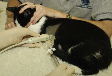
Needle thoracocentesis should be carried out early to obtain diagnostic specimens and to stabilize the patient. This will be tolerated in most cases without the need for sedation. Subcutaneous instillation of local anesthetic (e.g., 0.5 mL of 1 per cent lidocaine) at the thoracocentesis site facilitates the procedure. Diagnostic thoracocentesis should be performed, preferably under ultrasound guidance, at the ventral third of the sixth, seventh, or eighth intercostal space with the cat positioned in sternal recumbency (Figure 1-3). Care should be taken to avoid intercostal vessels and nerves located near the caudal rib margin. A 21- or 23-gauge butterfly needle with extension tubing and three-way tap attached to a syringe can be used for this purpose (Figure 1-4). Once a sample has been obtained for diagnostic evaluation, thoracocentesis is continued to remove as much pleural exudate as possible before general anesthesia for placement of thoracostomy tubes. Thoracocentesis should be carried out bilaterally unless imaging indicates a unilateral effusion.
Stay updated, free articles. Join our Telegram channel

Full access? Get Clinical Tree


