Chapter 14 Tumors of the Urinary System
Renal Neoplasia
I. Primary renal tumors are uncommon, comprising approximately 1% of all neoplasms in dogs and 1% to 1.5% of all neoplasms in cats.
II. Most are of epithelial origin (approximately 85%), metastasize, and are associated with a poor prognosis.
III. Problems at presentation:
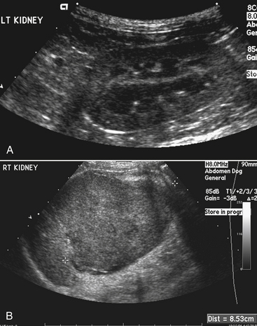
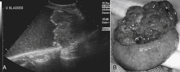
C. Mass effect: Based on abdominal palpation or finding on imaging with ultrasound or radiographs (Figure 14-1).
D. Paraneoplastic syndromes: Polycythemia, extreme leukocytosis, cachexia, hypoglycemia, hypertrophic osteopathy.
Clinical Signs
Nodular Dermatofibrosis With Renal Cystadenocarcinoma in German Shepherd Dogs
I. Autosomal dominant syndrome; 6 % of German shepherd dogs in Norway are affected with renal tumors.
II. Cystadenocarcinoma is usually bilateral and can metastasize to lymph nodes, peritoneum, liver, spleen, lung, and bone.
III. All breeds are potentially at risk but most commonly reported in German shepherds. No sex predisposition.
IV. Lesions of the skin and subcutis are the main reason for presentation. Some dogs are lame as a consequence of lesions on the paws that frequently are overlooked. Ulceration and inflammation can be associated with the skin lesions.
V. Fibrosis may occur both in skin and kidney. Tubular obstruction and cyst formation occur in the kidneys. The lesion may progress from cyst to cystic adenomatous hyperplasia to cystadenoma and ultimately to cystadenocarcinoma.
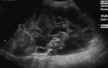
VI. Affected kidneys may be enlarged and abnormally shaped on abdominal radiographs. Cysts of varying size are identified on renal ultrasonography, which also confirms changes in renal size and shape.
VII. Computed tomography identifies multiple cysts and tumor masses of varying size bilaterally. The earliest lesions have been detected between 4 and 5 years of age, with the smallest cysts being 2 to 3 mm in diameter. Small amounts of tumor tissue often can be identified inside the cyst wall or renal parenchyma.
VIII. In one study, mean age at first detection of nodular dermatofibrosis was 6.4 years, renal cystadenocarcinoma with nodular dermatofibrosis was 8.2 years, and death occurred at 9.3 years.
Nephroblastoma (Wilms Tumor, Embryonal Nephroma)
IV. Extremely large in size, causing abdominal enlargement. There may be multiple tumors in one kidney; occasionally they are bilateral.
VI. Prognosis is good if tubular or glomerular differentiation is observed; prognosis is poor if categorized as a sarcoma.
Other Tumors
II. Squamous cell carcinoma of the renal pelvis is uncommon, but has been associated with renal pelvic calculi.
IV. Metastatic tumors are common.
A. Primary pulmonary adenocarcinomas are difficult to distinguish from primary renal adenocarcinoma with pulmonary metastases.
B. LSA is common, and is the most common renal neoplasm in cats.
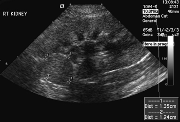
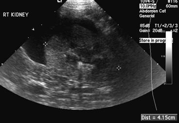
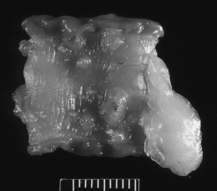
4. A recent study showed that hypoechoic subcapsular thickening of the kidney is associated with renal LSA in the cat (Figure 14-4).
5. Can occur in feline leukemia virus (FeLV)-negative or feline immunodeficiency virus (FIV)-negative cats, but retrovirus status still should be checked because these viral infections can be associated with LSA.
6. Renal LSA usually is bilateral in cats. It can be associated with renal failure if infiltration of the kidneys is extensive.
Treatment
I. If renal neoplasia is unilateral with no metastases, nephrectomy is the treatment of choice. Efforts should be made before surgery to ensure adequate function of the contralateral kidney in patients with marginal renal function before surgery. Renal scintigraphy with determination of individual kidney glomerular filtration rate (GFR) is ideal to make this assessment.
Bladder Neoplasia
V. Canine bladder neoplasia.
G. Rhabdomyosarcoma.
6. Special immunohistochemical staining may be needed to confirm the skeletal muscle origin of the tumor. Desmin, sarcomeric actin, and vimentin are likely to be positive, whereas cytokeratin and smooth muscle actin should be negative on immunohistochemistry. Cross-striations of skeletal muscle are sometimes seen on routine hematoxylin and eosin–stained specimens.
Etiology and Risks
I. Etiology is multifactorial. Risk factors are related to chemical carcinogen exposure. The bladder is the storage site for eliminated waste products, and chronic exposure of bladder tissue to carcinogenic compounds appears to play a role.
II. Obesity. Fat tissue acts as a storage site for lipophilic chemicals and pesticides allowing for prolonged exposure in the body.
III. Previous treatment with the chemotherapeutic agent, cyclophosphamide. Cyclophosphamide frequently causes sterile hemorrhagic cystitis. Chronic irritation of the bladder by this drug may play a role in development of neoplasia.
IV. Bladder tumors are more common in industrialized parts of the world.
A. A direct correlation was found between level of industrialization and manufacturing in the United States and parts of Canada and the occurrence of bladder cancer in dogs.
V. Chemical Exposure.
A. Topical flea and tick dips, shampoos, powders, sprays and collars, and the dose applied were directly correlated with disease in one study.
B. The use of spot-on flea and tick control products containing fipronil and imidacloprid did not cause increased risk of TCC in Scottish terriers.
C. Scottish terriers exposed to phenoxy herbicide lawn treatments had increased risk of developing TCC. Dogs with seasonal or year-round exposure had higher risk than those with only sporadic exposure.
VI The incidence of bladder neoplasia is unrelated to second-hand cigarette smoke exposure or to chronic drinking of chlorinated water (known risk factors in humans).
VII. Neutered female dogs are at higher risk for TCC than males, whereas neutered male cats are at increased risk as compared with females. Potential reasons for this association have been proposed.
VIII. Bladder neoplasia is more common in dogs than cats.
A. Tryptophan metabolism differs between cats and dogs. Dogs metabolize this amino acid by forming ortho-amino-phenol, which is similar in structure to chemicals known to induce bladder tumors in dogs.
IX. Vegetable Consumption.
A. A study of TCC in Scottish terriers found a significant correlation between vegetable ingestion and risk of TCC.
1. Consumption of any type of vegetable at least three times per week decreased the risk of developing TCC by 70%.
2. Green, leafy vegetables and yellow-orange vegetables consumed at least three times weekly decreased risk of TCC by 70% to 90%.
Canine Transitional Cell Carcinoma (TCC)
Signalment
C. Breed predispositions have been identified (Table 14-1). The reason for these associations is unknown but, genetic predisposition is suspected. Differences in metabolic activation and detoxification pathways could account for increased risk in selected breeds.
1. Scottish terriers have the highest prevalence of TCC and are 18 times more likely to develop TCC than mixed breed dogs.
TABLE 14-1 Breed Risk for Diagnosis of Transitional Cell Carcinoma in Dogs
| Breed | Odds Ratio | 95% Confidence Interval |
|---|---|---|
| Mixed breed | 1.0 | — |
| All purebreds | 0.74 | 0.62-0.88 |
| Scottish terrier | 18.09 | 7.30-44.86 |
| Shetland sheepdog | 4.46 | 2.48-8.03 |
| Beagle | 4.15 | 2.14-8.05 |
| Wirehaired fox terrier | 3.20 | 1.19-8.63 |
| West Highland white terrier | 3.02 | 1.43-6.40 |
| Miniature Schnauzer | 0.92 | 0.54-1.57 |
| Miniature poodle | 0.86 | 0.55-1.35 |
| Doberman pinscher | 0.51 | 0.30-0.87 |
| Labrador retriever | 0.46 | 0.30-0.69 |
| Golden retriever | 0.46 | 0.30-0.69 |
| German shepherd | 0.40 | 0.26-0.63 |
Modified from Knapp DW, Glickman WN, DeNicola DB, et al: Naturally-occurring canine transitional cell carcinoma of the urinary bladder: A relevant model of human invasive bladder cancer. Urol Oncol 5:47-59, 2000. Data in this table provides a summary from 1290 dogs with transitional cell carcinoma (TCC) and 1290 institution and age-matched control dogs without TCC in the Veterinary Medical Data Base.
Clinical Signs
Physical Examination
Diagnostic Tests
Laboratory Evaluation
3. Urinalysis.
e. Neoplastic transitional cells are identified in 30% of cases.
(2) Neoplastic cells are difficult to distinguish from reactive epithelial cells that result from chronic inflammation.
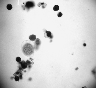
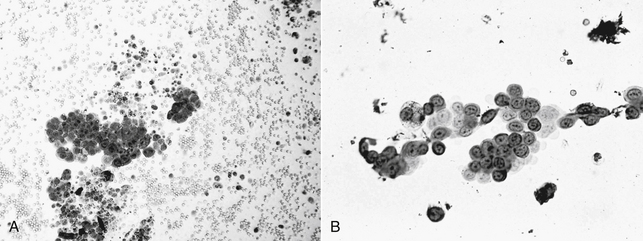
(3) Results may be highly suggestive for TCC (e.g., anisocytosis, anisokaryosis, binucleate cells, mitotic figures, clumps or rafts of epithelial cells) (Figure 14-7).
< div class='tao-gold-member'>
Only gold members can continue reading. Log In or Register to continue
Stay updated, free articles. Join our Telegram channel

Full access? Get Clinical Tree


