CHAPTER 69 Tumors of the Ear
Benign tumors occur less often than malignant tumors in the ears of cats; however, benign masses including inflammatory polyps, basal cell tumors, papillomas/fibropapillomas (feline sarcoids), and ceruminous or apocrine gland adenomas are diagnosed with some frequency. In descending order, the ear tumors diagnosed most commonly in cats are inflammatory polyps, SCC of the pinna, and ceruminous gland adenocarcinomas.1
INCIDENCE AND ETIOLOGY
There are many possible etiologies for the various ear tumors diagnosed in cats. Chronic inflammation has been associated with malignant transformation of normal cells of the ear.2 Ear tumors typically are diagnosed in older cats, although mast cell tumors and inflammatory polyps are seen commonly in younger cats. No gender predilection has been associated with any type of feline ear tumor.
TUMORS OF THE PINNA
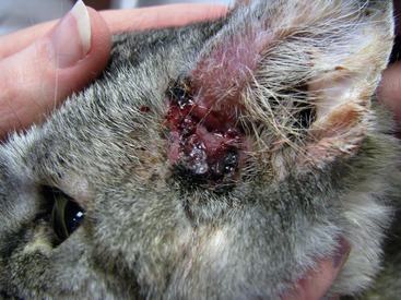
SCC is the most commonly diagnosed neoplasm of the feline pinna. These tumors often are actinically induced as a result of DNA damage secondary to solar radiation. Thin-haired and light-color coated cats are at an increased risk for developing cutaneous SCC. White cats are 13.4 times more likely to develop SCC than cats with other coat colors.1 These tumors occur frequently on the face where the hair is thin and exposure to sunlight is common. Lesions are found commonly on the inner ear pinna, at the ear base, preaurally, periocularly, and on the nasal planum (Figure 69-1). Indoor cats also are at risk. Any cat who sunbathes (whether on the patio or in the skylight) is exposed to solar radiation. The incidence of cutaneous SCC is highest in areas of the country receiving the highest levels of solar radiation annually (up to 26.9 cats out of 10,000).3
Mutations of the tumor suppressor gene p53 have been identified in nine of 11 cats in one study and in two of four cats in another study who were diagnosed with SCC of the ear pinna.4,5 This mimics the same pathway of malignant transformation as seen in human beings. It is often associated with head and neck cancer in people. Retroviral status plays no known role in this disease process; however, the two may represent dual risk factors because outdoor cats have a higher risk of exposure to both sunlight and retroviruses.
Other tumors of the ear pinna in cats also have been associated with sunlight exposure. Basal cell tumors, hemangiomas, and hemangiosarcomas have been associated with UVB exposure in cats. Additionally, specific breeds, such as Siamese, Persians, and Himalayans, may be predisposed to developing basal cell tumors.6
PAPILLOMAS AND FIBROPAPILLOMAS (FELINE SARCOIDS)
Papillomas are rare tumors that occur most commonly on the face (typically on the nose, lip, and pinna) of cats. They also can be found within the external ear canal and on the digits.7 These tumors frequently are pedunculated and rarely ulcerated. Although the causative factor has not been described definitively, a viral etiology most certainly is the culprit. In a study of 19 feline sarcoids, a papilloma virus most similar to bovine papilloma virus I was identified positively in 17 of the lesions.8 The prevalence of feline sarcoids increases in cats with exposure to cattle.
TUMORS OF THE EXTERNAL EAR CANAL
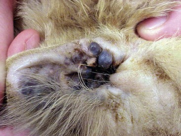
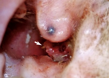
External ear canal tumors are more aggressive in cats than in dogs. More than 85 per cent of these tumors are malignant in cats.9 They arise from the epithelial and adnexal structures of the auditory meatus. Ceruminous gland adenocarcinomas (CGA) are the most common malignant tumors; however, SCC, sebaceous gland adenocarcinomas (SGA), and carcinomas of unknown origin have been described. These tumors may be associated with chronic inflammation of the external ear canal. Benign tumors such as ceruminous gland adenomas and sebaceous gland adenomas also occur. These tumors may be pigmented or nonpigmented (Figures 69-2 and 69-3).
INFLAMMATORY POLYPS
Inflammatory polyps do not represent a neoplastic process; however, they are one of the most commonly identified benign ear masses of cats. These polyps arise from the mucosal lining of the middle ear, Eustachian tubes, or pharynx. They can extend into the external ear canal where they may be visualized with an otoscope, or they may protrude into the nasopharynx. A definitive etiology has not been elucidated, but several causes are suspected to play a role in the development of this disease. Chronic inflammation of the upper respiratory tract or chronic otitis media are commonly accepted factors. Polyps may be congenital in origin or may be secondary to a chronic ascending infection from the nasopharynx. Viruses such as feline herpesvirus-1 (FHV-1) and feline calicivirus (FCV) also have been incriminated, but this possible etiology is controversial. One study of 41 polyps found no virus particles in any of the lesions, whereas other studies have found FHV-1 or FCV in some of the polyps studied.10–13 Although these viruses probably are not necessary for tumor formation, a possible role in development can not be excluded.
MIDDLE EAR TUMORS
Middle ear tumors are quite rare in cats, and very little information exists on their true incidence and possible etiologies. The few reports of middle ear tumors in cats include two carcinomas, three lymphomas (LSA), small numbers of SCC, and one fibrosarcoma. Of the cats with LSA, only one was tested and it was found to be negative for retrovirus infection. Retroviral status of the other cats was not reported.1,14,15
BIOLOGICAL BEHAVIOR AND PATHOGENESIS
Ear tumors in cats tend to be aggressive. Their behavior often is characterized by rapid local invasion, and lymphatic or vascular spread. Distant metastasis is uncommon. Five of 56 cats with various carcinomas of the ear canal had evidence of metastasis to local lymph nodes; none had evidence of lung metastasis during the study, although lung metastases were found in one cat during a postmortem examination.9 Benign tumors are less likely to be ulcerated and tend to have a narrow base of attachment. Additionally, they are more likely to be lobulated and typically do not invade into the cartilage like their malignant counterparts.9
TUMORS OF THE PINNA
SCCs of the pinna tend to start as hyperemic lesions, and progress to erythematous crusts and ulcerated lesions. Premalignant lesions often are described as actinic keratosis on histopathological examination. These neoplasms tend to be locally invasive with a low metastatic rate. If these tumors metastasize, they usually spread first to the local lymph nodes, then to the lungs. Approximately 15 per cent of affected cats will have multicentric actinically induced lesions involving the pinna, nasal planum, and/or the periorbital region.1
Mesenchymal tumors of the pinna are rare. Rhabdomyomas are benign striated skeletal muscle tumors that have been reported in four cats.16 No association with sun exposure or other etiological factors has been identified. Fibrosarcomas of the pinna are non–vaccine-related soft-tissue sarcomas that are locally invasive with a low metastatic rate of approximately 15 per cent.2 Non–vaccine-associated fibrosarcomas tend to occur on the head and extremities and usually are less aggressive than their vaccine-induced counterparts. Vascular tumors tend to occur in the inguinal region and on the ears of white cats. An association with UVB radiation has been proposed.17
Hemangiosarcomas tend to be locally invasive, although metastasis is rare. However, full staging is still recommended for cats with malignant vascular tumors.6,17
Melanomas (MSA) are rare in cats. They occur most commonly as ocular tumors (see Chapter 70); however, dermal MSAs have been diagnosed on the pinnae of cats. One study of 23 cats with dermal MSA included four cats with MSA of the pinna. Three of these cats were cured with surgery alone, whereas one cat experienced recurrence with evidence of distant metastasis.18 These tumors can be considered to have a benign behavior, although a subset exists with a more aggressive phenotype.
Cutaneous mast cell tumors are uncommon in cats; however, when they occur, they are commonly located on or near the ear (see Chapter 67). These tumors have a benign behavior and often are cured with surgery alone. These tumors rarely recur even with incomplete surgical margins.
Papillomas/fibropapillomas (feline sarcoids) do not metastasize and generally have a benign behavior. However, they are likely to recur after surgery. This behavior is consistent with the proposed viral etiology, because these tumors behave similarly to virally-induced papillomas of other species.7
TUMORS OF THE EXTERNAL EAR CANAL
Tumors of the external ear canal are often slow to metastasize and are locally invasive. Occasionally cats present with a very aggressive phenotype with early spread to local lymph nodes and lungs. The degree of invasion is prognostic for these tumors.9 Ceruminous gland carcinomas often destroy the adjacent soft tissues and are capable of distant metastasis. They have the longest median survival time of the malignant external ear canal tumors when treated aggressively.9
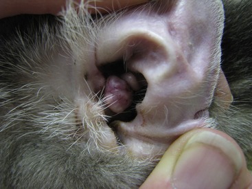
SCC of the external ear canal is a very aggressive tumor. These tumors often are ulcerated, painful, and highly invasive into the local structures. The lesions are difficult to excise completely, and they respond poorly to adjunct therapies such as radiation or chemotherapy (see Figure 69-4). Carcinomas of unknown origin have a similar prognosis to SCC of the external ear canal.9
Two cats reportedly have been diagnosed with meningeal carcinomatosis secondary to SCC of the ear. Both patients had been diagnosed previously with SCC of the pinna and had recurrence within the external ear canal or along the previous pinnectomy site. Both cats presented with neurological signs, with one patient being slowly progressive over several months whereas the other presented with an acute onset of neurological abnormalities.19
CLINICAL SIGNS ASSOCIATED WITH EAR TUMORS
TUMORS OF THE PINNA
SCCs often start as small hyperemic lesions that progress to erythema. Eventually crusting is seen when the lesion progresses to ulceration and then to an invasive lesion that is highly destructive, painful, and often open and draining. These tumors can invade deep into the ear canal and even affect the facial nerves, causing hearing loss, Horner’s syndrome, or facial nerve paralysis. Eventually these tumors can invade the brain stem or the brain itself.1
Basal cell tumors are dermal, well-circumscribed, pigmented masses that may be filled with a brown fluid. They may be nonpigmented or hyperpigmented. They are nonpainful, but occasionally can ulcerate. They have been reported to range from 0.5 to 3 cm in size and are slow growing.1 These tumors almost always are benign but rare carcinomas do occur. Invasiveness is prognostic, and a mass that appears to invade into underlying structures should be treated more aggressively.
Mast cell tumors typically are well circumscribed. They are dermal in location with intact overlying skin. They may be erythematous and edematous, but rarely are ulcerated. These masses tend to be slow growing with little impact on the cat’s quality of life. Young Siamese cats are at an increased risk for developing multiple mast cell tumors of the head. These tumors may resolve on their own with time.4
TUMORS OF THE EXTERNAL EAR CANAL
SCC in this location is more likely to invade deep into normal structures and cause neurological signs, when compared with other tumors of the external ear canal (Figure 69-4). All external ear canal tumors can cause otorrhea with dark, bloody, and often foul smelling discharge. Secondary infections due to the breakdown of the natural protective barriers also are common. Most cats demonstrate pain and often will guard the affected ear. Sedation may be needed for a thorough otoscopic examination.
Stay updated, free articles. Join our Telegram channel

Full access? Get Clinical Tree


