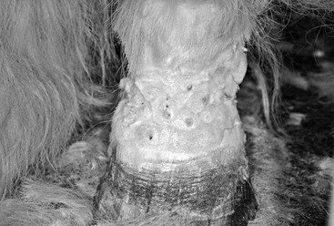
Rosanna Marsella Ticks can cause dermatologic disease in a variety of ways, most commonly by inducing a nodular reaction at the site of the bite. The inflammatory response is determined by the interaction between the tick and the immune response of the host. In first exposures, the main reaction is a toxic one, which manifests as necrotic changes in the epidermis and dermis and a subsequent inflammatory response to the tissue damage. In animals that have already been exposed and have developed sensitization, the inflammatory response to the bite is more severe and persistent. Pyogranulomatous reactions are common and are manifested clinically by the development of hard nodules at the site of the bite that can open and drain a purulent exudate. Pruritus is variable. Different types of hypersensitivities may be developed against tick bites. Some individuals can build type I hypersensitivity and, when challenged, develop a generalized pruritic papular reaction. Others develop generalized urticaria that may persist for weeks, and even angioedema. Ticks can also trigger type III hypersensitivity, which leads to development of vasculitis-type lesions. Vasculitis can present as punctuate ulcerated lesions that, in severe cases, coalesce and form large necrotic areas. Body sites that are more prone to vasculitis are the extremities (e.g., tip of the ears and tail) and the lower limbs. Generalized malaise may be observed, as well as fever and edema. Secondary infections are common and should be treated aggressively. By their ability to transmit various viral, rickettsial, and bacterial diseases, ticks are additionally able to trigger vasculitis through the development of those infections. With regard to classification, ticks are divided into soft ticks and hard ticks. An example of soft tick (argasid) is Otobius megnini, also called the spinous ear tick. This tick lays eggs in crevices in the environment and the larvae invade the ears of the host, where they can cause severe otitis. Clinical signs include severe inflammation of the ear canal, head shaking, and ear rubbing. In severe cases, head tilt and muscle spasm have been described. Diagnosis is made by detection of the tick. Treatment involves physical removal of the ticks and cleaning of the exudate. Secondary skin infections should be properly diagnosed and treated. Cytology may aid in the initial assessment of the infection by providing information on the presence and type of bacteria or yeasts. Hard ticks are ixodids, and examples include Ixodes, Dermacentor, and Amblyomma species. Ixodes can transmit Lyme disease, which is caused by infection with Borrelia burgdorferi (see Chapter 91). Such infection is common in horses and ponies from the New England and mid-Atlantic regions of the United States. Although horses appear to be less predisposed to development of the disease than humans, they can still develop clinical signs. In horses, clinical signs include shifting lameness, poor performance, personality changes, laminitis, anterior uveitis, arthritis, fever, edema, and encephalitis. In humans, the early signs may also include a characteristic circular skin rash called erythema chronicum migrans. This skin lesion develops at the site of the tick bite a few days to several weeks later. The area is erythematous and warm but is generally painless. These circular macules of erythema show some clearing in the center, developing the appearance of a bull’s eye. In horses, cutaneous lesions are possible but are typically missed because of the hair coat. Lyme disease is diagnosed on the basis of a combination of clinical signs and blood tests to detect antigen-specific antibodies. Even if antibodies are detected, however, this does not necessarily mean that clinical signs are caused by Lyme disease. Vaccinated horses, as well as those that have been exposed to the Lyme disease bacterium but do not have the disease, will develop an antibody titer against Borrelia. In a recent study, luciferase immunoprecipitation systems for profiling antibody responses against three different antigenic targets for the diagnosis of equine B burgdorferi infection were evaluated and appeared to be promising for evaluation of antibody responses during the course of Lyme disease in horses. In another study, infections with B burgdorferi were detected in 8% of equine samples in New York State. In an experimental model of Lyme disease in ponies, skin changes consisting of lymphohistiocytic nodules up to 2 mm in diameter scattered about the middle and deep layers of the dermis were reported. A case of pseudolymphoma associated with Borrelia has also been described in a horse that developed multiple dermal papules following removal of a tick from the same site 3 months earlier. Histologic examination of a papule biopsy specimen was suggestive of either a T-cell–rich B-cell lymphoma or cutaneous lymphoid hyperplasia. Several mites can cause dermatologic disease in horses, but at present, none of these conditions is reportable, according to the U.S. Department of Agriculture. Regulations in individual states may be more rigorous and should be followed. Spreading scabies in the horse population would have major consequences. For this reason, if scabies is strongly suspected, it is prudent to contact the local regulatory office, regardless of the regulations. Chorioptes infestation is a common cause of dermatitis in horses. This is a superficial mite that completes its cycle entirely on the host animal. The mite can survive for many weeks in the environment, so environmental disinfestations are an important part of therapy. Higher numbers of mites infest horses in the colder months, and clinical signs are typically worse in the winter. Chorioptic mange is most commonly seen in feathered horses. The mites cause a primary papular eruption that involves the pastern and fetlocks, giving rise to a condition with the name leg mange. Pruritus is variable and can particularly be aggravated by the secondary development of skin infections. Thus, chorioptic mange should be always considered as a differential diagnosis in cases of pastern dermatitis affecting draft horses (Figure 130-1). Because this disease is contagious, other horses in the herd are at risk for becoming infested, although the severity of clinical signs varies among individuals. Skin scrapings are recommended to confirm the clinical suspicion. These mites are fast movers, and it is helpful to apply fly spray before performing the skin scraping to increase the likelihood of detecting mites on the scraping.
Tick- and Mite-Associated Dermatologic Diseases
Ticks
Mites
Chorioptes

< div class='tao-gold-member'>
![]()
Stay updated, free articles. Join our Telegram channel

Full access? Get Clinical Tree


Tick- and Mite-Associated Dermatologic Diseases
Chapter 130
Only gold members can continue reading. Log In or Register to continue
