Thoracolumbar Spine

Chapter 52Thoracolumbar Spine


Back problems are a major cause of altered gait or performance (see Chapters 50, 51, and 97). Both identification and documentation of vertebral lesions are difficult in horses; therefore treating back pain is a challenge for the equine clinician. The equine back is a large area covered by thick muscles. Therefore assessment of the bony elements is limited. Every joint in the thoracolumbar region has restricted mobility; therefore detecting changes in restricted movement when the horse is working is difficult. Diagnostic imaging of the thoracolumbar region is also limited, and radiological assessment requires special equipment. Specific treatment of back pain can only be performed after complete identification of the site and nature of the lesions.


Anatomy and Function


Bones


The thoracolumbar spine is basically composed of 18 thoracic vertebrae (T1 to T18), six lumbar vertebrae (L1 to L6), and five fused sacral vertebrae (S1 to S5). Some horses have individual (congenital) variations at the cervicothoracic, thoracolumbar, and lumbosacral junctions. The most common is the sacralization of the last lumbar vertebra (sacralization of L6), which can be seen by ultrasonography. Transitional vertebrae, with a rib on one side and a transverse process on the other side, can be found at the thoracolumbar junction. Intervertebral ankylosis alters the biomechanical behavior of the involved part of the spine, especially at the lumbosacral junction, which in normal horses is the most mobile joint between T2 and S1. Ankylosis of this joint puts more stresses on the caudal lumbar intervertebral joints and may predispose to osteoarthritis (OA) of the facet joints and intervertebral disk lesions. Fusion of the lumbar transverse processes (see the following discussion) is also sometimes seen, and this has similar consequences on adjacent intervertebral joints, but the consequences are fewer because little movement usually occurs in these joints. Transitional transverse processes, or ribs that do not involve the vertebral body, have less biomechanical significance.


The first 10 thoracic vertebrae have long spinous processes. These have a dorsocaudal orientation and provide insertion for the strong but elastic supraspinous ligament. The anticlinal vertebra, the vertebra with a spinous process perpendicular to the vertebral axis, is usually T15. Caudal to the anticlinal vertebra the spinous processes are orientated obliquely dorsocranially. The spinous processes are higher between L2 and L5. Therefore lumbar muscular atrophy results in a kyphotic appearance of the lumbar area.


In most horses the spinous processes of L6 and S1 are divergent, allowing a range of flexion and extension movements at the lumbosacral joint. The transverse processes from L5 (sometimes L4) to S1 articulate through intertransverse synovial joints, which limit lateral flexion in this area. The lumbar vertebral bodies are bigger than the thoracic ones and have a ventral crest for the insertion of the diaphragm.


Joints


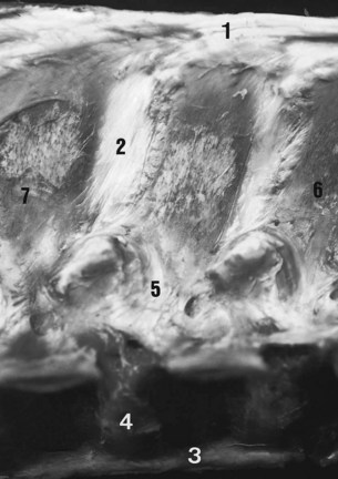
Stability of the thoracolumbar vertebrae is provided by the supraspinous and interspinous ligaments, the joints between the cranial and caudal articular processes (the facet joints), the joints between the vertebral bodies, and the dorsal and ventral longitudinal ligaments. Stability of the spinous processes is provided by the supraspinous and interspinous ligaments (Figure 52-1). These ligaments are wider and more elastic in the cranial and middle thoracic areas, permitting more movement than in the caudal thoracic and lumbar regions.


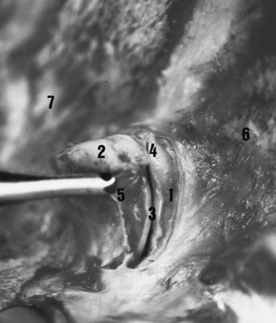
The caudal and cranial articular processes articulate via synovial intervertebral articulations (the facet joints). At the base of the spinous processes these joints are symmetrically placed on each side of the median plane (Figure 52-2). These are typical synovial (diarthrodial) joints with articular cartilage, a closed synovial cavity containing synovial fluid, a synovial membrane, and a fibrous capsule. They have a single flat articular facet in the cranial thoracic area (up to T12) and two angulated articular facets between T12 and T16. From T17 to S1 the articular facets are congruent, with a cylindrical shape aligned on a paramedian axis. These regional variations are correlated with the limited mobility of the lumbar spine and the wider range of movement in the thoracic region, including flexion and extension in the median plane, lateral flexion in the horizontal plane, and rotation. The vertebral bodies are stabilized by joints composed of a fibrous intervertebral disk and two longitudinal ligaments. The ventral longitudinal ligament is replaced by the longissimus cervicis muscle in the cranial thoracic area. The dorsal longitudinal ligament is located in the vertebral canal and adherent to the dorsal border of each intervertebral disk.


Muscles


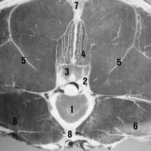
The vertebral column is moved by wide muscles (Figure 52-3). The strong epaxial muscles, located dorsal to the vertebral axis, have an extensor effect when contracted bilaterally. Unilateral contraction induces lateral flexion and contributes to rotation of the vertebral column. Electromyographic studies show that the epaxial muscles limit flexion and stabilize the vertebral column during the suspension phase at the trot.1,2


The epaxial muscles include the following:


The longissimus dorsi is the strongest muscle. The iliocostalis muscles are small but have a greater role in lateral flexion because of their eccentric location. Caudally, these muscles fuse to form the erector spinae muscle. The multifidus muscle lies under the spinosus muscle and is in close contact with the vertebrae (juxtavertebral muscle). This muscle plays a major role in the stability of the vertebrae and in the proprioceptive adjustment of the spine.


The hypaxial muscles, located ventral to the vertebral axis, have a flexor effect on the spine when contracted bilaterally. Unilateral contraction induces lateral flexion and contributes to rotation of the vertebral column. The hypaxial muscles include psoas minor and major, rectus abdominis, and rectus oblique.


The psoas minor and major muscles insert on the ventral aspect of the lumbar and caudal three thoracic vertebrae (juxtavertebral muscles). They act mainly at the lumbosacral junction but are also able to flex the thoracolumbar junction and the lumbar spine. The rectus abdominis muscle is an effective flexor of the complete thoracolumbar spine because of its eccentric insertions on the pubis, sternum, and ventral part of the ribs. The oblique muscles can create lateral flexion and rotation of the thoracolumbar spine because of eccentric insertions on the tubera coxae and ribs. Electromyographic studies show that the rectus abdominis muscles act to limit extension and stabilize the vertebral column during each diagonal stance phase at the trot.1,2


Diagnosis of Back Pain


The objectives of clinical examination of the horse’s back3-7 are to determine whether back pain is present, the site or sites of pain, and the potential lesions responsible for the pain. Acute back pain can arise after traumatic injuries, such as a fall, or after an awkward jump. Acute pain and muscle spasm may be identified and may be the primary lesion, but a complete radiographic examination of the thoracolumbar spine may be indicated to assess for vertebral fractures or long-standing bony lesions that may be responsible for the accident. These lesions may become clinically significant because of overstress on the intervertebral structures. For example, a long-standing spondylosis lesion may cause acute pain on landing after a jump.


A poor rider or an ill-fitting saddle may cause back pain, but correction of these problems may not be easy. It is important to manage these causes of back pain before proceeding with more advanced imaging techniques.


Back pain may also be manifest by abnormal behavior, for example, bucking (see page 992).


Physical Examination


Physical examination is essential in diagnosing back pain. Only the main criteria for each step or procedure are discussed. It is obviously important to perform a comprehensive evaluation of the whole horse to identify any other potential problems that may contribute to gait abnormalities or poor performance.


Inspection


The most commonly described abnormal curvatures of the back are lumbar kyphosis and thoracic lordosis. These can be seen in the same horse. Atrophy of the epaxial muscles in the lumbosacral regions results in prominence of the normal summits of the spinous processes and apparent kyphosis of the lumbar spine. Detection of atrophy of the epaxial muscles is a key finding in a horse with potential back pain because atrophy reflects the reduction of movement in the painful areas. The finding can help provide information on the possible location of the lesions. However, one should bear in mind that muscular development also reflects the horse’s previous work history, and if the horse has never worked properly through its back, the epaxial muscles will be poorly developed.


Thoracic lordosis may be seen in clinically normal horses and does not necessarily imply the presence of back pain. However, a short coupled horse with lordosis is more likely to have impinging spinous processes, a reflection of the horse’s normal conformation.


Abnormal swellings in the saddle area or abnormal hair loss may reflect a poorly fitting saddle or the position and balance of the rider. A rider who sits crookedly predisposes to excessive movement of the saddle and asymmetrical hair loss. However, it should be noted that hindlimb lameness may also induce abnormal movement of the saddle.


The stance of the horse may also be important; some horses with back pain may stand either camped out or camped under. However, a normal stance certainly does not preclude thoracolumbar region pain.


Mobilization


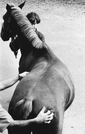
Stimulation of movement of the thoracolumbar spine (Figure 52-4) is important to assess the amount of movement tolerated by the horse and any signs of pain, such as flexion of the limbs, alteration of facial expression, tension of the back muscles, movement of the tail, and alteration of behavior (kicking, rearing, bucking, and grunting).


The following protocol is recommended:


The clinician should try to determine which movements are restricted or not tolerated so as to determine potential sources of pain. These movements can be induced by skin stimulation of the dorsal and lateral aspects of the trunk and hindquarters. Although some horses respond to soft digital stimulation, in others a stronger stimulus is required, for example, using the tips of a pair of artery forceps. Firm stroking with a hard instrument displaced craniocaudally and inducing spectacular wide extension and flexion movements may lack sensitivity and specificity in determining the site of back pain, but firm stroking may be necessary in extremely stoical cob-type horses to induce any movement. A normal, relaxed horse is able to flex and extend the thoracolumbar spine smoothly and repeatedly. The degree of movement reflects in part the type of horse. Cob-type horses naturally tend to have much more restricted movement than TBs or Warmbloods. The clinician will find it useful to keep one hand resting on the midback region during these maneuvers to detect induced muscle spasm or abnormal cracking of the muscles or ligaments (a crepitus-like feeling as the epaxial muscles or ligaments contract and relax). Further descriptions of assessment of thoracolumbar movement are provided in Chapter 51. Pressure algometry has been used to more objectively quantify back pain.8,9


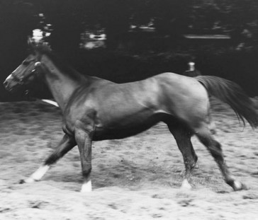
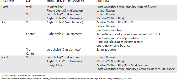
Examination during Movement


Evaluation of the horse moving at walk, trot, and canter is essential to assess whether pain is present and to identify functional disorders, such as limitation of regional intervertebral mobility (Figure 52-5). The clinician should always bear in mind that impinging spinous processes can be present asymptomatically. Therefore the clinical significance of impinging spinous processes should not be overinterpreted, unless clinical signs of back pain are evident. The horse should be assessed moving in straight lines and in small circles at a walk and trot on a hard surface and moving at a trot and canter on the lunge to determine whether any reduction of back mobility is apparent (Table 52-1).


In vivo kinematic studies have quantified dorsoventral flexibility of the back in sound horses trotting10,11 and at various gaits on the treadmill.12 Horses with vertebral lesions showed a reduction of passive flexibility of the back at trot,13-15 with reduced flexion and extension, lateral flexion, and rotation. Thus the horse may appear to hold its back rather stiffly. This can be caused by mechanical problems (partial or complete ankylosis) or pain. Back pain may also influence stride length and limb flight, resulting in a more restricted and less animated gait. On the lunge the horse may show loss of balance and a tendency to lean the body rather than bend the trunk toward the direction of circle. However, such clinical signs may also be seen in association with lameness, which, if bilateral, may not be obvious.


Examination of the Horse Being Ridden or in Harness


The presence and the degree of back pain may be underestimated unless the horse is evaluated under its normal working conditions, that is, ridden or in harness. The influence of a rider on mobility of the horse’s back has recently been quantified.16 The clinician should watch carefully as the tack or harness is applied, particularly as the girth is tightened. However, the clinician should bear in mind that cold-back behavior (see Chapter 97) is not necessarily a reflection of back pain, although it may be. The fit of the saddle for the horse and the rider should be evaluated. Back mobility,3 the movements that the horse finds difficult, and the horse’s attitude toward work should all be assessed (see Chapter 97). The clinician should pay attention also to the observations of the rider because the horse may feel considerably worse than it appears. The rider may describe lack of hindlimb power, lateral stiffness of the back to the left or right, unwillingness of the horse properly to take the bit, or loss of fluidity in the paces. The rider may complain of back pain induced by riding the horse.


Examination of the horse while it is ridden also gives the clinician the opportunity to assess the rider because back pain is readily induced by poor riding, a situation that may not be easy to handle diplomatically.


Jun 4, 2016 | Posted by in EQUINE MEDICINE | Comments Off on Thoracolumbar Spine

Full access? Get Clinical Tree

Get Clinical Tree app for offline access