The Urinary System

CHAPTER 38 The Urinary System


The urinary tract of the dog and cat consists of the kidneys, ureters, urinary bladder, and urethra. The kidneys play an important role in the regulation of the body’s water balance, electrolytes, and acid-base status. The kidneys are also involved in hormone metabolism and the elimination of toxins and wastes. Diseases of the urinary tract of puppies and kittens may be caused by congenital or acquired disorders. Congenital disorders are the result of abnormal development of the structures within the urinary tract, whereas acquired disorders may result secondary to congenital disorders or when the normally developed urinary tract becomes diseased. To further understand the congenital and acquired diseases of the urinary tract, a review of embryology and developmental physiology has been provided.


Normal Development


The development of the kidney is complex. There are three stages of development: the pronephros, mesonephros, and metanephros (Table 38-1). A schematic diagram of this process is illustrated in Figure 38-1. Nephrogenesis continues for at least 2 weeks postnatally in the dog. In newborns, the inner glomeruli are larger than those in the outer cortex. However, all cells within the proximal convoluted tubules have a significantly smaller membrane area than the adult. These differences gradually disappear as the kidney grows.


TABLE 38-1 Developmental embryology of the kidney
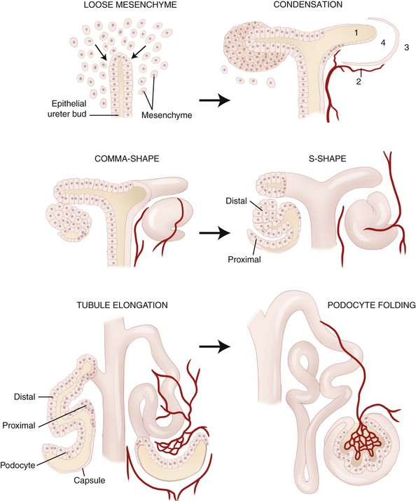


Stage Description
Pronephros


Mesonephros


Metanephros


In cats, normal renal development can be permanently altered by taurine deficiency. In addition to a decreased size, which is apparent by 8 weeks of age, kittens from dams with taurine deficiency have ureteral dilatation, large sclerosed glomeruli, proximal tubular flattening, epithelial atypia, reduced mitochondria at the apex of the tubule, and simplification of the tubule compared with age-matched controls.


The epithelial lining of the urinary bladder and urethra is endodermal in origin. The urinary bladder originates from the cloaca. The urorectal septum forms in the cloaca, which separates the rectum dorsally from the urogenital sinus ventrally. The cranial urogenital sinus becomes the vesicourethral canal and urinary bladder. The caudal urogenital sinus becomes the penile urethra in the male and urethra and vestibule in the female. The mesonephric ducts enter the dorsal aspect of the urogenital sinus in what becomes the trigone.


Physiology


Excretion and Reabsorption


During development, urine and wastes excreted by the fetal kidneys pass from the developing urinary bladder through the urachus into the allantoic cavity of the placenta. Waste products are then absorbed into maternal circulation. At birth, the neonatal puppy and kitten kidney is immature in both structure and function. Functionally the newborn has a lower glomerular filtration rate (GFR), renal plasma flow, and filtration fraction compared with the adult. The GFR increases with age postnatally. Glomerular capillary surface area and pore density increase between the first and sixth weeks after birth. Studies suggest that GFR and renal blood flow increase up to 11 weeks of age in the puppy and up to 9 weeks of age in the kitten before reaching adult levels.


There is limited evidence that puppies are capable of concentrating and diluting their urine at birth. When measuring urine-specific gravity, the urine may not be maximally concentrated, particularly in puppies less than 4 weeks of age, but this may be a reflection of the water content in their diet rather than an inability to concentrate their urine. Puppies also produce a higher daily urine volume and have an increased extracellular fluid volume compared with adult dogs. The urine and plasma osmolality in puppies and kittens increases with age and reaches adult values by 11 weeks of age in the puppy and 13 to 19 weeks of age in the kitten.


Neonatal renal handling of water and electrolytes differs from adults. The fractional reabsorption of water in dogs is constant, whereas sodium excretion increases during the first 3 weeks. Increases in renal arterial blood pressure are associated with increases in both absolute and fractional sodium excretion by adult and newborn dogs. In puppies, the fractional excretion of potassium and phosphorus decreases between 9 and 27 weeks of age, whereas the fractional excretion of chloride and calcium increases in the developing puppy. Despite these findings, the fractional excretion of all electrolytes in the puppies in these studies was within the normal range established for adult dogs. When the fractional excretion of sodium, potassium, chloride, phosphorus, and calcium was evaluated in kittens from 4 to 30 weeks of age, the only significant change observed during the study period was an increase in fractional excretion of potassium, but all values were within adult reference ranges.


Postnatal excretion of uric acid decreases from 83% at birth to 51% by 90 days of age. Similar to human infants, the decrease in uric acid excretion with age appears to be unrelated to binding of uric acid to plasma proteins or to urine flow rate. The amount of protein in the urine is higher in newborn puppies but decreases within the first few months of life. Possible contributors to proteinuria include colostral proteins absorbed within the first few days of life, increased filtration by immature and fewer glomeruli, decreased renal tubular reabsorption, alkaline urine, urine concentration, and protein from cells in the urinary tract.


Examination of the Urinary System


Physical examination of the patient with urinary tract disorders may reveal urinary incontinence, lower urinary tract symptoms, or clinical signs of renal insufficiency or failure. Urinary incontinence may be intermittent and only noticed after the animal lies down for prolonged periods, or the incontinence may be more persistent, resulting in urine dribbling. When urine dribbling is noted, it is important to differentiate true incontinence from dribbling caused by overdistention, such as can be seen with urethral obstruction or neurologic bladder.


Lower urinary tract signs include pollakiuria, dysuria, stranguria, and hematuria. Lower urinary tract signs in young dogs and cats are seen most frequently with infections or calculi as a result of a congenital disease. Hematuria may be the only historic finding in diseases such as renal telangiectasia and idiopathic hematuria. If hemorrhage is severe, blood clots may form and lead to urinary tract obstruction. Anemia may also occur in dogs with idiopathic hematuria and renal telangiectasia.


Clinical signs of renal insufficiency include lethargy, poor growth, poor haircoat, weight loss, polydipsia, polyuria, and nocturia. With progression to renal failure, signs of uremia are seen, including anorexia, dehydration, pale mucous membranes, oral ulceration, halitosis, vomiting, hematemesis, diarrhea, melena, and death. Kidneys may be palpably enlarged with polycystic kidney disease. In most dogs and cats with primary renal disease, kidneys may be normal or small in size. Signs related to renal secondary hyperparathyroidism are more common in young dogs because their bones are more metabolically active. These signs include enlargement of the maxilla and mandible, a pliable mandible, bone pain, and pathologic fracture.


Hematologic and biochemical abnormalities are most often seen in animals with renal failure and include azotemia, hyperkalemia, hyperphosphatemia, and metabolic acidosis. Regenerative anemia is occasionally found with renal failure as a result of gastrointestinal ulceration and hemorrhage. Regenerative anemia also occurs in cases of significant blood loss through the urinary tract as seen with idiopathic hematuria and renal telangiectasia. In cases of chronic renal failure, nonregenerative anemia may be present.


Examination of the urine may be normal or reveal isosthenuria, proteinuria, glucosuria, hematuria, crystalluria, pyuria, and/or bacteriuria. Urinary fractional excretion of electrolytes may be abnormal with some tubular disorders. Urine culture is indicated in cases of suspected infection of the upper or lower urinary tract.


Diagnostic imaging of the urinary system includes plain radiography, contrast radiography, ultrasonography, and cross-sectional imaging. Plain radiographs may be used to evaluate the size and position of the kidneys and urinary bladder. This may be difficult in emaciated animals or in the presence of peritoneal or retroperitoneal fluid. Soft tissue mineralization and certain types of calculi may also be visualized on plain radiographs.


Contrast studies of the urinary tract include excretory urography, antegrade pyelography, and retrograde contrast administration. Typically this is done with iodinated contrast agents. Excretory urography can be used as a crude measure of renal function because excretion of the contrast agent depends on renal blood flow, GFR, and tubular reabsorption of water. Most commonly, excretory urography is used to evaluate the kidneys and ureters for position and filling defects. With antegrade pyelography, iodinated contrast is injected directly into the renal pelvis in an attempt to identify lesions of the ureter.


Retrograde contrast studies involve placement of a balloon catheter distal to the area of interest, followed by administration of an iodinated contrast agent. This technique can be used to evaluate lesions in the vagina, urethra, urinary bladder, and distal ureters. With double-contrast cystography, iodinated contrast agent and air are administered through a urinary catheter. This is typically done to evaluate the wall of the urinary bladder for thickening or mass lesions, as well as its contents (e.g., calculi). However, care must be taken when placing any type of urinary catheter in a pediatric patient because of the possible risk of iatrogenic urethral perforation.


Transabdominal ultrasound is used primarily to evaluate the kidneys, urinary bladder, and proximal urethra. Ultrasound is a useful determinant of size and echotexture of the kidneys, as well as a means of identifying filling defects. The renal pelvis can be evaluated for dilation, calculi, and mineralization. Normal ureters are poorly visualized with ultrasound because of their small size, but if they are dilated, as can occur with an obstruction, they can usually be visualized with ultrasound. Occasionally the opening of the ureters into the urinary bladder can be seen as the expulsion of urine from the ureters into the urinary bladder. The urinary bladder wall can be assessed for abnormalities and the vesicular contents seen. Although transabdominal ultrasonography can be used for the proximal urethra in females and prostatic urethra in males, transrectal ultrasonography is necessary for the more distal portion of the female urethra and prostatic and pelvic portions of the male urethra in the dog.


Magnetic resonance imaging and computed tomography are occasionally used when developmental abnormalities are suspected. Computed tomography using intravenous urography is believed to be superior to traditional contrast radiography for the diagnosis of ectopic ureters.


Flexible and rigid endoscopy has both diagnostic and therapeutic implications in diseases of the urinary tract. Endoscopy allows visualization of the vagina, urethra, and urinary bladder of dogs and cats. Endoscopy can also be used to aid in obtaining biopsies, in performing urethral injections of collagen in cases of urinary sphincter incompetence, and in performing urethral and vesicular lithotripsy and stone removal.


Biopsy is necessary to definitively diagnose many primary renal disorders such as glomerulonephropathies, amyloidosis, and renal dysplasia. Biopsy is also indicated for definitive diagnosis of mass lesions within the urinary tract. In cases of recurrent urinary tract infections, biopsy of the urinary bladder wall may be indicated to identify deeper tissue infections.


Congenital Disorders


Some congenital diseases are incompatible with life, and clinical signs of renal failure develop within the first few weeks of life. However, most of the diseases are not immediately life threatening, and clinical signs might not be apparent for several months or even years. The majority of congenital diseases manifest with urinary incontinence, lower urinary tract signs, renal insufficiency, or chronic renal failure.


Congenital disorders of the urinary tract may be caused by the presence of one or more abnormal genes. The end result may be production of an abnormal protein, failure to produce a normal protein, or production of excess amounts of a normal protein. In some instances, these mutations result in clinical syndromes that are sometimes identified in related animals. Familial or hereditary genetic diseases occur when a disease-causing gene is passed from one or both parents to offspring. The mode of inheritance is known for some of these diseases but not for others. Heritable diseases of the urinary tract have been identified in several breeds of dogs (Table 38-2) and cats (Table 38-3) and should be suspected when any of these breeds present with signs consistent with urinary tract disease. Newly affected breeds continue to be identified, and some of these conditions have been found in mixed-breed dogs and cats; therefore it is important that heritable diseases are not ruled out exclusively by signalment.


TABLE 38-2 Canine familial or heritable urinary tract disorders


Breed Disorder Trait
Alaskan Malamute Renal dysplasia  
Basenji Fanconi’s syndrome Familial
Bernese Mountain Dog Membranoproliferative glomerulonephritis Autosomal recessive
Beagle Renal agenesis Familial
Membranoproliferative glomerulonephritis Familial
Border Terrier Renal dysplasia  
Fanconi’s syndrome  
Boxer Renal dysplasia Familial
Brie Sheepdog Renal dysplasia  
Brittany Spaniel Membranoproliferative glomerulonephritis Autosomal recessive
Bull Terrier Glomerulopathy Autosomal dominant
Polycystic kidney Autosomal dominant
Bulldog Renal dysplasia  
Bullmastiff Glomerulonephropathy Autosomal recessive
Renal dysplasia  
Cavalier King Charles Spaniel Renal agenesis  


Chinese Shar-Pei Amyloidosis  
Chow Chow Renal dysplasia Familial
Cocker Spaniel Renal dysplasia–related?  
Dachshund Xanthinuria  
Dalmatian Glomerulopathy Autosomal dominant
Uric aciduria Recessive
Doberman Pinscher Renal agenesis Familial
Glomerulopathy Familial
Dutch Kooiker Renal dysplasia Familial
English Bulldog Renal and ureteral duplication  
Uric aciduria  
Ectopic ureter  
Urethrorectal fistula  
English Cocker Spaniel Glomerulopathy Autosomal recessive
English Foxhound Amyloidosis  
Finnish Harrier Renal dysplasia Familial
Fox Terrier Ectopic ureter  
German Shepherd Dog Multifocal renal cystadenocarcinoma Autosomal dominant
Golden Retriever Renal dysplasia Familial
Ectopic ureter  
Great Dane Renal dysplasia  
Labrador Retriever Ectopic ureter  
Lhasa Apso Renal dysplasia Familial
Miniature Poodle Urethrorectal fistula, urethroperineal fistula, urethral duplication  
Miniature Schnauzer Renal dysplasia Familial
Fanconi’s syndrome  
Newfoundland Glomerulopathy Familial
Cystinuria Autosomal recessive
Ectopic ureter  
Norwegian Elkhound Fanconi’s syndrome  
Pekingese Renal agenesis  
Pembroke Welsh Corgi Ectopic ureter  
Renal telangiectasia  
Poodle Ectopic ureter  
Rhodesian Ridgeback Renal dysplasia  
Rottweiler Glomerulopathy Unknown
Samoyed Renal dysplasia  
Glomerulopathy X-linked
Scottish Terrier Cystinuria Autosomal recessive
Shetland Sheepdog Renal agenesis Familial
Fanconi’s syndrome  
Shih Tzu Renal dysplasia Familial
Siberian Husky Ectopic ureter  
Skye Terrier Ectopic ureter  
Standard Poodle Renal dysplasia Familial
West Highland White Terrier Ectopic ureter  
Soft-Coated Wheaten Terrier Renal dysplasia Familial
Membranoproliferative glomerulonephritis Familial

TABLE 38-3 Feline familial or heritable urinary tract disorders


Breed Disorder Trait
Abyssinian Amyloidosis Autosomal dominant with variable penetrance?
Domestic Shorthair Renal agenesis  
Oriental Shorthair Amyloidosis  
Siamese Amyloidosis  
Persian Polycystic kidney Autosomal dominant


Ectopia


Ectopic kidneys are very rare in the dog and cat and occur as a result of failure of the metanephros to migrate from the sacral region of the embryo. They are typically located in the pelvis or inguinal region. One or both kidneys may be malpositioned and may be normal or small in size and are structurally and functionally normal. Diagnosis is made with radiography or ultrasonography.


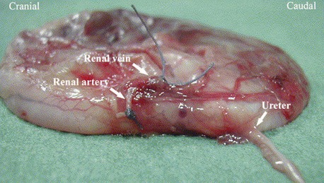
Ectopic ureters occur in both puppies and kittens as a congenital abnormal termination of the ureters with the urinary bladder. Ectopic ureters are described as intramural or extramural. Intramural ectopic ureters enter the bladder wall and run transmurally but fail to empty at the trigone and instead continue transmurally and empty in the urethra or vagina vestibule. Extramural ectopic ureters bypass the urinary bladder completely and enter the urethra, vagina vestibule, or uterus. Females are affected more often than males. Ectopic ureters empty into the urethra of most females and prostatic urethra in males. Ectopic ureters may occur alone or with other developmental abnormalities of the urinary tract. Hydronephrosis may occur if the ureter ends in a blind pouch or forms a functional stenosis. An increased incidence has been found in English Bulldogs, Fox Terriers, Golden Retrievers, Labrador Retrievers, Newfoundlands, Poodles, Siberian Huskies, Skye Terriers, and Welsh Corgis. Clinical signs are related to variable degrees of urinary incontinence. Diagnosis is made with contrast radiography (excretory urogram with retrograde urethrogram or vaginogram), endoscopy, or cross-sectional imaging (Figure 38-2). Surgery is the treatment of choice with transection and reimplantation or unilateral ureterectomy and nephrectomy. In some dogs incontinence persists, and pharmacologic management with diethylstilbestrol or phenylpropanolamine may be necessary.


Stay updated, free articles. Join our Telegram channel

Sep 11, 2016 | Posted by in SMALL ANIMAL | Comments Off on The Urinary System

Full access? Get Clinical Tree

Get Clinical Tree app for offline access