Chapter 76The Tarsal Sheath
Functional Anatomy
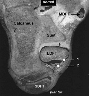
The tarsal sheath is enclosed at the level of the sustentaculum tali by a thick, transversely oriented ligament, the plantar retinaculum (Figure 76-1), and in the distal tarsus by a superficial fascia. The plantar nerves and vessels run within the retinaculum, in the plantar two thirds of its width, that is, plantar and plantaromedial to the lateral digital flexor tendon. The sheath is lined by a parietal synovial membrane with few villi, except distally. This membrane reflects plantarly to wrap around the tendon, leaving a thin but continuous membrane, or mesotendon, along the plantaromedial aspect of the lateral digital flexor tendon. This membrane carries vessels to the tendon and therefore should be preserved during surgery.
Causes of Tarsal Tenosynovitis
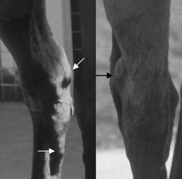
Distention of the tarsal sheath (Figure 76-2) is commonly termed thoroughpin, or true thoroughpin,14 but the condition may have distinct causes (see Figure 6-30).
Stay updated, free articles. Join our Telegram channel

Full access? Get Clinical Tree


