The Suspensory Apparatus

Chapter 72The Suspensory Apparatus


Anatomy and Pathophysiology


The suspensory ligament (SL) is correctly called the third interosseous muscle, but it is referred to as the SL throughout this text. The anatomy in the forelimbs and hindlimbs is similar, and separate mention of the hindlimb is made only when substantial differences exist.


The SL can be divided into three separate regions that are subject to injury: the proximal part, the body, and the branches. For clinical purposes, in the forelimb the proximal part extends from 4 to 12 cm distal to the accessory carpal bone, and in the hindlimb, from 2 to 10 cm distal to the tarsometatarsal joint. The distal sesamoidean ligaments are considered individually.


In the forelimb the SL originates from two differently shaped lobes that rapidly fuse. In the hindlimb this division is less obvious. The SL contains a variable amount of muscular tissue (2% to 11%), which tends to be bilaterally symmetrical.1 In the forelimb the SL originates from the palmar carpal ligament and the proximal aspect of the third metacarpal bone (McIII), whereas in the hindlimb it originates principally from the proximoplantar aspect of the third metatarsal bone (MtIII). There is an accessory ligament of the SL that extends proximally and originates on the plantar aspect of the fourth tarsal bone. The SL in the forelimb is approximately rectangular in cross-section, but it is more rounded in the hindlimb. There are also fibers that arise from the axial aspect of the fourth metacarpal bone (McIV).


The body of the SL descends between the second metacarpal or metatarsal bone (McII, MtII) and the McIV or the fourth metatarsal bone (MtIV) and divides into two branches at a variable site in the midmetacarpal (midmetatarsal) region. The level of division is usually bilaterally symmetrical. Each branch inserts on the abaxial surface of the corresponding proximal sesamoid bone (PSB). Each branch detaches a thin extensor branch dorsodistally that courses obliquely across the pastern to join the dorsal digital extensor tendon just above the proximal interphalangeal joint. Each extensor branch also blends with the corresponding collateral sesamoidean ligament.


The distal sesamoidean ligaments are the functional continuation of the SL in the digit and consist of the straight sesamoidean ligament, oblique sesamoidean ligaments, cruciate sesamoidean ligaments, and short sesamoidean ligaments. All attach proximally to the base of the PSBs and the proximal scutum. The straight sesamoidean ligament is a flat trapezoidal band that inserts via the scutum medium onto the proximal aspect of the middle phalanx. The oblique sesamoidean ligaments are triangular structures that converge to insert on the palmar aspect of the proximal phalanx. The cruciate sesamoidean ligaments form the palmar wall of the distal palmar synovial recess of the metacarpophalangeal joint. They consist of two thin layers of tissue that cross each other and insert on the proximal palmar tuberosities of the proximal phalanx. The short sesamoidean ligaments insert on the proximal palmar aspect of the proximal phalanx and are difficult to separate from the dorsal aspect of the oblique sesamoidean ligaments.


The palmar ligament of the fetlock, also referred to as the intersesamoidean ligament, is a thick collagen structure that completely covers the palmar and axial surfaces of the PSBs and is strongly attached to them. Together with the PSBs, the palmar ligament forms the proximal scutum. The concave palmar surface of the proximal scutum provides a smooth surface over which the digital flexor tendons glide.


In the forelimb the SL is innervated by the palmar metacarpal nerves, derived from the lateral palmar nerve, which receives fibers from the ulnar and median nerves.2 The hindlimb SL is innervated by the plantar metatarsal nerves, branches from the deep branch of the lateral plantar nerve, which is derived from the tibial nerve. The proximal aspect of the SL is closely related to the distal palmar outpouchings of the carpometacarpal joint in the forelimb3 and the plantar outpouchings of the tarsometatarsal joint in the hindlimb.4


The principal function of the SL is to prevent excessive extension of the fetlock joint.5 During weight bearing the relative tension in the SL and digital flexor tendons regulates the stresses applied to different aspects of the McIII. When a limb is fully load bearing, the distal parts of the SL branches are apposed closely to the abaxial aspects of the metacarpal condyles and then move to the palmar aspect as the fetlock drops. During hyperextension the PSBs move distally and dorsally, so the branches of the SL act as articular surfaces to balance the position of the McIII. If the limb is loaded asymmetrically, so that torque is on the fetlock, the SL branches contribute to joint stability on the side opposite compression of the joint.


Some evidence exists that training increases the strength of the SL; the mean absolute load to failure in a single load-to-failure compression test was significantly higher in horses that were in race training compared with those that were confined to box or paddock rest.6 In the trained group failure was most likely to be by fracture of a PSB, whereas in the untrained group the SL failed. However, when six 2-year-old Thoroughbred (TB) fillies underwent an 18-month controlled exercise program including galloping and were compared with six fillies that were restricted to walking exercise, no differences in the collagen fibril mass-average diameter in the body of the SL were found.7 Mass-average diameter is correlated with ligament strength.


Proximal Suspensory Desmitis in the Forelimb


Proximal suspensory desmitis (PSD) is a common injury in the forelimbs8-12 of athletic horses and may occur unilaterally or bilaterally. Some confusion has occurred about what constitutes PSD, and many clinicians have used this term for lameness that is worse with the affected limb on the outside of a circle and that is alleviated by analgesia of the proximal palmar metacarpal region but in which radiological and ultrasonographic findings have been negative. In our experience this case scenario is relatively unusual. Ultrasonographic abnormalities of the proximal SL are usually detectable, and the absence of detectable structural abnormality should alert the veterinarian to search for an alternative diagnosis. However, in a small proportion of horses abnormalities of the SL that were not apparent using ultrasonography have been identified using magnetic resonance imaging (MRI).13,14


PSD usually results in sudden-onset lameness, which can be remarkably transient, resolving within 24 hours unless the horse is worked hard. In horses with more chronic desmitis, lameness may be persistent. Lameness varies from mild to moderate and is rarely severe, unless the lesion is extensive. Lameness in Standardbred (STB) racehorses may be apparent only at high speeds. Bilateral PSD may result in loss of action rather than overt lameness, which occurs more commonly in flat racehorses than other sports horses, probably because of failure to recognize earlier, subtle unilateral lameness. However, dressage horses and event horses may be similarly affected. In a jumping horse the rider’s complaint may be of failure of a horse to land with a specific forelimb leading. While ground reaction force is greater in the nonlead forelimb when landing from a fence, the lead forelimb has greater extension of the fetlock, resulting in potentially greater stress on the suspensory apparatus. So with SL injury the horse may be reluctant to land with the lame forelimb leading. Lameness is usually worse on soft ground, especially with the affected limb on the outside of a circle, and when subtle may be more easily felt by a rider than seen by an observer. In some horses lameness is never detectable either in hand or on the lunge and is only apparent when the horse is ridden. Lameness may not be apparent at the working trot but may be detectable at the medium or extended trot. Presumably this reflects increased stress on the SL because of increased extension of the fetlock at extended trot compared with working trot. Recognition of these features in the history may be important, because acute lameness often resolves rapidly, and working the horse hard to reproduce lameness, with the inherent risk of worsening the injury, may be undesirable. Lameness is often transiently accentuated by distal limb flexion. We believe that this is because the suspensory apparatus is relaxed during flexion and undergoes sudden stretching when the limb is loaded.


In the acute phase slight edema in the proximal metacarpal region, localized heat, and distention of the medial palmar vein may occur, but these features may be transient or absent. Pressure applied to the SL against the palmar aspect of the McIII or forced extension and protraction of the limb may elicit pain, but the absence of pain does not preclude the presence of PSD.


The feet should be evaluated carefully, because frequently foot imbalance is a predisposing factor. Back-at-the-knee and tied-in below the knee conformations also may be predisposing factors.


PSD is a common compensatory injury; therefore the whole horse should be evaluated to ensure that other causes of lameness are not missed. There is a relationship between front foot pain and PSD. PSD can occur in forelimbs and hindlimbs simultaneously.


Diagnostic Analgesic Techniques


If PSD is suspected, perineural analgesia of the lateral palmar nerve using lateral3 or medial15 approaches (2 mL mepivacaine) or the medial and lateral palmar metacarpal nerves (2 mL per site) is indicated (see Chapter 10). This should result in substantial improvement in, or alleviation of, lameness within 10 minutes, assuming PSD is the only cause of lameness. However, neither technique is necessarily specific. Blockade of the lateral palmar nerve also has the potential to alleviate pain associated with a lateral source of pain in the more distal aspect of the limb (e.g., a splint). The risks of influencing middle carpal joint pain are less than with the subcarpal approach, but with the lateral approach local anesthetic solution may diffuse and improve lameness associated with the middle carpal joint16 or with the carpal canal. Perineural analgesia of the palmar metacarpal nerves may alleviate pain associated with the middle carpal or carpometacarpal joints because of local diffusion or inadvertent deposition of local anesthetic solution into the distopalmar outpouchings of the carpometacarpal joint capsule. There is no right or wrong method, but it is important to be aware of the limitations of whichever technique is used. One author (SJD) usually blocks the medial and lateral palmar metacarpal nerves using a lateral approach, with the limb non–weight bearing. In a difficult, potentially dangerous horse that strikes out, the lateral palmar nerve is blocked with the limb bearing weight. It is easy to hit the lateral palmar nerve using the medial approach, causing the horse sudden severe pain; therefore this technique is rarely used. A false-negative result may be achieved because of inadvertent injection into the carpal sheath or failure of the local anesthetic solution to diffuse proximally to the most proximal extent of a lesion. Although the SL receives innervation from fibers from the median and ulnar nerves, perineural analgesia of the ulnar nerve usually resolves or substantially improves lameness associated with PSD. However, in a minority of horses perineural analgesia of the median and ulnar nerves is required to abolish lameness completely.


Intraarticular analgesia of the middle carpal joint may result in partial improvement or complete alleviation of pain associated with the proximal aspect of the SL in some horses (15 [60%] of 25 horses).16 Using a dorsal approach to the middle carpal joint rather than a palmarolateral approach should theoretically reduce the risks of diffusion of local anesthetic solution to the proximal aspect of the SL and palmar metacarpal nerves; however, in practice the difference seems minor. Comparison of the relative responses to middle carpal analgesia (6 mL mepivacaine; assessed 10 minutes after injection) and perineural analgesia of the lateral palmar nerve or the palmar metacarpal nerves is potentially useful but can be highly misleading. Generally a horse with lameness caused by PSD responds better to perineural analgesia than intraarticular analgesia, but this is not universal. Similarly, primary middle carpal joint pain usually is improved best by intraarticular analgesia, but this is not always the case. Middle carpal joint pain and PSD may occur concurrently, especially in STB and TB racehorses. The clinician should evaluate the response to these diagnostic analgesic techniques in light of the following:


Perineural analgesia of the palmar nerves at the level of the base of the PSBs often results in lameness because of PSD appearing worse. We believe that this reflects some loss of proprioceptive function of the distal aspect of the limb so that the horse is less able to protect the painful SL. Perineural analgesia of the palmar nerves (at midmetacarpal level) and palmar metacarpal nerves (distal to the button of the McII and the McIV) (four-point or low palmar block) often results in some improvement in pain associated with PSD, possibly because of proximal diffusion of local anesthetic solution via lymphatic vessels or along fascial planes. However, a recent contrast study indicated that this may not occur.17 Perhaps improvement may be due to pain extending further distally in the SL than the site of detectable injury.


More than one source of pain may be contributing to lameness. PSD and concurrent foot pain occur commonly. Hindlimb lameness also may be present, especially in the contralateral hindlimb, so it is important to assess and to reevaluate the whole horse.


Differential Diagnosis


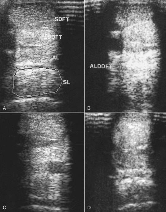
PSD should be differentiated from middle carpal joint pain, being aware that especially in young TB racehorses and STB racehorses lesions may occur in both locations simultaneously. Osteoarthritis of the carpometacarpal joint occasionally occurs (see page 417). Horses with pain associated with palmar cortical fatigue fractures or stress reactions of the McIII11,18-20 respond similarly to diagnostic analgesic techniques; however, in horses with fracture, lameness tends to be more severe and worse on firm ground and often deteriorates the farther the horse trots (see page 413). Avulsion fractures of the McIII at the origin of the SL (see page 417) occur less frequently and tend to be associated with more persistent and severe lameness.1,21 Pain associated with the carpal sheath or carpal retinaculum also should be considered (see Chapter 75). Perineural analgesia of the deep branch of the lateral palmar nerve or the palmar metacarpal nerves alone should not alleviate pain associated with the deep digital flexor tendon (DDFT) or its accessory ligament (ALDDFT), the superficial digital flexor tendon (SDFT), or the fetlock region, without simultaneous blockade of the palmar nerves. However, horses with proximal lesions of the SDFT or ALDDFT may show partial improvement in lameness.


Diagnostic Ultrasonography


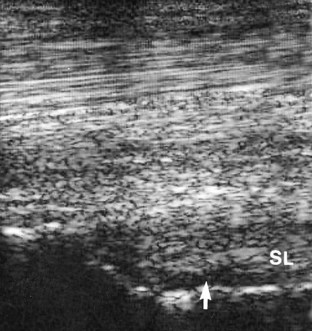
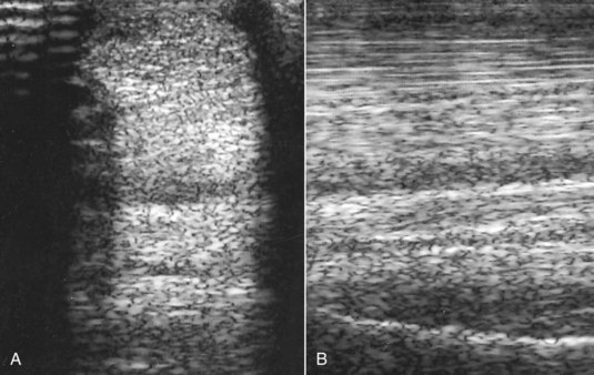
Diagnostic ultrasonography is essential for accurate diagnosis of PSD. The limb should be evaluated in transverse and longitudinal planes, and careful comparison should be made with the contralateral limb. High-quality images are required, because lesions can be subtle and easily missed if the gain controls are too high or if the transducer is not focused on the SL. Artifacts are readily created if the transducer is not in complete contact with the limb. The contours of the proximal palmar aspect of the metacarpal region can make obtaining longitudinal images difficult, especially because the proximal palmar aspect of the McIII slopes backward. Therefore creating hypoechoic artifacts at the enthesis of the SL on the McIII is easy. Cross-sectional area measurements may be extremely valuable, especially in horses with acute PSD, because enlargement of the ligament may be the only detectable ultrasonographic abnormality. Bear in mind that muscular tissue appears less echogenic than does ligamentous tissue and that proximally the SL originates in two halves. The entire cross-section of the SL cannot be seen from a palmar approach, and marginal lesions may be missed unless oblique images are also obtained. A convex transducer or virtual convex transducer allows a greater proportion of the proximal abaxial aspects of the SL to be evaluated in transverse images compared with a linear transducer. Previous injuries to the SL may not resolve fully to restore normal, uniform echogenicity. Be aware that poor diagnostic analgesic technique may result in aspiration of air, which creates artifacts. This usually resolves in 24 hours. A thin band of the SL passes proximally from the enthesis on the McIII to blend with the palmar carpal fascia. The anechogenic space seen dorsal to the SL at this level is fluid in the palmar recess of the carpometacarpal joint and should not be mistaken for a lesion (Figure 72-1).


Abnormalities associated with PSD include the following22 (Figures 72-2 and 72-3):


In a horse with bilateral PSD an obvious lesion may be detectable in the lamer limb, but abnormalities may be much more subtle and occasionally not apparent in the less lame limb. In a 3-year-old TB that sustains PSD at 2 years of age, mild lameness may be recurrent, and discerning any structural abnormality other than enlargement of the SL may not be possible.


The degree of ultrasonographic abnormality (cross-sectional area involved and proximodistal extent of the lesion) usually reflects the severity of the lameness. In horses with acute PSD the ultrasonographic abnormalities may be subtle, although if lameness is unilateral, slight enlargement of cross-sectional area may be detectable. Care should be taken to compare measurements in the contralateral limb at the same distance distal to the accessory carpal bone. Ultrasonographic abnormalities may worsen over the next 10 to 14 days, and reevaluation may be useful to confirm the diagnosis.


In horses with an avulsion fracture of the McIII at the origin of the SL, the fracture fragment is usually readily identifiable and generally is associated with only a focal lesion in the SL itself, usually restricted to the dorsal aspect (see page 417).


Treatment


Most horses with acute forelimb PSD respond well to box rest and controlled walking exercise for 3 months.29,30 Uncontrolled turnout is contraindicated. Attention to correct foot balance is important. Elevation of the heel is contraindicated because it increases load on the SL. A premature resumption of work usually results in recurrent injury. Approximately 90% of horses resume full athletic function without recurrent injury.1 In the early period after return to work circumstances that create hyperextension of the fetlock should be avoided. For example, dressage horses should avoid medium and extended trot. Attention should also be paid to the footing on which the horse works. Horses with chronic PSD may require more prolonged rehabilitation, and in a small proportion lameness is persistent. If low-grade residual lameness persists after 3 to 6 months but serial ultrasonographic examinations reveal no change in echogenicity, then introduction of trotting exercise sometimes stimulates further repair and resolution of lameness. Some TB racehorses with chronic lesions have been able to be maintained in training with judicious use of phenylbutazone, without clinically significant deterioration of the lesion. No fatalities associated with PSD occurred in 630 TB racehorses examined post mortem because of musculoskeletal injuries.29 Some STB racehorses with acute lesions have been managed by slightly reducing the training schedule, administering local injections of corticosteroids and hyaluronan, using symptomatic antiinflammatory therapy (local icing, liniments such as dimethyl sulfoxide [DMSO] and corticosteroid paints, and phenylbutazone as necessary), and shortening the toes and increasing hoof angle. Although a few treated horses were able to race successfully, prognosis after rest was better.29


Extracorporeal shock wave treatment or radial pressure wave therapy (three treatments at 2-week intervals) was successful in some horses with chronic lesions that had failed to respond to conservative management.31,32 Ten (50%) of 20 horses with chronic forelimb PSD with lameness of greater than 3 months’ duration were in full work 6 months after treatment.32 Repeated long-term treatments are sometimes required. Local infiltration with 4 to 6 mL of 2% iodine in almond oil has been used successfully.8


In some horses the lesions disappear completely ultrasonographically. In others echogenicity may increase, but uniform echogenicity is never restored. Rest should be continued until the ultrasonographic appearance remains stable.


Surgical splitting of the SL has been used in some horses with PSD with successful results in some horses; however, the results have been unpredictable.14,33 There are currently no long-term, peer-reviewed studies of the results of injection of pigs’ urinary bladder matrix, mesenchymal stem cells, or other biological agents such as platelet-rich plasma, although anecdotally successful results have been reported. In sports horses with chronic injuries that have failed to respond to conservative management, one author (SJD) has successfully used either mesenchymal stem cells or desmoplasty combined with injection of platelet-rich plasma.


Proximal Suspensory Desmitis in the Hindlimb


PSD in the hindlimb results in either an insidious or a sudden-onset lameness that may be mild or severe. Some horses show poor performance rather than a recognized lameness. In contrast to the forelimb, lameness may persist and remain severe, despite restriction to box rest. Such persistence probably is caused by a compartment-like syndrome and pressure on the adjacent plantar metatarsal nerves.34,35 In view of the chronicity of some lesions when first identified and the finding of secondary radiological changes in sound horses, some lesions likely exist subclinically or are associated with a low-grade lameness that goes unrecognized. The incidence of bilateral lesions is higher than in forelimbs.


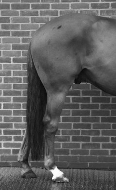
PSD in the hindlimb occurs in horses in all athletic disciplines and of all ages and is a particular problem in dressage horses.36 There is an association between straight hock conformation and hyperextension of the metatarsophalangeal joint and hindlimb PSD (Figure 72-4). Such conformational abnormalities were identified in nine (21%) of 42 horses with hindlimb PSD but in only four (8%) of 50 horses examined consecutively with hindlimb lameness unrelated to the suspensory apparatus.37image Straight hock conformation may predispose to PSD or develop secondarily; hyperextension of the metatarsophalangeal joint may develop as a sequela to PSD, probably as the result of progressive degeneration of the SL. A long-toe and low-heel conformation also may be a predisposing factor, especially if associated with abnormal orientation of the distal phalanx, with the plantar aspect lower than the toe.29


PSD in the hindlimb in a STB racehorse is common and usually results in an abnormal gait at high speeds, which may or may not be apparent at the trot. Unilateral left hindlimb lameness may manifest as the horse drifting to the right shaft and being on the left line and vice versa.


Horses with acute hindlimb PSD may have localized heat and swelling and pain on pressure applied to the SL, but frequently no localizing clinical features are apparent.


Lameness is often characterized by a reduced height of arc of foot flight, with or without intermittent catching of the toe. There may be reduced extension of the metatarsophalangeal joint unless the functional integrity of the SL is compromised. The cranial phase of the stride may be shortened. Lameness may be accentuated by proximal or distal limb flexion. Bilateral lesions may result in poor hindlimb action rather than obvious hindlimb lameness. Performance horses may not be overtly lame in hand, but when ridden show reduced hindlimb impulsion, difficulties in transitions, stiffness, resistant behavior, evasions such as bolting, difficulties in turning or stopping, reluctance to perform certain dressage movements, or reduced power when jumping. Lameness may be more obvious on a circle on the lunge, but unlike in forelimb PSD the lameness is not necessarily worse with the lamer limb on the outside. Approximately 50% of horses with hindlimb PSD are lamer with the affected limb on the outside or inside of a circle. As with many horses with hindlimb lameness, lameness is often more obvious when the horse is ridden, especially when the rider sits on the diagonal of the lame or lamer limb.


Diagnostic Analgesic Techniques


Perineural analgesia of the plantar nerves (midmetatarsal level) and plantar metatarsal nerves may result in slight improvement in lameness because of proximal diffusion of the local anesthetic solution or distal extension of pain in the SL, or concomitant injury of a SL branch. Lameness usually is improved substantially by perineural analgesia of the medial and lateral plantar metatarsal nerves (2 mL of mepivacaine 2% per site) or the deep branch of the lateral plantar nerve distal to the tarsus, but lameness may not be alleviated fully. Improvement is usually seen within 10 minutes of injection. Critical evaluation of the degree of improvement is considered essential if considering treatment by neurectomy of the deep branch of the lateral plantar nerve. If there is a component of entheseous pain, this may result in only partial improvement in lameness, which is further improved by deep infiltration of local anesthetic solution toward the plantar aspect of the MtIII. PSD may occur together with pain associated with the tarsometatarsal joint, and if lameness is improved but not abolished by subtarsal analgesia, addition of intraarticular analgesia of the tarsometatarsal joint may abolish the lameness. False-negative results also may be obtained because of inadvertent injection into the tarsal sheath or the tarsometatarsal joint capsule. Subtarsal analgesia can influence tarsometatarsal joint pain, and occasionally (two [8%] of 24 horses34

Stay updated, free articles. Join our Telegram channel

Jun 4, 2016 | Posted by in EQUINE MEDICINE | Comments Off on The Suspensory Apparatus

Full access? Get Clinical Tree

Get Clinical Tree app for offline access