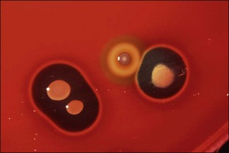
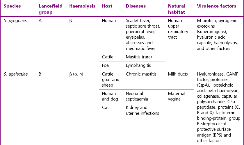
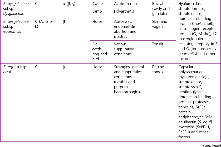
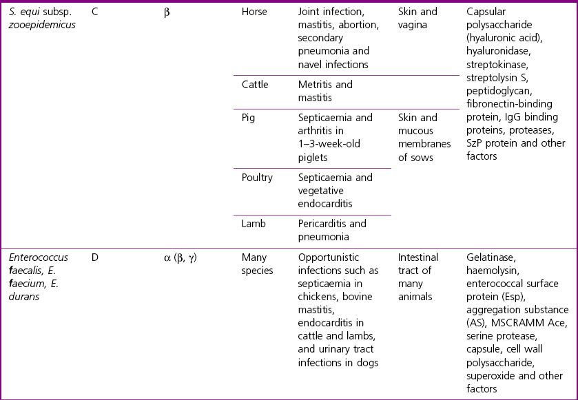
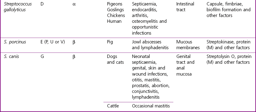
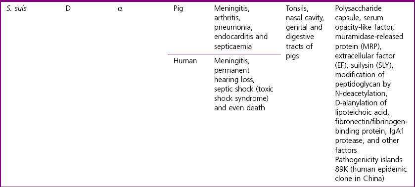
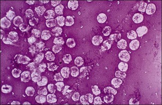
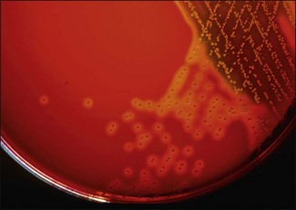
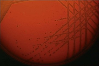
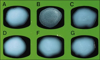
Chapter 8 • Haemolysis: the type of haemolysis produced by a streptococcal species can be variable. The main types of haemolysis are: Generally the beta-haemolytic streptococci tend to be the most pathogenic for animals. Lancefield Group C streptococci from horses usually produce large zones of clear, beta-haemolysis (Fig. 8.3). Figure 8.3 Close-up of colonies of Streptococcus equi subsp. equi (beta-haemolytic) after four days’ incubation compared with the greening-type haemolysis of an alpha-haemolytic Streptococcus (contaminant). • Lancefield Groups: The serological Lancefield grouping scheme is based on group-specific carbohydrate cell wall antigens, with groups from A to H and K to V. Some isolates are not groupable. The methods for Lancefield grouping include: 1. Conventional method: the C-substance (antigen) is extracted either by autoclaving or by acid extraction (hydrochloric acid). A ring precipitation test is conducted by layering the extracted antigen over known antisera that can be obtained commercially for some Lancefield Groups. 2. Latex agglutination test: This method is the most commonly used and kits are available commercially for identifying Lancefield Groups A, B, C, D, F and G (Fig. 8.4). Streptococci are pyogenic bacteria that are commonly associated with suppuration and abscess formation while enterococci are opportunistic pathogens. The polysaccharide capsules of S. pyogenes, S. pneumoniae (Fig. 8.5) and some strains of S. equi subsp. equi and S. agalactiae are anti-phagocytic. The principal diseases and associated virulence factors of the streptococci and enterococci of veterinary importance are given in Tables 8.1 and 8.2. Table 8.1 Streptococci of veterinary importance, the diseases they produce and their principal virulence factors Table 8.2 General functions of the main virulence determinants of streptococci and enterococci Figure 8.5 Streptococcus pneumoniae in a blood smear showing the distinctive capsule. (Nigrosin stain, ×1000) Streptococcus equi subsp. equi is the agent of equine strangles, a contagious upper respiratory tract disease with abscessation of the local lymph nodes. Clinical signs include fever, nasal discharge and swelling of the mandibular and retropharyngeal lymph nodes and intermandibular region. Dissemination of the organism may occur with abscesses forming in the lungs or other locations. The source of the infection is usually pus from abscesses or nasal discharges from infected horses. However, feed and/or water can also be contaminated. A small proportion of animals fail to clear the infection from the guttural pouches and these healthy carriers are likely to be responsible for spread of infection between epidemics. Depending on the immunity status, the disease can take an acute or mild course with most horses recovering relatively quickly. Streptococcus equi subsp. zooepidemicus is a commensal organism with a large host range, which opportunistically causes disease following viral infections or tissue injury. It is a frequent cause of pneumonia and joint infections in horses. There have been several reports of haemorrhagic pneumonia in dogs associated with this organism in recent years (Priestnall & Erles 2011). It is also a zoonotic agent with infection following close contact with horses or contaminated milk products. Streptococcus dysgalactiae subsp. equisimilis may be a more significant pathogen of horses than previously thought as a recent study showed that it was frequently isolated from cases of infection of the equine genital tract (Erol et al. 2012). Streptococcus suis is associated with septicaemia, meningitis, polyarthritis and pneumonia in pigs. It is also a zoonotic pathogen capable of causing severe invasive disease in humans exposed to pigs or pork products. Most pigs are healthy carriers with S. suis located in their tonsils, nasal cavities, genital or digestive tracts. Disease development is incompletely understood but is believed to be related to the strain and immune status of the host. Some virulence factors appear to be associated with more severe, invasive disease (Beineke et al. 2008, Wei et al. 2009). Stress and intensive management practices are also likely to predispose to disease occurrence. Most cases of disease in both pigs and humans are caused by S. suis serotype 2. In both species the main clinical manifestations of S. suis are meningitis and septicaemia although some serotypes are commonly associated with pneumonia. Streptococcus suis outbreaks in pigs are common while most reports in humans describe sporadic cases (Gottschalk et al. 2007). However, S. suis has recently emerged as an important zoonotic agent in some Asian countries, and has been reported as the primary cause of adult meningitis in Vietnam (Mai et al. 2008). Streptococcus porcinus causes a variety of pathological conditions in pigs including contagious cervical lymphadenitis, ‘porcine strangles’, endocarditis, and abortion. It is believed that the organism enters its host through the mucosa of the pharyngeal or tonsillar surfaces. Organisms are then carried to the lymph nodes, primarily of the head and neck region, where abscesses are formed. Streptococcus porcinus is also an opportunistic agent in many other hosts. It has rarely been implicated as a human pathogen with only a few cases reported. Streptococcus dysgalactiae subsp. equisimilis is a commensal of the porcine tonsil and a cause of suppurative athritis in piglets. Acquisition of this microorganism is usually via the tonsils, umbilicus or a skin abrasion. Streptococcus canis is an opportunistic pathogen found as part of the normal flora of the oropharyngeal, anal and genital mucosa in both dogs and cats. A variety of infections have been reported associated with this organism. Contagious lymphadenitis is possibly the most frequent manifestation in cats. In dogs, sporadic necrotizing fasciitis and toxic shock syndrome have been associated with S. canis. This syndrome may be related to fluoroquinolone usage, which was found to induce a lytic bacteriophage encoding a homologue of pokeweed mitogen (Ingrey et al. 2003). Streptococcus pneumoniae is an important human pathogen which causes invasive disease in adults and children but is rarely isolated from clinical disease in animals. It has been reported to infect and cause pneumonia in horses. Equine isolates of S. pneumoniae are reported to represent a tight clonal group and to be genetically distinct from human isolates (Whatmore et al. 1999). The agent of streptococcosis in pigeons is Streptococcus gallolyticus subsp. gallolyticus, which was formerly classified as S. bovis biotype I (Devriese et al. 1998). Streptococcosis is an important septicaemic disease in pigeons. Clinical signs include lameness, emaciation, inability to fly, polyuria, production of green and slimy droppings and sudden death. Extensive and well-circumscribed areas of necrosis in the pectoral muscles and arthritis of the knee, hock and shoulder joints are observed at necropsy. Streptococcus gallolyticus is an opportunistic pathogen of humans and S. gallolyticus subsp. gallolyticus is the subspecies most often linked with human endocarditis-associated colonic cancer.
The streptococci and related cocci
General Differentiation of the Streptococci
Pathogenesis and Pathogenicity
Virulence determinant
Functions
Beta-haemolysin
Cytotoxic for bacterial and eukaryotic cells by pore formation in the target cell membrane
CAMP factor (ceramide-binding protein) of S. agalactiae
Enhances the haemolysis of staphylococcal sphingomyelinase beta-toxin, cytotoxic and lethal for cell cultures, possibly cytotoxic for mammary tissues
Capsular polysaccharide (polysaccharide type-specific antigen, hyaluronic acid)
Antiphagocytic, type-specific antibodies can be protective
Collagenase
Extracellular enzyme which disrupts collagen and likely contributes to endothelial cell damage, tissue destruction, and haemodynamic derangement
Streptococcal C5a peptidase (SCPA)
Highly specific endopeptidase which acts primarily to eliminate C5a chemotactic signal from inflammatory foci
Exotoxins (such as SePE-H, SePE-I, SePE-M, SePE-G)
Pyrogenic exotoxins, mitogens (superantigens): non-specific T cell stimulation and cytokine release
Hyaluronidase
Promotes tissue dissemination, increased activity in S. equi subsp. zooepidemicus compared to S. equi subsp. equi may explain the more frequent dissemination of the former throughout the body
Equibactin of S. equi subsp. equi
Siderophore, iron acquisition
IgA protease
Cleaves immunoglobulin A in its hinge region, evades host responses
IgG binding protein (Protein G)
Evades host responses, likely to reduce phagocytosis
Leukocidal toxin of S. equi subsp. equi
Damages polymorphonuclear leukocytes, imparing phagocytosis and killing
L2 macroglobulin receptor
Binds L2 macroglobulin
Fibronectin-binding protein (such as fnbA and fnbB)
Evades host responses, likely to reduce phagocytosis
Muramidase-released protein (MRP) of the cell wall of S. suis
Unknown functions but contributes to protection
Extracellular factor (EF) of S. suis
Unknown functions but associated with virulence
Peptidoglycan
Potent activator of alternative complement pathway
Plasminogen receptor
Binds plasminogen, may reduce phagocytosis
Proteins C and R of S. agalactiae
C: Unknown functions but promotes protective and opsonophagocytic antibodies
R: Unknown functions but likely to enhance epithelial colonization
Proteins M-like (SzPSe protein) of S. equi subsp. equi
Binds equine fibrinogen, may reduce phagocytosis
Proteins (M)
Antiphagocytic
SeM protein of S. equi subsp. equi
Binds equine fibrinogen, antiphagocytic and protective antigen
Serine protease (EspA) of S. agalactiae
Cleaves fibrinogen, and blocks phagocytosis
Streptokinase
Plasminogen activation (active plasmin hydrolyses fibrin which may promote dispersion)
Streptodornase (DNase)
Escape killing in neutrophils
Streptolysin S and O
Beta-haemolysis, a bacteriocin-like cytotoxin
Suilysin of S. suis
Thiol-activated haemolysin that is either secreted or lightly cell-attached which produces transmembrane pores in target cells
Opp proteins
Involved in the active transport of solutes, essential for growth in milk
SUAM
Mediates adherence to and invasion of epithelial cells
MSCRAMM Ace of enterococci
Microbial surface component recognizing adhesive matrix molecules, adhesion of collagen
Gelatinase
Protease capable of hydrolysing collagen, casein, haemoglobin and other peptides
Haemolysin
Cytolytic protein capable of lysing human, horse and rabbit erythrocytes
Esp
Cell wall associated protein involved in biofilm formation
![]()
Stay updated, free articles. Join our Telegram channel


Full access? Get Clinical Tree


