CHAPTER 41 The Skin and Ear
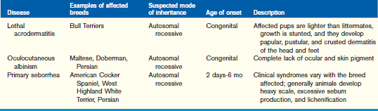
The normal pediatric integument of newborn dogs and cats undergoes significant change between birth and 6 months of age. The thicknesses of the epidermis and dermis increase twofold to threefold as the skin matures. During the same period, the integument as a percentage of body weight decreases from 24% to 12% in the dog. Meanwhile, there is a rapid replacement of reticulum fibers composed of type III collagen by mature type I collagen fibers in the dermis. These collagen fibers, as well as elastic fibers, increase in size and number during the first months of life.
The coat of kittens and puppies consists principally of fine hairs. At 12 to 16 weeks of age, hairs begin to thicken and decrease in curvature in a breed-specific manner, giving rise to the adult-type coat. Pigment of the skin and hair continues to develop into the adult phenotype until approximately 3 to 6 months of age.
Genodermatoses
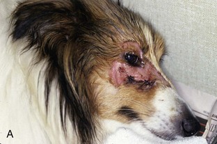
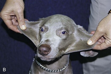
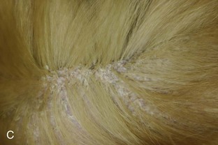
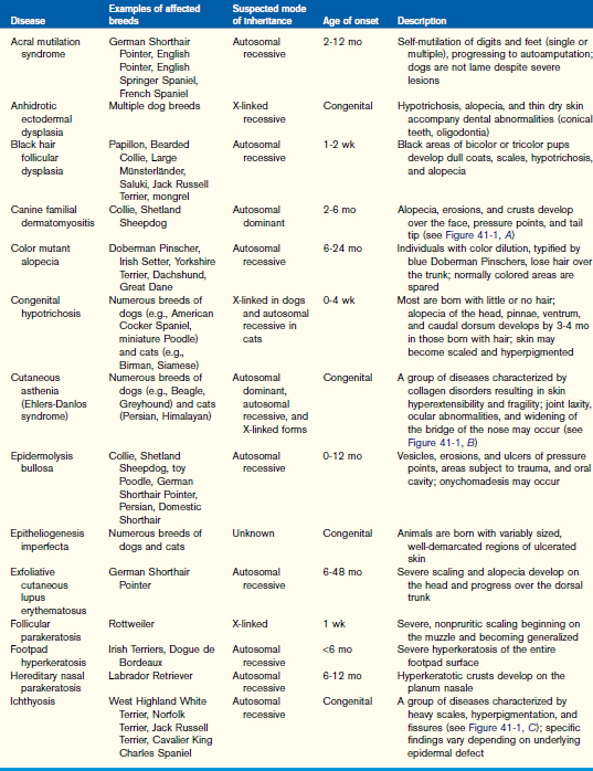
Signs of congenital and hereditary skin disorders are most often observed during the first 2 to 3 months of age (Figure 41-1). In many cases, the genetic defect resulting in the disorder has not been fully characterized. Some of the described genodermatoses of dogs and cats are summarized in Table 41-1.


Figure 41-1 Examples of congenital and hereditary dermatoses. A, Canine familial dermatomyositis in a Shetland Sheepdog. B, Cutaneous asthenia (Ehlers-Danlos syndrome) in a 5-month-old Weimaraner. C, Ichthyosis in a Golden Retriever.
(B, from Medleau L, Hnilica KA: Small animal dermatology: a color atlas and therapeutic guide, ed 2, St Louis, 2006, Saunders/Elsevier. C, courtesy Candace Sousa.)
Infectious Diseases
Young animals may be predisposed to infectious skin diseases as a result of either an immature immune system or a hereditary primary immunodeficiency. As the epidermal thickness increases and the protective function of the skin improves, puppies and kittens become less susceptible to viral, bacterial, and fungal infections.
Papillomas
Mucocutaneous viral papillomas are common in puppies. Smooth white lesions quickly progress to gray verrucous nodules that are often pedunculated (Figure 41-2). They are seen most often in the oral cavity and on the lips but may also occur on haired skin and conjunctiva. It is believed that various papilloma virus types have site predilections. The virus appears to be spread most often by direct contact but can survive for 2 months in the environment. There is a 1- to 2-month incubation period. Diagnosis can most often be made by clinical recognition in a puppy. Histopathology, if performed, reveals a hyperplastic and hyperkeratotic epidermis.
Papillomas typically regress in 2 to 3 months without therapy. In cases that fail to resolve or when treatment is otherwise warranted, cryotherapy is the treatment of choice. At least two freeze-thaw cycles are suggested. As long as the majority of lesions are frozen, all generally resolve, presumably by stimulating a host immune response.
Impetigo
Staphylococcus sp. impetigo is a nonpruritic superficial infection recognized commonly in puppies and rarely in kittens. Impetigo is characterized by nonfollicular pustules that occur most commonly in the nonhaired skin of the ventral abdomen. In contrast, in the adult dog, impetigo often results in larger pustules that span follicular units and is commonly associated with immunosuppression.
Cytology of the pustules will demonstrate neutrophils and intracellular cocci. Bathing every 3 to 7 days with a gentle shampoo containing an antibacterial ingredient such as chlorhexidine or triclosan is often sufficient to resolve impetigo. In severe cases that fail to respond to topical therapy, a 14-day course of a systemic antibiotic (amoxicillin-clavulanic acid or cephalexin) is indicated.
Dermatophytosis
Young animals carry an increased risk of developing dermatophytosis, reflecting their immature immune status and potential for exposure to carriers. Microsporum canis is the most frequent cause of ringworm in kittens and puppies. Entire litters can develop lesions, typically multifocal alopecic, mildly erythematous patches that progressively develop papules, scales, and hyperpigmentation (Figures 41-3 and 41-4). Frequently affected areas of kittens and puppies include the head, muzzle, pinnae, and distal limbs.

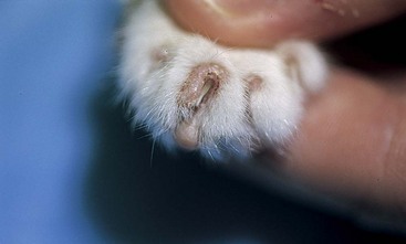
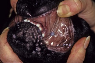
Figure 41-3 Paronychia in a cat caused by Microsporum canis.
(From Medleau L, Hnilica KA: Small animal dermatology: a color atlas and therapeutic guide, ed 2, St Louis, 2006, Saunders/Elsevier.)

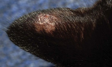
Figure 41-4 Focal alopecia and erythema on the ear pinna of a cat with dermatophytosis.
(From Medleau L, Hnilica KA: Small animal dermatology: a color atlas and therapeutic guide, ed 2, St Louis, 2006, Saunders/Elsevier.)
Approximately 50% of M. canis infections will cause hairs to fluoresce with ultraviolet (Wood’s) light examination. Microscopic examination of plucked hairs for ectothrix fungal spores also provides a rapid method of confirming the diagnosis. Incubating the hair for 15 to 30 minutes in KOH digests the keratin and may aid in visualization of the spherical spores. Fungal cultures should be performed to identify the species of ringworm.
In general, treatment of pediatric patients should be limited to topical products until they are approximately 16 weeks of age. Topical application of 2% lime-sulfur dip every 5 to 7 days is suggested. A protective collar is applied until the patient is dry to prevent excessive ingestion through grooming. Care should be taken to ensure that body temperature is maintained after applying a full-body dip to a pediatric patient. Small, localized lesions can also be treated with topical clotrimazole or terbinafine creams applied daily. When possible, affected animals should be isolated and the patient’s environment cleaned with 0.5% bleach. Caretakers should be educated regarding the zoonotic potential of dermatophytosis.
Treatment is continued until there is both a clinical resolution and a microbiological cure, as determined by follow-up fungal culture. Once the patient reaches 16 weeks of age, systemic therapy can be considered, following recommendations for adult dogs and cats. Oral itraconazole (5 to 10 mg/kg/day), terbinafine (30 to 40 mg/kg/day), or microsized griseofulvin (50 or 100 mg/kg/day) is commonly prescribed.

Stay updated, free articles. Join our Telegram channel

Full access? Get Clinical Tree

