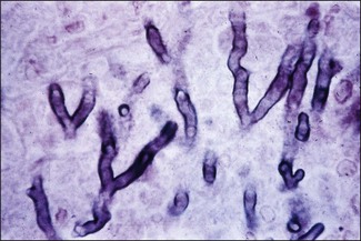
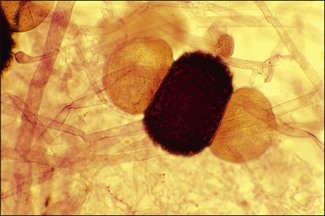
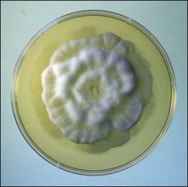
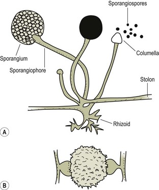
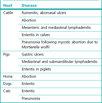
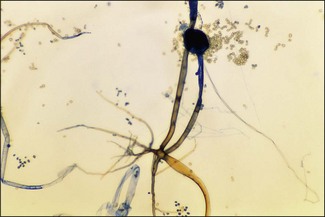
Chapter 42 Zygomycosis (older term phycomycosis) is an all-embracing term for an infection due to a fungus in the taxonomic Class Zygomycetes, phylum Zygomycota. Most of these fungi are saprophytes and widespread in the environment but some of them can be opportunistic pathogens. Clinical disease usually occurs if the host’s defences are lowered or if large numbers of spores are ingested or inhaled. The hyphae, in cultures and in animal tissues, are wide (5–15 µm diameter), irregular and ballooning (Fig. 42.1). They lack septa except near fruiting structures and if there has been damage to hyphae or in older cultures. The sexual spores are thick-walled zygospores (Fig. 42.2) produced through fusion of gametangia, often from two different strains of the species. The mucoraceous zygomycetes characteristically have rapidly growing colonies with greyish, woolly mycelium that fills the Petri dish in three to five days and often reaches and raises the lid of the plate (Fig. 42.3). An exception to this is Mortierella wolfii that has a white, low, velvety growth (Fig. 42.4). The aerial hyphae (sporangiophores) arise from stolons and usually end in a columella that is enclosed in the sac-like sporangium. Within the sporangium the asexual spores (sporangiospores) are formed. These spores can be colourless, yellowish or brown and the sporangia, packed with spores, often appear as dark, pinhead-sized dots within the woolly grey mycelium. As the spores mature, the wall of the sporangium becomes fragile and ruptures releasing myriads of spores (Fig. 42.5). The root-like rhizoids are produced by several genera and promote anchorage to the substrate (Fig. 42.6). Figure 42.6 Rhizopus species: sporangiophore, collapsed sporangium, sporangiospores and a rhizoid. (LPCB, ×100) Mortierella wolfii causes abortion in cattle and in about 5% of these animals an acute, fulminating pneumonia followed by death occurs within 48 hours of the abortion. Lesions are characteristic of mycotic abortion and resemble those seen in Aspergillus fumigatus infections. The placenta is thickened and ‘wooden’ in appearance (Fig. 42.7) and the foetus may have ringworm-like fungal skin plaques (Fig. 42.8). In fatal pneumonia cases the lungs are heavy, red and wet with most lobes affected (Fig. 42.9). A variable amount of fluid is present in the thoracic cavity. Other zygomycetes have been isolated from cases of abortion in cattle but, as these other fungi are more widely distributed than M. wolfii, fungal involvement should be confirmed by histological examination of cotyledons and foetal lesions. Table 42.1 summarizes the main hosts and diseases caused by the mucoraceous zygomycetes. Zygomycosis has also been reported in mink, guinea pigs, mice, poultry and exotic birds.
The pathogenic Zygomycetes
The mucoraceous zygomycetes (orders mucorales and mortierellales)
Pathogenesis
< div class='tao-gold-member'>
![]()
Stay updated, free articles. Join our Telegram channel

Full access? Get Clinical Tree


The pathogenic Zygomycetes
Only gold members can continue reading. Log In or Register to continue
