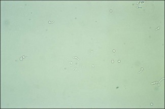
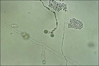
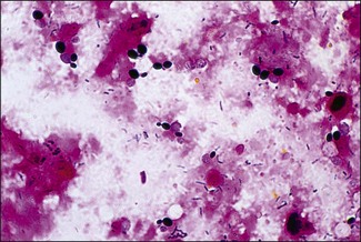
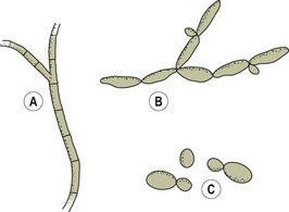
Chapter 40 Neuraminidase and proteases may play a part in virulence and cell wall glycoproteins have an endotoxin-like activity. Candidal surface proteins facilitate adhesion to matrix proteins. The production of extracellular, cytotoxic phospholipases and proteases is thought to aid tissue invasion and correlate with virulence. Phospholipase activity is concentrated at the growing tips of hyphae. Infections caused by C. albicans frequently involve overgrowth of resident C. albicans on mucous membranes. The yeast form is thought of as the form responsible for colonization of epithelial surfaces whereas tissue penetration and invasion is mediated by transition to the hyphal form. Granulomatous lesions are rare and inflammatory responses are predominantly neutrophilic. In severe, chronic infections, such as infection of the crop in chickens, the wall of the crop becomes thickened and covered by a corrugated pseudomembrane of yellowish-grey necrotic material giving it the characteristic ‘terry-towelling’ effect. Infections due to C. albicans have been given several names including moniliasis, candidosis and candidiasis. Table 40.1 lists the diseases caused by C. albicans in animals. There have been reports of mastitis in cattle being caused by other Candida species including C. tropicalis, C. pseudotropicalis, C. parapsilosis, C. guilliermondii, C. krusei and C. rugosa. Table 40.1 Diseases and main hosts of Candida albicans Candida albicans can be demonstrated in specimens by Gram-stained smears (Fig. 40.1), 10% KOH preparations, or in tissue sections stained by PAS-haematoxylin or methenamine silver stains. Candida albicans stains purple-blue with the Gram-stain. In tissue sections it appears as thin-walled, oval, budding yeast cells, hyphae and/or pseudohyphae (Fig. 40.2). Colonies of C. albicans usually appear in one to three days. They are white to cream, shiny, high-convex and have a pleasant ‘beery smell’. They can attain a diameter of 4–5 mm (Fig. 40.3). A small inoculum from an isolated colony is suspended in 0.5 mL of sheep, bovine, rabbit or human serum and is incubated at 37°C for two to three hours. A drop of the preparation is examined under phase contrast or the high-dry objective of the light microscope (with the condenser slightly lowered). Small tubes will be seen projecting from some of the yeast cells (Fig. 40.4). This is characteristic of C. albicans (Fig. 40.5). Some strains of C. tropicalis can occasionally produce pseudo-germ-tubes but only after three hours’ incubation or more. Figure 40.4 Candidia albicans forming germ tubes following two hours’ incubation at 37°C in serum. (Unstained, ×400) A plate of cornmeal Tween 80 or chlamydospore agar is inoculated by making three parallel cuts in the medium 1.0 cm apart. The cuts are made at 45° to the surface to facilitate later microscopic examination. Subsurface inoculation is made as chlamydospore production is enhanced by lowered oxygen tension. The inoculated plates are incubated at 30°C for two to four days. A thin cover slip is placed on the surface of the agar and the preparation examined under the low and high-dry objectives for the thick-walled chlamydospores (8–12 µm) borne on the tips of pseudohyphae (Fig. 40.6). Clusters of smaller blastospores may also be present (Fig. 40.7). Figure 40.6 Candidia albicans: thick-walled terminal chlamydospores, pseudohyphae and two clusters of smaller blastospores. (Unstained, ×400) Bismuth sulphite glucose glycine yeast (BiGGY) agar can be used for the isolation and/or presumptive identification of Candida species. Most bacterial contaminants are inhibited by the bismuth sulphite. Candida albicans, C. kruseri and C. tropicalis strongly reduce the bismuth sulphite to bismuth sulphide. Candida albicans gives smooth, circular, brownish colonies with a slight white fringe and no colour diffusion into the surrounding medium (Fig. 40.8). The colonies of C. tropicalis are similar but there is diffuse blackening of the medium after 72 hours. Candida krusei gives large, flat, wrinkled, silvery, brown-black colonies with a brown periphery and yellow diffusion into the surrounding medium.
The pathogenic yeasts
Candida albicans
Pathogenesis
Hosts
Diseases
Birds
Crop mycosis (‘thrush’) may affect the crop, mouth or oesophagus causing stunting and high mortality in young birds
Horses
Gastric ulceration in foals. Genital infections have been described in adults
Cattle
Pneumonic, enteric (gastro-oesophageal ulcers, rumenitis) and generalized candidiasis. Infection seen in calves following prolonged antibiotic therapy. A mild and self-limiting mastitis may occur in cows. Abortion has also been reported
Pigs
Gastro-oesophageal ulcers
Dogs
Mycotic stomatitis occurs in pups. Genital tract infections in bitches. Cutaneous candidiasis has been described. Generalized infections occur rarely
Cats
Mycotic stomatitis in kittens. Pyothorax, rare cases due to C. albicans
Laboratory Diagnosis
Direct microscopy
Identification
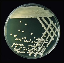
Colonial appearance
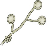
Demonstration of germ tubes
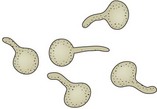
Chlamydospore production
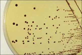
BiGGY agar
< div class='tao-gold-member'>
![]()
Stay updated, free articles. Join our Telegram channel

Full access? Get Clinical Tree


The pathogenic yeasts
Only gold members can continue reading. Log In or Register to continue
