Chapter 12. The Optic Nerve
CHAPTER CONTENTS
Congenital and hereditary diseases 400
Optic nerve swelling, trauma and degeneration 405
NORMAL ANATOMY (Fig. 12.1)
Optic nerve head, intraocular optic nerve (optic disc, optic papilla)
• There is a central depression within the optic nerve head, called the physiologic cup and this area is supported by a thickening of the inner limiting membrane, i.e. processes of native glial cells, called the supporting meniscus of Kuhnt
• Peripheral to these glial cells, blood vessels enter from the orbit and spread out to form the retinal vessels. Unlike primate species, dogs and cats lack a central retinal artery. The canine and feline optic nerve and retina receive their blood supply from the short posterior ciliary arteries, derived from anastomosis of the large external ophthalmic artery and the smaller internal ophthalmic artery
• In dogs, a small dark spot within the center of the optic nerve head is known as the physiologic pit, which is considered a remnant of the hyaloid artery. An exaggeration of the normal thickening of glial cells over the optic nerve is also considered to represent a remnant of the hyaloid artery, and is termed ‘Bergmeister’s papilla’. More pronounced vascular remnants of the fetal hyaloid artery often persist in cattle, as tubular structures projecting anteriorly from the optic nerve head into the vitreous
• In dogs myelination of retinal ganglion cell axons in the optic nerve begins at the optic chiasm and extends to the optic nerve head, with variable myelination extending within the peripapillary nerve fiber layer, accounting for the very irregular shape of the optic nerve head seen in many dogs
• In cats, myelination stops at the lamina cribrosa and the optic nerve head appears relatively small, dark and round
• In horses, the optic nerve head is typically a horizontally oriented oval with a very well-developed lamina cribrosa structure.
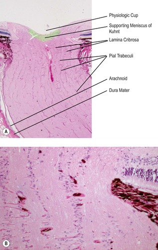 |
| Figure 12.1 Normal optic nerve. (A) Photomicrograph showing the normal morphological features of the canine optic nerve and optic nerve head. (B) Higher magnification photomicrograph showing the optic nerve tissue within the lamina cribrosa. |
The intrascleral optic nerve
• In glaucoma, physical distortion resulting from elevation in intraocular pressure leads to outward bowing of the lamina cribrosa and physical distortion and misalignment of the laminar plates, with resulting compression of axons (see Ch. 13).
The intraorbital optic nerve
• The intraorbital optic nerve has an S-shaped bend to accommodate for globe movement within the orbit
• The optic nerve may be considered to represent a white matter tract of the brain and is ensheathed by the meninges
▪ Dura mater
– Collagen-rich layer farthest from the nerve bordering the orbital tissue
– This tough, outermost dural sheath fuses with the orbital periosteum at the entrance to optic canal (optic foramen) and is also continuous with the lining of the cranial vault
▪ Arachnoid mater
– The arachnoid mater is a highly cellular layer with scant collagen poorly or unattached to the dura mater. The cells of the arachnoid are often large and form epithelial-like clusters which can be very numerous immediately adjacent to the globe
– The cerebral spinal fluid circulates in the space between the arachnoid and the innermost pia mater
– Arachnoid cap cells are clusters of epithelial-like cells which extend through the dura mater and form clusters in the soft tissue of the orbit
○ It is from these arachnoid cap cells that canine orbital meningioma arises
▪ Pia mater
– The collagenous and vascular layer closest to the optic nerve and continuous with the pial septae which penetrate the neuropil of the nerve and divide the tissue into columnar subunits.
The intracanalicular optic nerve
• Posterior to the orbit, the nerve enters the bony optic canal.
The intracranial optic nerve
• Represents a small portion of the nerve which merges into the optic chiasm, where a proportion of the axons cross over, or decussate, to the contralateral side before projecting to the lateral geniculate nuclei as the optic tracts
▪ The percentage of optic nerve axons that decussate at the optic chiasm ranges from about 50% in the primate, 65% in cats and 75% in dogs, to 100% in avian species.
Comparative Comments
In general, the human optic nerve conforms to the description given earlier for the canine and feline optic nerve. The human optic nerve contains approximately 1 million fibers and is about 5 cm long. A branch of the ophthalmic artery gains access to the nerve through the dura approximately 1 cm posterior to the globe, and pial branches provide the blood supply posteriorly.
CONGENITAL AND HEREDITARY DISEASES
Canine optic nerve hypoplasia (Figs 12.2, 12.3)
There are 15 canine cases of optic nerve hypoplasia in the COPLOW collection, 10 of which are bilateral.
• Although no clear breed predilection is evident in the COPLOW collection, optic nerve hypoplasia is suspected to be inherited in a number of breeds including Dachshunds, Poodles and Shih Tzu
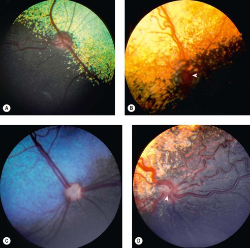 |
| Figure 12.2 Optic nerve hypoplasia, fundus. (A) Lhasa Apso, 8 months old: the small optic disc is slightly depressed. (B) Miniature Poodle, 6 months old: the very small, depressed disc (arrow) is located in the non-tapetal area. (C) Bernese Mountain Dog, 8 weeks old: the optic disc looks cat-like as the retinal vessels drop over the edge of the disc. (D) Collie, 4 months old: the optic disc is poorly discernible (arrow), as the major venules appear to be confluent. |
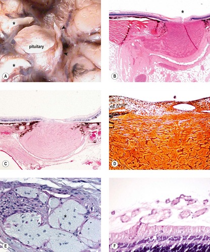 |
| Figure 12.3 Optic nerve hypoplasia, pathology. (A) Gross photograph of the ventral aspect of a dog brain with optic nerve hypoplasia showing vestigial remnants of optic nerve tissue (arrows). (*oculomotor nerves) (B) Low magnification photomicrograph of a canine hypoplastic optic nerve showing a narrow disc (*) and meandering nerve tissue. (C) Low magnification photomicrograph of a young Shih Tzu optic nerve showing hypoplasia. (D) Photomicrograph of the same optic nerve as (C), stained with Bielschowsky silver stain for axons, showing a lack of axons within the neuropil. (E) High magnification photomicrograph of a canine optic nerve showing vestigial remnants of optic nerve neuropil (*) within peripheral nerve tissue. (F) Photomicrograph of the inner retina from an affected dog showing blood vessels extending from the retina into the vitreous. Peripheral to this point the retina becomes avascular. |
Morphologic features of canine optic nerve hypoplasia
• The neuropil is densely gliotic
▪ This feature can be surprisingly hard to recognize unless one is very familiar with the normal appearance of nerve tissue
• A careful search often reveals vestigial remnants of optic nerve glial tissue in orbital tissues outside the optic nerve proper. The most common place to find these remnants is within peripheral nerve tissue
• The retina always has markedly decreased numbers of ganglion cells and there may be segments of retina with more profound atrophy
• Several cases within our collection have retinal blood vessels which leave the retina itself and extend into the vitreous
▪ This change is peripheral and segmental
▪ The far peripheral retina beyond the vascular anomaly is avascular.
Canine optic nerve aplasia (Fig. 12.4)
• There are six canine cases in the COPLOW collection, all unilateral
• There is no particular bred predilection
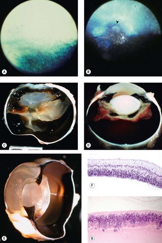 |
| Figure 12.4 Optic nerve aplasia. (A) American Shorthair, 2 months old: only a hyperreflective tapetum is present in the area that should contain the optic disc and retinal vessels. (B) DSH, 9 months old: the tapetum is hyperreflective. White striae are present inferior to a gray amorphous mass (arrow), which supplants the area that should contain the optic disc. (C,D) Gross photographs of canine eyes in which the retina is stretched across the back of the lens and never makes contact with the posterior pole of the globe, because there is no optic nerve. (E) Gross photograph of a dog eye viewed obliquely showing the same feature as (C) and (D). A remnant string of tissue extends to where the optic nerve and optic disc should be. (F,G) Photomicrographs of the retinas from two dogs with optic nerve aplasia showing no ganglion cells or blood vessels. |
Morphologic features of canine optic nerve aplasia
• No optic nerve tissue is detectable grossly or microscopically, except for the rare appearance of vestigial remnants of glial tissue within peripheral nerve tissue
• The retina is stretched across the back of the lens and makes no contact with the posterior pole of the globe
• The retinal tissue is totally devoid of ganglion cells and there is disorganization of the retinal layers
• The retinal tissue is totally avascular.
Achiasma and congenital nystagmus
• There is a line of black Belgian Sheepdogs with a recessive mutation leading to a chiasmatic optic nerves and congenital nystagmus, that has been studied extensively by vision researchers.
Optic nerve coloboma (Fig. 12.5)
• Colobomas of the optic nerve are rarely submitted to the COPLOW service
• Optic nerve head colobomas may be classified according to their location as either ‘typical’, at the 6 o’clock position in the location of the fetal fissure, or ‘atypical’ if away from this location
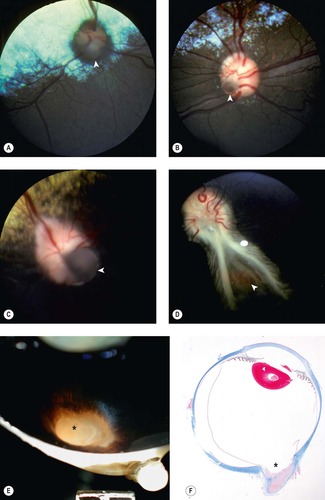 |
| Figure 12.5 Coloboma of the optic nerve head. (A) Basenji, 4 years old: the arrow points to a typical coloboma in the inferior optic disc. (B) Cairn Terrier, 3 years old: a small coloboma is identified by the arrow. (C) Cocker Spaniel, 3 years old: this large coloboma (arrow) involves about a quarter of the optic disc. (D) Great Dane, 1.5 years old: this is the right optic disc in a bilateral condition. The arrow points to a coloboma that extends into the disc. Abnormal myelination extends on both sides of the coloboma. (E) Gross photograph showing an equine optic nerve head coloboma (*). (F) Subgross photomicrograph of the same eye as (E) showing optic nerve head coloboma (*) (trichrome stain). |
Morphologic features of optic nerve colobomas
• The nerve head is widened and there is an outward bulging of vitreous
• A segmental defect in the lamina cribrosa and ectasia of the posterior sclera.
Comparative Comments
As in the dog and cat, the major congenital anomalies of the optic nerve in humans are hypoplasia, colobomatous defects, and pits of the optic nerve head. Minor congenital anomalies include persistence of the hyaloid system on the disk, projection of vascular loops from the disk, myelination of the nerve fibers extending onto the retina, and pigmentation of the disk.
OPTIC NERVE SWELLING, TRAUMA AND DEGENERATION
Papilledema (optic disc swelling, edema) (Fig. 12.6)
• Globes with documented papilledema are seldom brought to the attention of a pathologist when the globe is available at necropsy. There are no examples of papilledema in the COPLOW collection.
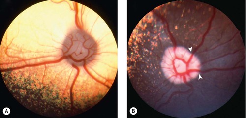 |
| Figure 12.6 Papilledema, clinical. (A) Chinese Crested Powderpuff dog, 2 years old: the optic disc is ‘full’ and extends vitreal. The borders of the disc are indistinct. Hydrocephalus was diagnosed based on CT scan. (B) Shetland Sheepdog, 4.5 years old: this dog developed severe ataxia. The retinal vessels have a characteristic bend (arrows) as they extend over the edge of the elevated disc. |
Papilledema and optic nerve degeneration in Vitamin A deficient in cattle
The COPLOW collection contains a small series of cases of vitamin A deficient optic neuropathy in a one small group of cattle.
• Vitamin A deficiency optic neuropathy is well-documented in the veterinary literature
• In addition to causing night-blindness and progressive retinal degeneration in adult cattle, vitamin A deficiency in calves and young, growing cattle, is associated with papilledema and optic nerve degeneration
• Narrowing of the optic canals, due to excess bone deposition, thickening of the dura and increased CSF pressure all contribute to compression, edema and ischemia of the optic nerves
• Malacia and demyelination of the optic nerve axons is associated with blindness and widely dilated pupils.
Glaucomatous optic neuropathy
• Initial swelling of the optic nerve head often precedes optic nerve degeneration in acute glaucoma, particularly in dogs
• Glaucomatous optic neuropathy is considered in detail in Chapter 13.
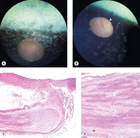
Optic nerve trauma and malacia in horses (Fig. 12.7)
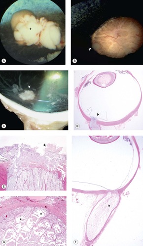 |
| Figure 12.7 Equine traumatic optic neuropathy (equine optic neuritis/neuropathy) (A) Fundus photograph of an equine eye showing a plume of necrotic nerve tissue extruding into the vitreous from the disc (*). (B) Fundus photograph showing a similar disc to (A), but less affected (arrow). (C) Gross photograph of an equine globe showing similar extrusion of necrotic nerve tissue in optic nerve malacia (arrow). (D) Subgross photomicrograph of an affected equine globe showing similar extrusion of necrotic material from the disc (arrow). (E) Higher magnification of the disc from (D). The extruded material (arrow) is a combination of granular necrotic neuropil and macrophage cells (gitter cells). (F) Subgross of another equine globe showing a swollen and pale necrotic optic nerve (*). (G) Cross-section from the same nerve as (F) showing cavitations within the nerve tissue (*) filled with protein-rich remnants. |
• This condition is often preceded by a dramatic traumatic event, most commonly a backward fall and head trauma followed by vision loss
▪ If the traumatic event is not witnessed, or vision loss is not immediately noticed at the time of optic nerve damage, it may be hard to establish the relationship between the optic nerve lesion and the traumatic event
• Morphologic features of equine optic nerve trauma and malacia
▪ There will be total or segmental necrosis followed, very rapidly by gitter cells
▪ Gitter cells from the damaged nerve tissue migrate into the vitreous and are seen as white nodules/excrescences protruding anteriorly into the vitreous from the optic nerve head (exudative optic neuropathy)
– The clinical appearance of pale extrusions of optic nerve material into the vitreous is considered a poor prognostic indicator for recovery of vision after a traumatic event
▪ The end-stage optic nerve lesion involves profound atrophy and fibrosis of the neuropil (Fig. 12.8)