CHAPTER 5The Normal Uterus in Estrus
Once the mare has entered the ovulatory season, the estrous cycle is on average 22 days long. The follicular phase, or estrus, typically lasts 5 to 7 days, and the luteal phase, or diestrus, lasts 14 to 16 days. Enormous variability, however, has been recognized in the number of days during which mares exhibit estrus, particularly early in the breeding season, when standing heat can be prolonged for weeks. The variation between mares is mostly due to differences in duration of estrous behavior, rather than in diestrus—depending on the mare and the time of the year, estrous behavior can last from 24 hours to several days.
Circulating concentrations of estrogen and progesterone have been well documented for the estrous cycle of the mare and reviewed by Ginther.1 Most of the cyclic changes in the reproductive tract of the mare are under the control of the ovarian steroids. The changes during estrus and diestrus are due to the presence of the ovarian steroid that predominates during a given phase of the cycle—that is, estrogen and progesterone, respectively. In mares, estradiol is thought to be responsible for estrous behavior,2 and estrogen concentrations during estrus correlate well with sexual behavior and grossly observable changes in the reproductive tract. During diestrus, when circulating levels of progesterone are greater than 2 ng/ml, estrus is inhibited, and the mare is aggressive toward the stallion. During estrus, progesterone concentrations in plasma are less than 1 ng/ml.1 The effects of progesterone on behavior and morphologic characteristics of the uterus are reported to be dominant over the effects of estrogen.3
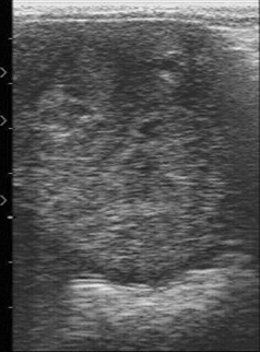
For the most part, estrus in mares that are at breeding farms is detected by exposing the mare to a stallion (teasing) and observing the typical squat and wink of estrus. The introduction of transrectal ultrasonography to visualize the reproductive tract in mares4 has allowed study of cyclic changes in tissue morphology. Both the ovaries and the uterus need to be examined thoroughly at every examination of a mare. One of the most striking characteristics visualized at ultrasonography is the appearance of the endometrium during the estrous cycle. During diestrus in a normal mare, individual endometrial folds are not routinely detected, and the uterus has a homogeneous echotexture. The lumen of the body of the uterus often is visible as a white line formed by specular reflections at the closely apposed luminal surfaces, in marked contrast with the features typically seen during estrus, when individual endometrial folds can be visualized.5 It is thought that endometrial folds become edematous during estrus as a result of increased concentrations of circulating estrogen.6 These changes give a heterogeneous image, with the dense central portions of the folds appearing echogenic and the edematous portions of the folds represented by nonechogenic areas. When the uterine horn is imaged in cross section, the appearance suggests a sliced orange or wagon wheel (Figure 5-1).
Occasionally, free estrous fluid can be imaged. It also is possible to grade the degree of endometrial edema; this concept was introduced by Ginther and Pierson5 and later developed by Squires et al.7 The endometrial edema scores have been found to reflect changes in circulating estrogen and progesterone concentrations and to correlate with sexual behavior scores.6 Endometrial folds first become visible at the end of diestrus, become more prominent as estrus progresses, and generally diminish from approximately 2 days from ovulation until the day of ovulation, when edema may have disappeared. This sequence is supported by three studies that used Thoroughbred and Standardbred or pony-type mares.5,8,9 However, one recent study using Belgian Draft mares reported that peak edema was detected at 6.6 days before ovulation and abruptly regressed at 5.5 days before ovulation.10 Breed differences may therefore be important for veterinarians to take into account.
Stay updated, free articles. Join our Telegram channel

Full access? Get Clinical Tree


