The Neurologic Examination

Chapter 20 The Neurologic Examination


NEUROLOGIC EXAMINATION—OVERVIEW


This chapter provides a complete description of the neurologic examination in one place. It is located near the end of this textbook because it represents the culmination of your understanding of the neuroanatomy of your patients. Most of this information has been described at some point in the various chapters that precede this one. The emphasis of this description is based on what can be performed in a cooperative dog. Features of the examination that apply specifically to the horse or food animal will be noted. Our opinion is that if you learn to perform this examination well on a cooperative dog systematically, you can adapt it to any other species. You would be amazed at how much you can determine in a confined wild animal such as a cheetah by just studying the animal through the barrier that confines it.


Examination of a patient with signs of a neurologic disorder should include a review of the history, a complete physical examination, a neurologic examination, and appropriate ancillary procedures. The purpose of the neurologic examination is to determine the neurologic abnormalities and, based on that, the location of the lesion or lesions responsible for causing these abnormalities. The location is the anatomic diagnosis. The continual use of a routine systematic procedure will provide you with the experience and confidence to make an accurate anatomic diagnosis.


The differential diagnosis must be based on the anatomic diagnosis, and the order of significance of these disorders will depend on your evaluation of the signalment and history. Be sure to consider all five major kinds of lesions in your differential diagnosis or a list of these with which you are comfortable. Our list is described in Chapter 1 and includes malformation, injury, inflammation, neoplasia, and degeneration (MIIND). Your experience with the anatomic diagnosis together with the signalment and history will often lead to a presumptive clinical diagnosis. Further examination of the patient with ancillary procedures must depend on your differential diagnosis and what you consider to be the most likely clinical diagnosis. Many factors will be considered in this selection, including the cost to the patient’s owner.


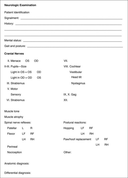
Be sure to record carefully all your observations from the neurologic examination, and never rely on your memory. Many forms are available for recording your observations. Some of these forms list in detail every possible response that is present or absent, with numbers to estimate the level of response. The considerable variation between individual patients of the same species makes the recording of the degree of a response less reliable and often misleading. We prefer a less time-consuming form that is easier to follow and adaptable to all species (Fig. 20-1).


Signalment


The signalment of the patient provides the examiner with the age, sex, breed, and use of the patient. When considered together with the chief complaint, this information may help direct the line of questioning as you take the history. For example, canine patients younger than 1 year that are presented for seizures are more likely to have an inflammatory lesion than a neoplasm, and lead poisoning is more common in dogs younger than 1 year. Toy breeds with functional hypoglycemia usually have seizures when they are younger than 6 months. Hypoglycemic seizures caused by a functional neoplasm of the pancreatic islet beta cells are rarely seen before 4 years of age. Neoplasms of the nervous system usually occur in the older patient, except for lymphosarcoma, which can occur at any age, and a spinal cord nephroblastoma, which occurs in young dogs. Exceptions are common. Intervertebral disk extrusion-protrusion is a concern in the chondrodystrophic breeds anytime after 1 year of age, whereas in the nonchondrodystrophic breeds, it is rarely a concern before 5 years of age.


The sex of the patient is an important consideration when the differential diagnosis includes brain neoplasms, given that mammary gland adenocarcinomas of the female patient rank high among the more common neoplasms that metastasize to the brain. Estrus may also lower the seizure threshold in animals that have or are at risk for idiopathic epilepsy.


Many disorders of the nervous system are restricted to one or more specific breeds. You must consider these breed-related disorders in your differential diagnosis. This practice is especially true for the inherited degenerative diseases such as the storage diseases, abiotrophies, and movement disorders. Many neurology texts provide a list of these breed-related disorders, and their recognition is a continual process. These disorders can be readily searched for each breed on the Internet.


History


The line of questioning followed in taking the history of the patient depends on the chief complaint. In all cases, this review should include a summary of any medical and surgical history unrelated in time to the present complaint.


If the chief complaint is an injury, the questioning will focus on the authenticity of the trauma, when the clinical signs first appeared, and how they have changed to the present time. By 24 to 48 hours after an injury, the clinical signs usually remain static or improve. Progressive neurologic signs are not usually the result of a single episode of trauma. Be aware that owners will often blame the neurologic signs on an injury from a fall when the fall was actually caused by the initial neurologic signs that were not recognized by the owner. This scenario is especially common when a dog falls down a set of stairs.


When a patient is brought in with the complaint of seizures, you need to obtain as thorough a description of the seizure event as possible to be certain of its authenticity and to determine the kind (classification) of seizure. The majority of seizures seen in domestic animals are generalized seizures. This type of seizure is the one that most commonly occurs in idiopathic epilepsy, most intoxications, and many prosencephalic structural disorders. Complex partial seizures (psychomotor) are more common in lead poisoning and diseases that affect the limbic system. Descriptions of these seizures often include episodic activities of the patient that the owner will describe as bizarre behavior or hysteria. Simple partial seizures may occur with or without confusion but with no loss of consciousness. The episodic activity is limited to groups of skeletal muscles such as the facial muscles or the muscles of one or both limbs on one side. Distinguishing between a simple partial seizure and a movement disorder may be difficult. The latter is described in Chapter 8. It is very useful to have the owner provide a video of the episodic event for you to study, given that these seizures rarely occur in your hospital. A thorough description or a video evaluation of a dog that is exhibiting episodes of collapsing will help distinguish among a seizure, cataplexy, movement disorder, syncope, or neuromuscular disorder. Episodic behavioral disorders are also best evaluated on videos.


Be sure to evaluate the history of each neurologic clinical sign that the owner observed to determine its validity, its progression, and whether these clinical signs represent a lesion that is focal or disseminated through the nervous system. For example, a patient that initially exhibited a spastic paresis and ataxia of the pelvic limbs that progressed and then developed a head tremor, head tilt, and abnormal nystagmus requires lesions in more than one location to explain these clinical signs (i.e., a lesion in the thoracolumbar spinal cord segments and a lesion in the cerebellomedullary region). Such a multifocal distribution of lesions is characteristic for an inflammatory lesion, such as the encephalomyelitis caused by the canine distemper virus in dogs, toxoplasmosis or cryptococcosis in dogs and cats, or Sarcocystis neurona in horses. The clinical signs of inflammatory disease usually progress more rapidly than those from an inherited degenerative disorder. Careful questioning may determine that what an owner thought was a sudden onset was, in fact, a progressive disorder. “My 6-year-old male German shepherd fell down the stairs this morning and injured himself. Oh yes, I did notice him stumble once yesterday.” As a rule, clinical signs that are precipitous at onset and not progressive after 1 to 2 days are caused by an injury or vascular compromise. The outdoor dog that is found in the morning unable to use its pelvic limbs is a candidate for an extrinsic spinal cord injury, ischemic or hemorrhagic myelopathy caused by fibrocartilaginous emboli or an acute intervertebral disk extrusion. Be aware that neoplasms occasionally cause an acute onset of clinical signs that are followed by progression.


As a rule, malformative or inflammatory diseases that occur in utero will cause clinical signs that are observed at birth and are nonprogressive. These signs are readily observed in foals and the newborns of farm animals that are born on the trot. In small animals, these clinical signs may not be obvious until the puppy or kitten starts to stand and tries to walk. A good example of this circumstance is the kitten in which the feline panleukopenia virus destroyed its cerebellum in the perinatal period. The cerebellar ataxia may not be apparent until 3 to 4 weeks of age. As the kitten develops normally in its second month of life and becomes more active, the cerebellar ataxia may appear to be worse, but the disease is not progressive. Most puppies with inherited cerebellar cortical abiotrophy develop normally for a few weeks and then begin to exhibit progressive cerebellar ataxia. Be aware that the onset of clinical signs of a cerebellar disorder at 10 to 12 weeks of age might just as well be the onset of progressive canine distemper encephalomyelitis. In a cairn or West Highland white terrier, this sign might signal the onset of clinical signs of inherited globoid cell leukodystrophy.


If intoxication is suspected, a thorough search should be carried out to find a possible source of the toxic agent. Sources of lead include lead-based paints, linoleum, tarpaper, welding equipment, and batteries. Cattle that lick old vehicle batteries are at risk for lead poisoning. Cats seem to enjoy licking antifreeze, which is a source of ethylene glycol. Be aware of the various houseplants that can be toxic if eaten by small animals and pasture plants that large animals consume. Consumption of rotting garbage, compost piles, and animal carcasses left in the woods may also be the basis for the cause of neurologic signs.


Be sure to ask about the patient’s behavior in its home environment. The owner will be aware of subtle changes that you cannot appreciate in your hands-on examination.


Inquire about the health of other animals in the same environment. Canine distemper often affects more than one puppy in a litter, as do many of the inherited degenerative disorders. Equine herpesvirus may affect multiple animals on a property. A brain abscess caused by Streptococcus equi may occur in a horse that is associated with other horses that are exhibiting signs of a strangles infection caused by the same bacterium.


Always determine the vaccination history of your patient because many of these vaccines involve infectious agents that affect the nervous system (i.e., canine distemper, eastern equine encephalitis, rabies).


The following additional questions should be asked as part of the historical examination:


What diet do you feed? Raw or homemade diets may affect the nervous system.


Is your pet on any medications? Surprisingly, many owners forget to tell you that their pet is on metronidazole, which, at high doses or for prolonged usage, can cause profound neurologic clinical signs.


Is your pet confined indoors, or does it have access to the outdoors? Exposure to the outside environment is necessary for the development of Cuterebra spp. myiasis and ischemic encephalopathy.


Has your pet traveled anywhere recently? Certain geographic locations may have endemic diseases such as coccidioidomycosis in the southwest United States and tick paralysis in the middle eastern coastal states.


Does your pet exhibit any polyuria, polydipsia, or polyphagia? These clinical signs may reflect dysfunction of the diencephalon.


Has your pet exhibited any coughing, sneezing, vomiting, or diarrhea on a regular basis? Diseases such as myasthenia gravis can result in regurgitation and secondary aspiration pneumonia with a resultant cough.


NEUROLOGIC EXAMINATION— SPECIFICS


The neurologic examination can be divided into five parts:


The order in which parts are performed is usually determined by the degree of patient cooperation and your preference. We usually perform the examination in the order listed. If the patient is resting comfortably in its cage, performing the cranial nerve examination first may be preferable. If the patient is excited or apprehensive, performing the cranial nerve examination may be more convenient after the patient has been handled for the examination of its gait, postural reactions, and spinal nerve reflexes.


Remember that, as described in Chapter 5, spinal nerve reflexes require only the specific peripheral nerves that innervate the area being tested and the spinal cord segments with which they connect. Postural reactions depend on the same components as the spinal nerve reflexes plus the cranial projecting pathways in the spinal cord white matter to the brainstem, cerebellum, and frontoparietal portion of the cerebral hemisphere and the caudal-projecting upper motor neuron (UMN) pathways that return from the cerebrum and brainstem and comprise tracts in the white matter of the spinal cord that terminate in the cervical and lumbosacral intumescences. These postural reactions test the integrity of nearly the entire peripheral and central nervous systems. By themselves, the postural reactions are relatively less reliable for lesion location.


Sensorium—Mental Attitude


An assessment should be made and recorded of the patient’s sensorium, its mental attitude, and response to the immediate environment and attitude to being handled by you. The owner is the best judge of subtle changes in the patient’s behavior in its normal environment. Be sure to explore this issue when you obtain the history. Considerable patient variation exists in how alert and responsive the patient may be in the examination room of a veterinary hospital. Do not mistake a very laidback behavior for depression. Descriptive terms for this portion of your examination include alert and responsive, depressed, lethargic, obtunded, semicoma (stupor), and coma. These states are described with the discussion of the ascending reticular activating system (ARAS) in Chapter 19. Other descriptions include acting vague, disoriented, hyperactive, propulsive, and aggressive.


As a rule, alterations in the patient’s normal sensorium reflect disturbances in the ARAS and limbic system components of the cerebrum or rostral brainstem. Be sure to evaluate the sensorium of a recumbent patient thoroughly. Recumbency from diffuse neuromuscular disease or focal cervical spinal cord disease will not alter the patient’s sensorium. A horse that is recumbent as a result of botulism may appear to be severely depressed or lethargic because it has no voluntary movement to show a response. The quality of the tetraparesis or tetraplegia with a cervical spinal cord lesion is the same as that caused by a mid- to caudal brainstem lesion, but the latter circumstance will often alter the patient’s level of response to its environment. Be aware that horses that suddenly become recumbent from an aortic thromboembolism or spinal cord ischemia and hemorrhage from an equine herpesvirus infection may act delirious as they struggle to stand. Horses that become acutely recumbent from a pontomedullary lesion that involves the vestibular system may be extremely disoriented and thrash wildly and appear maniacal as they try to recover their balance. The behavior is often remarkably altered in horses that have acute encephalitis from infection with the rabies, eastern equine encephalomyelitis, or West Nile viruses, as well as from hepatic encephalopathy. In human neurology, the Glasgow scale is used to report the patient’s sensorium objectively. Scales of this nature are presently being modified and evaluated for animals but are not routinely used in veterinary medicine at this time.


Gait and Posture


Examination of the gait should be performed in a place where the patient can be walked with a leash or shank and where the surface is not slippery. Most hospital floors have a very slippery surface, which facilitates cleaning but is poor for evaluating a gait disorder and can be dangerous if the patient slips and falls, especially horses and cattle. A washable carpet is very useful for small animals. A rubberized floor is ideal for large animals. Be careful if you walk horses or cattle on a macadam surface because it is slippery. If you are constructing a small animal hospital, we recommend that you consider having a covered area with a specialized surface used in playgrounds that is relatively soft, provides excellent traction for the patient, and is easily cleaned. The material is Vitriturf and is available from Hanover Specialties, Inc., Hauppauge, New York. Whatever facility you adopt for this gait evaluation in patients with neurologic disorders will also be useful for orthopedic examinations.


Observe the patient while it is standing for a head tilt, lowered position of the neck, trembling, degree of tarsal extension, and its tail position. You should evaluate the gait both as you lead the patient and as an assistant leads the patient. Most deficits are best seen during a slow walk and as it turns. Walk the animal back and forth in a straight line and in circles in each direction. Observe the patient from all directions. In our opinion, most abnormalities are best seen from a side view. Trotting a horse is occasionally helpful. If it is difficult to determine an abnormality, evaluate the patient on a slope or turn the patient loose in a confined area such as a paddock or riding ring. Be aware of breed characteristics that influence the posture and gait. The overflexed tarsus in German shepherds and the excess flexion action in the thoracic limbs of the Paso Fino and Tennessee walking horse are examples.


Is your patient unwilling or unable to move normally? When you see a gait disorder, this question is the first one that you need to answer. This circumstance is especially true when the patient is short strided or does not support weight well on one or more limbs. A loss of support from a femoral or radial nerve disorder will mimic a severe painful disorder causing a reluctance to bear weight.


Pattern recognition is critical in evaluating gait disorders. With experience, clinicians recognize specific patterns in abnormal gaits that suggest an anatomic diagnosis. We can describe these patterns, but observing them on videos is the best way to learn them. These patterns have five components consisting of two qualities of paresis and three qualities of ataxia.


Paresis is defined as “weakness” in the dictionary, but in clinical neurology, it is defined as “a deficiency in the generation of the gait or the ability to support weight.” This definition includes the two qualities of paresis, which are lower motor neuron (neuromuscular) and upper motor neuron. Lower motor neuron (LMN) paresis reflects degrees of difficulty in supporting weight and varies from a short stride that is easily mistaken for a musculoskeletal lameness to complete inability to support weight, causing collapse of the limb whenever weight is placed on it. Animals with LMN disorders affecting both pelvic limbs will occasionally use them simultaneously. This action is described as bunny hopping. Be aware that bunny hopping can also be seen in orthopedic disorders, as well as spinal cord dysplasias. UMN paresis causes a delay in the onset of protraction, which is the swing phase of the gait. The stride will usually be longer than normal. Stiffness and spasticity may be apparent in the stride. You may hear the hoof slap the ground in horses that are spastic. Most of the UMN pathways necessary for gait generation are anatomically adjacent to the pathways of the general proprioceptive (GP) sensory system, and lesions usually affect both simultaneously. Therefore the gait that reflects UMN paresis also reflects ataxia caused by dysfunction in the GP system. It is unnecessary to recognize the separate clinical signs of dysfunction of these two systems. Therefore we recognize a pattern that reflects the combined dysfunction of UMN paresis and GP ataxia. To observe and compare these two forms of paresis, see Videos 5–14 and 10–36.


Ataxia is a synonym for incoordination, and we recognize three qualities of ataxia: (1) GP, (2) vestibular (special proprioception [SP]), and (3) cerebellar. GP ataxia reflects the lack of information reaching the central nervous system (CNS) that informs the CNS of where the neck, trunk, and limbs are in space and the state of muscle contraction at any time. Without this GP information, the onset of protraction of a limb may be delayed, and the stride may be lengthened. During protraction, the limb may swing to the side (abduct) or swing under the body (adduct), overflex during protraction, scuff or drag one or more digits, and in the support phase, stand on the dorsal aspect of one or more digits. Remember that these clinical signs overlap with those caused by dysfunction of the UMN. The gait pattern of a patient with a focal cervical spinal cord lesion between the C1 and C5 segments reflects dysfunction of the UMN and GP systems and is observed as spastic tetraparesis and ataxia. This cervical spinal cord pattern is often recognized by the overextension of the thoracic limbs creating an overreaching or floating action. This clinical sign can be augmented by holding the head and neck extended as the patient is led, especially in horses. This unique form of hypermetria must not be confused with cerebellar ataxia in which the limb is overflexed on protraction. At no time do we try to differentiate between conscious (cerebral) and unconscious (cerebellar) GP pathways! No examination will clearly differentiate these two pathways from each other or from the UMN pathways. No pure conscious proprioceptive deficit exists. This term should be dropped from the clinician’s vocabulary. See Video 10–36 for this combination of UMN and GP clinical signs. Vestibular ataxia reflects the loss of orientation of the head with the eyes, neck, trunk, and limbs, which results in a loss of balance. Lesions in this system cause the patient to lean, drift, or fall to one side. However, the patient’s strength and awareness of where its limbs are in space are normal with lesions confined to this system. This ataxia is usually accompanied by a head tilt and sometimes abnormal nystagmus. We will occasionally blindfold our patients with bandage material in small animals or a towel in horses and cattle to exacerbate vestibular ataxia. See Video 12-1. Cerebellar ataxia most commonly causes hypermetric ataxia characterized by sudden bursts of motor activity with a marked overflexion of the limbs on protraction. Vestibular system components exist in the cerebellum that, if dysfunctional, may cause loss of balance, head tilt, and abnormal nystagmus. Cerebellar ataxia in horses produces more hypertonia than hypermetria when compared with other species of domestic animal. See Videos 13-9 and 13–21.


Many clinicians use a grading system for the clinical signs observed in the gait caused by spinal cord lesions. These systems facilitate determination of a prognosis and provide a basis for monitoring their response to therapy. The grading systems used are different for small animals and large animals. In small animals, the system is designed primarily for lesions that involve the spinal cord segments between T3 and L3 and is a measure of the degree of pelvic limb strength (UMN) and coordination (GP) that is present. In large animals, the system is designed primarily for lesions between the C1 and C5 spinal cord segments and is a measure of the degree of gait deficit.


As you observe the gait of your patient, look carefully for any evidence of a head tilt (vestibular system), head and neck deviation or tendency to walk in circles (prosencephalon), neck flexion (neuromuscular), and tail movement.


Postural Reactions


The degree of functional limb deficit will determine the need for postural reaction testing. In a patient that is recumbent with tetraplegia or is paraplegic, you need not perform postural reactions in the affected limbs. However, in the paraplegic patient, you must test the thoracic limb postural reactions so as to prevent overlooking a focal cranial thoracic lesion or a multifocal disorder.


In small animals, we evaluate muscle size and tone just before our evaluation of the postural reactions. Be sure to talk to your patient continually, and use its name to gain its cooperation. Stand over the patient with both of you facing in the same direction. Simultaneously palpate the muscles of the neck and both thoracic limbs from proximal to distal for any evidence of atrophy. Flex and extend each limb for range of motion and to determine the degree of muscle tone. A short stride or stiffness in the gait may be caused by a joint disorder, limiting the range of motion. When you place the limb back on the ground surface, turn the paw over so that its dorsal surface bears the weight of the limb to determine how rapidly the paw is replaced. Most normal small animals immediately return the paw to its normal position. This response is known as the paw or hoof replacement reaction that requires many peripheral and central components to be normal. This test is not just for conscious perception of GP and should not be called the CP test. This term is a misnomer that we are well aware will be difficult to eliminate from the clinical language of veterinarians, even boarded veterinary neurologists. Moving on, palpate the thoracolumbar epaxial muscles and then the muscles of both pelvic limbs followed by flexing and extending those limbs and checking for paw or hoof replacement. Check for the degree of tail tone, and while extending the tail, evaluate the anal tone. In horses and farm animals, make the same evaluation for muscle atrophy. Some equine clinicians will place the hoof on its dorsal surface or place one limb in front of the other or laterally to the side and assess its rate of replacement.


Hopping responses, in our opinion, are the most reliable of the postural reactions that we test. While still straddling the patient, move back to the thoracic limbs, and while elevating the abdomen with one hand, pick up the thoracic limb on the opposite side with your other hand. With all the weight supported on the other thoracic limb, hop the patient laterally on that thoracic limb. Go as far as you can without moving your pelvic limbs. Then switch hands and hop the patient back on the other thoracic limb. Repeat this test many times until you are sure the thoracic limbs are normal or abnormal. As you stand over your patient and look down the lateral aspect of the limb that is being hopped, the limb should move as soon as you move the shoulder region laterally over the paw or hoof. Any delay in this response is abnormal. The hopping movements should be smooth and fairly rapid and not irregular or excessive. The paw or hoof should never drag or land on its dorsal surface. Carefully compare one thoracic limb with the other. To test the hopping responses in the pelvic limbs, stand beside the patient, and place your forelimb that is closest to the patient’s head between its thoracic limbs with your hand on its sternum. Lift up on the thorax just enough to take the weight off of the thoracic limbs. With the other hand, pick up the pelvic limb on the side where you are standing and push the patient toward the pelvic limb that is bearing the weight. This action will force the patient to hop on that pelvic limb in a direction away from you. After a few hops, switch sides, and hop the patient back on the other pelvic limb. Keep repeating this test until you have determined that the response is normal or abnormal. The responses should be brisk and smooth but will not be quite as rapid as in the thoracic limbs. While you are hopping the patient, you will also be aware of the degree of tone in the limb that is bearing all the weight. In large dogs or small farm animals that are too heavy to lift for this testing, the same observations can be made while the patient is walked on one side (hemiwalking). Hemiwalking is performed by standing on one side of the patient. Grasp each limb on that side and lift the limbs off the ground surface and push the patient toward its opposite side. The patient will hop with both limbs on that side. Switch sides and repeat the hemiwalking performance on the opposite limbs. Be sure to compare one thoracic limb with the other and one pelvic limb with the opposite pelvic limb.


These hopping response evaluations can be performed on foals and calves and other farm animals that are not too large. Some clinicians will try to evaluate hopping in the adult horse by picking up one limb and using their shoulder to push the horse toward the opposite side, forcing it to hop on that limb. We find this method difficult to evaluate reliably, and the danger of self-injury for questionable value is not worth the effort.


In small animals in which the hopping responses are equivocal or difficult to interpret, testing the placing responses may be useful. Pick up the patient and bring its thoracic limbs to the edge of a shelf, table, or chair so that the dorsal surface of the paw contacts the front surface of the object. The normal patient will immediately place its paws on the horizontal surface of the object. Test both thoracic limbs while holding the patient from both sides. For some unknown reason, occasionally the normal patient will not respond on the side on which it is being held. Blocking the vision of the patient while conducting this test may also be useful. Do this by extending its head and neck so that it cannot see the protruding surface of the shelf, table, or chair. Whether the lack of vision or the position of the head and neck helps exacerbate the placing deficits is unclear.


In cooperative patients in which you are not sure of the thoracic limb function, you can wheelbarrow the patient while holding its head and neck in extension. With one of your forelimbs, elevate the abdomen of your patient, and with the other hand, hold the head and neck in extension and force the patient to walk forward. With this posture, vision is compromised, and the need for GP is increased. In large animals, elevate the head and neck as much as you can and still be able to lead them. With mild cervical spinal cord or brainstem lesions, this test may cause some patients to scuff the dorsal surface of its paws or overreach on protraction.


In large animals in which these postural reactions cannot be performed, we rely on other maneuvers that require more neurologic function of the UMN and GP systems to be normal. These maneuvers may elicit abnormalities that are not seen on observing the patient walking in a straight line. They include the head elevation described previously plus circling, backing, and swaying. Circling is the most useful of these maneuvers. Walk the patient in a tight circle for 8 to 10 times in each direction. The leader should be standing in the center of the circle as this test is conducted. The normal patient will step around briskly with relatively short strides and will not pivot on one hoof or step on itself. Patients with lesions that affect the UMN and GP systems, at most any level of the CNS, will appear awkward as they circle. The patient’s movements may be irregular, and the patient will often pivot on the inside limb, which is held in place and not protracted. The outside pelvic limb may flex and abduct excessively as it is protracted. This action is known as circumduction. The excessive flexion on protraction appears occasionally like a stringhalt action. The outside thoracic limb may overextend and cross over the standing limb. During this circling, the abnormal patient may step on itself. Be careful when you circle a patient with a severe deficit because you may cause it to fall. Backing up may be useful in a cooperative patient. Look for the tendency to be reluctant to back with one or both limbs. They may drag the limb or just collapse on one or both pelvic limbs and sit in a dog-like fashion. Sway the standing and walking patient by pulling its tail toward you and then releasing it. The degree of resistance may help you assess the patient’s strength, and releasing the tail may alter its balance and precipitate a brief ataxia. Some clinicians will place one limb cranial to the other or as much on the opposite side of the other limb as possible and determine how rapidly it is replaced. A delayed replacement suggests a neurologic deficit similar to paw replacement in small animals. In small animals with suspected spinal cord disease, palpation of the vertebral column may elicit an area of discomfort. In large animals, firm hand compression of the vertebral column that induces some extension may reveal a degree of paresis if the patient tends to collapse from this vertebral extension.


When the clinical signs are subtle, you might want to walk, circle, back up, and sway the equine patient on a gentle slope. Walking the horse back and forth over a road curb may also be helpful. When turned loose in a paddock, a horse with mild UMN and GP dysfunction may exhibit clinical signs only when it has to change directions at the fence corner. If you are suspicious of a subtle dysfunction of the vestibular system, blindfolding the patient may exacerbate the vestibular system signs; this measure has no effect on horses with spinal cord disease. Use a towel and tuck it under the halter where it can be readily grasped and removed. Never tie the blindfold onto the halter!


Remember that patients with LMN disease that still have some voluntary movements will hop or circle rapidly if their weight is supported because their GP is unaffected. This observation may help distinguish between subtle UMN and LMN paresis. When we are presented with a patient that we suspect has LMN paresis, we will hop the patient with and without supporting all the weight on the affected limb or limbs. As already stated, the patient with LMN paresis should know exactly where the limb is located during these postural reactions.


When presented with a recumbent patient, you should pick it up and hold it in a standing position. Get help if the patient is too large for you to handle alone. Use a sling in horses and large farm animals. By holding them in this position and lifting them up and down, you can readily determine the quality of the muscle tone, whether they are hypotonic-flaccid from LMN disease, or hypertonic from spasticity of UMN disease. You can determine whether any voluntary movements are present in the limbs, and while supporting the patient, you can determine the presence and quality of the hopping responses. Muscle tone in recumbent adult horses and cattle is difficult to evaluate, especially in the recumbent limbs.


One might conclude that these postural reactions are relatively nonspecific. This conclusion is absolutely correct. Why then are they useful? First, testing postural reactions acts as a screen for detecting abnormalities in the nervous system. Abnormal responses will be the first clinical sign of any progressive lesion in any part of the central or peripheral nervous system that is involved in limb movement. One or more of these postural reactions may be abnormal before any detectable abnormality in the gait is observed. Second, their importance in localizing lesions is dependent on the results of the rest of the neurologic examination. If the gait is normal in the environment of your examination and one or more of the postural reactions are abnormal in the limbs on one side of the body, a contralateral prosencephalic lesion is strongly suggested. This test is the most useful of the three clinical tests that we use for prosencephalic disorders. If you have a patient with clinical signs of a unilateral vestibular system disorder with a normal gait but the postural reactions are abnormal, then the lesion is in the central components of the vestibular system.


Remember to use terminology that all clinicians can understand. Lesions that affect the UMN pathways also affect the GP pathways; thus when you describe a gait abnormality as paraparesis, also include pelvic limb ataxia. Using hemiparesis and ataxia is preferable when this is visible in the gait. If the gait is normal and the postural reactions are abnormal on one side, describe it that way, and do not refer to this abnormality as hemiparesis. Be sure your reader clearly can understand what you have observed.

Stay updated, free articles. Join our Telegram channel

Aug 26, 2016 | Posted by in INTERNAL MEDICINE | Comments Off on The Neurologic Examination

Full access? Get Clinical Tree

Get Clinical Tree app for offline access