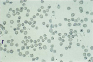
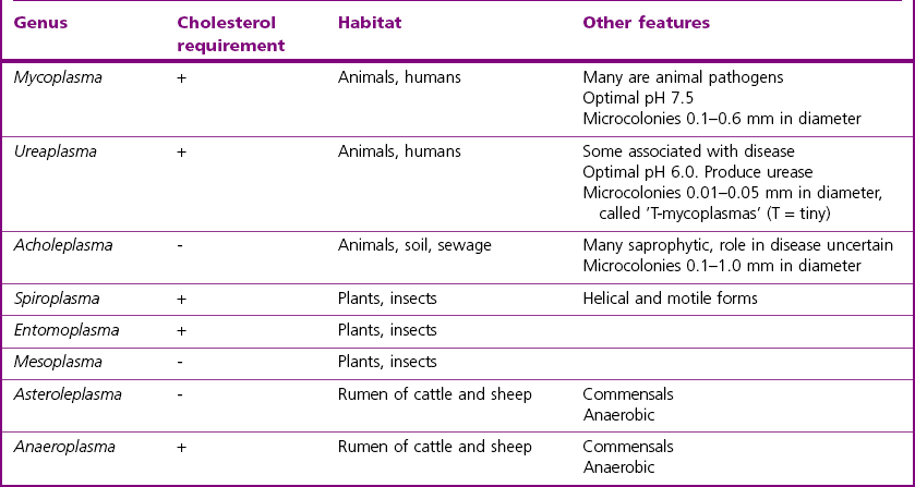
Chapter 35 Because of the inability of the mollicutes to synthesize peptidoglycan they stain poorly with the Gram stain. Better staining is obtained using Giemsa and other Romanowsky stains. The first organism in the class Mollicutes to be isolated was Mycoplasma mycoides subsp. mycoides, the cause of contagious bovine pleuropneumonia (CBPP). As a result the collective term pleuropneumonia-like organisms (PPLOs) was once used for Mycoplasma organisms. The differential features of the genera are shown in Table 35.1. Species of the genera Mycoplasma and Ureaplasma are of significance in animals. Members of the genus Acholeplasma may be found in clinical specimens but are not considered to be pathogenic for animals. Organisms in the genera Haemobartonella and Eperythrozoon, which were previously grouped with the rickettsial organisms, have been reclassified as members of the Mycoplasma genus. These organisms parasitize red blood cells (Fig. 35.1) and are known as the haemotropic mycoplasmas or are sometimes referred to by the trivial name ‘haemoplasmas’. Figure 35.1 Leishman-stained blood smear from a sheep with Mycoplasma (Eperythrozoon) ovis parasitizing the red cells. (×1000) The most important species of mycoplasmas and the diseases that they cause in poultry, other domestic animals and laboratory animals are listed in Table 35.2. The mycoplasmas tend to be fairly host-specific although it has been suggested that interspecies transmission can occur and may be of pathogenic importance in immunosuppressed hosts (Pitcher & Nicholas 2005). In addition to the mycoplasmas shown in Table 35.2, there are many species isolated from birds and animals, whose disease status is at present uncertain. These are listed in Table 35.3. Table 35.2 Mycoplasmas causing significant disease in domestic and laboratory animals The parasitic mycoplasmas tend to adhere firmly to the mucous membranes of the host and some species have been shown to affix to cells by specific attachment structures. A number of surface proteins play a role in adhesion, including the P26 antigen and variable surface proteins of M. bovis (Caswell & Archambault 2007), variable lipoprotein haemagglutinin of the avian mycoplasmas (Noormohammadi 2007) and P116 of M. hyopneumoniae (Seymour et al. 2010). Although the production of a cytotoxin (CARDS toxin) has been identified in the human pathogen, Mycoplasma pneumoniae (Kannan & Baseman 2006, Techasaensiri et al. 2010), production of specific toxins has not been demonstrated for pathogenic mycoplasmas of animals. It is considered that many of the toxic effects on host cells result from the action of toxic metabolic products which diffuse into the tissues of the host following adherence of the organisms. These metabolic products include H2O2 and other reactive oxygen species. Enzymes such as proteases and haemolysins may also be involved in the production of tissue damage. Variable surface proteins (Vsps) have been identified in many mycoplasmal species and these proteins play a role in adhesion and colonization and, most importantly, in the evasion of the humoral response of the host. The production of a number of different Vsps has been demonstrated in many of the major mycoplasmal pathogens of animals, including Mycoplasma mycoides subspecies mycoides (Persson et al. 2002), M. bovis (Sachse et al. 2000) and the avian pathogens M. gallisepticum and M. synoviae (Noormohammadi 2007). The similarity between some mycoplasmal antigens and host cell antigens may also contribute to persistence of mycoplasmas in the host because of the inability to induce an effective immune response due to a failure in antigen recognition. However, the sharing of antigenic determinants between the organism and host cells may also lead to the development of autoimmune disease. It is thought that immunological mechanisms may be involved in the destruction of red cells in cats infected with Mycoplasma haemofelis although direct damage by the organism may also play a role. Important virulence factors of Mycoplasma species are listed in Table 35.4. Table 35.4 Virulence attributes of pathogenic Mycoplasma species (the production of every pathogenic factor listed has not been demonstrated for all the pathogenic species)
The Mycoplasmas (class: mollicutes)
Pathogenesis
Species
Disease
Poultry
Mycoplasma gallisepticum
Chickens: chronic respiratory disease
Turkeys: infectious sinusitis
Infection in game birds and imported Amazon parrots
M. synoviae
Chickens and turkeys: infectious synovitis
M. meleagridis
Turkeys: Mycoplasma meleagridis disease (MM disease), air sacculitis and bursitis in young birds
M. iowae
Turkey poults: air sacculitis, stunting and leg deformities. Mortality of turkey embryos can occur
M. anatis
Ducks: sinusitis
Pigs
M. hyorhinis
Chronic progressive arthritis and polyserositis in three- to 10-week-old pigs
M. hyosynoviae
Mycoplasmal polyarthritis in 12–24-week-old pigs
M. hyopneumoniae
Enzootic (‘virus’) pneumonia of pigs
M. suis
Mild anaemia, poor growth rates
Cattle
M. mycoides subsp. mycoides (small colony type)
Contagious bovine pleuropneumonia (CBPP)
M. bovis
Mastitis, arthritis, pneumonia, genital infections, abortion
M. bovigenitalum
Vaginitis, arthritis, mastitis, seminal vesiculitis
Ureaplasmas including U. diversum
Vulvovaginitis, pneumonia
M. dispar
Pneumonia (calves)
M. californicum
Mastitis
M. canadense
Mastitis
M. bovoculi
A predisposing cause of infectious bovine keratoconjunctivitis (Moraxella bovis infection)
M. wenyonii
Mild anaemia
Goats
M. capricolum subsp. capripneumoniae
Contagious caprine pleuropneumonia (CCPP)
M. mycoides subsp. capri
Mastitis, arthritis, keratitis, pneumonia and septicaemia syndrome
M. putrefaciens
Mastitis, arthritis
Sheep
M. ovipneumoniae
Pneumonia
M. ovis
Haemolytic anaemia of varying severity
Sheep and goats
M. agalactiae
Contagious agalactia
M. conjunctivae
Keratoconjunctivitis
M. capricolum subsp. capricolum
Mastitis, arthritis, keratitis, pneumonia and septicaemia syndrome
Acholeplasma oculi
Keratoconjunctivitis
Horses
Mycoplasma felis
Pleuritis (a commensal that can enter the pleural cavity after severe exercise)
Dogs
M. cynos
Pneumonia (part of ‘kennel cough’ complex)
M. haemocanis
Usually mild or subclinical anaemia but more severe signs in splenectomized animals
Cats
M. felis
Conjunctivitis
M. haemofelis
Feline infectious anaemia
Rats and mice
M. neurolyticum
Rolling disease
M. pulmonis
Pneumonia
M. arthritidis
Polyarthritis
Virulence factor
Comments
Capsular polysaccharide
Exact role unknown, aids in persistence and dissemination of the organism
Lipoproteins
Function in adhesion, stimulate the release of pro-inflammatory cytokines
Adhesins
Protein adhesins have been identified in some mycoplasmal species. Sialyl moieties important in adhesion also
Variable surface proteins
Role in evasion of host antibody response, in colonization and adhesion and in modulation of the host immune response
Toxic metabolic pathway products
These products include H2O2 and reactive oxygen species which induce toxic damage to host cells
Heat shock proteins
Role in adhesion to host cells
Biofilm
Some Mycoplasma species have the ability to produce biofilms although the genes commonly associated with biofilm formation in other bacterial species are lacking
![]()
Stay updated, free articles. Join our Telegram channel

Full access? Get Clinical Tree


