Chapter 16 The horse
The horse appeared in its earliest form 55 million years ago as Eohippus, a small, multitoed mammal about 30 cm high. It had four toes on the fore limb and three on the hind limb and a weight- bearing pad under the central toe on each foot. Its teeth were capable of chewing succulent leaves. During its evolution over a period of many millions of years the number of toes reduced and the central third toe became encased in a simple hoof. This species was sequentially replaced by several others with similar skeletal structures and increasingly efficient teeth suitable for eating grass.
The skeletal system
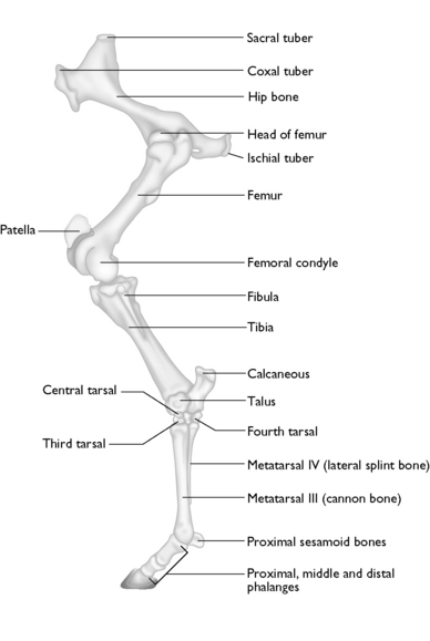
The equine skeleton (Fig. 16.1) consists of two separate sections:
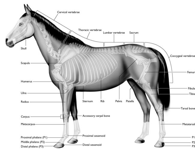
Fig. 16.1 The skeleton of the horse.
(With permission from Aspinall V 2006. The complete textbook of veterinary nursing. Butterworth- Heinemann, London, p 134.)
The skull
The equine skull (Fig. 16.2) is made up of approximately 37 fused bones providing a rigid structure with minimal movement; the only moving part is the temporo-mandibular joint, which is essential for chewing. The major components of the skull are:
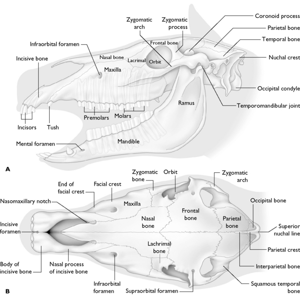
Fig. 16.2 The equine skull. A Lateral view. B Dorsal view.
(With permission from Aspinall V 2006. The complete textbook of veterinary nursing. Butterworth-Heinemann, London, p 135.)
The functions of the skull are:
The vertebral column
The vertebral column extends from the base of the skull to the tip of the tail and consists of approximately 54 individual vertebrae (Fig. 16.1).
The function of the vertebral column is:
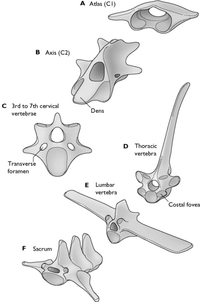
The vertebrae in the horse (Fig. 16.3) are grouped into regions as they are in the cat and the dog, although the numbers of each vertebrae may be different.
The vertebrae Fig. 16.3 in each region vary in size and shape, which relates to their movement and function.
The ribs and sternum
The horse has 18 ribs, although this may vary in some individuals (Fig. 16.1). The ribs play an essential role in housing and protecting the vital internal organs of the thorax. The ribs may be separated into two groups:
The appendicular skeleton
The forelimb
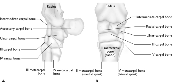
The bones of the forelimb (Fig. 16.4) are:
 Upper row – running from the medial to lateral surfaces – radial carpal, intermediate, ulnar and accessory carpals. The accessory carpal bone lies behind the ulnar carpal and does not directly bear weight.
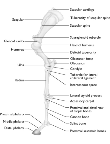
Fig. 16.4 The equine forelimb.
(With permission from Aspinall V. 2006. The complete textbook of veterinary nursing. Butterworth-Heinemann, London, p 136.)
Pelvis
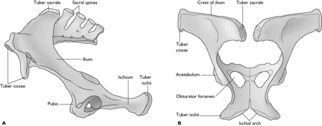
The pelvic girdle (Fig. 16.7) links the spine and the hindlimb and is composed of three large flat bones the pubis, the ischium and the ilium. The pubis forms the floor of the pelvis with the ischium lying caudal to it. The tuber ischii can be felt at the point of buttock. The ilium is the largest bone in the pelvis and is the upper, almost vertical portion. The wings or tubera sacrales of the ilium can be felt at the croup and the tuber coxae forms the points of the hip. All three bones meet to form the acetabulum or hip socket. The hip joint is formed by the head of the femur and the acetabulum.
The muscular system
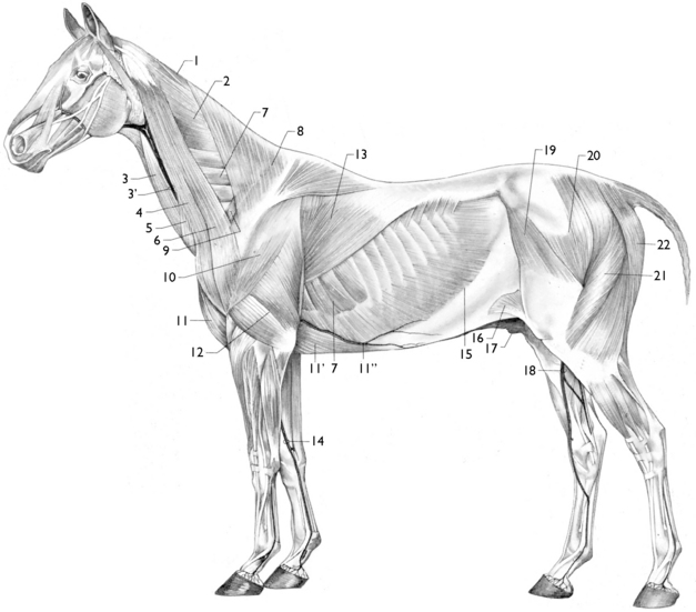
The arrangement of the superficial muscles covering the neck, thorax and abdomen (Fig. 16.8) is similar to that in the dog and cat, with the obvious difference that many are better developed to bring about the rapid locomotion characteristic of the horse.
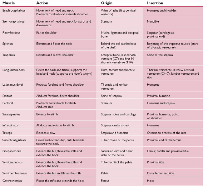
The limbs of the horse are superbly adapted for speed, although at rest both the fore- and hindlimbs support the body. The forelimbs, which should be more or less straight, carry about 60% of the weight and absorb most of the shock during locomotion, especially when landing from a jump. The hindlimbs are more angled and provide the main propulsive force. Details of the most significant muscles are shown in Table 16.1.
Soft tissues of the equine lower leg
The horse has evolved from a three- to four-toed, dog-like animal into the large animal taking its weight on one digit that we recognise today. The central digit is encased in a hoof while the outer toes are reduced to vestigial appendages that no longer reach the ground. This arrangement adds to the horse’s ability to run fast.
Ligaments and tendons
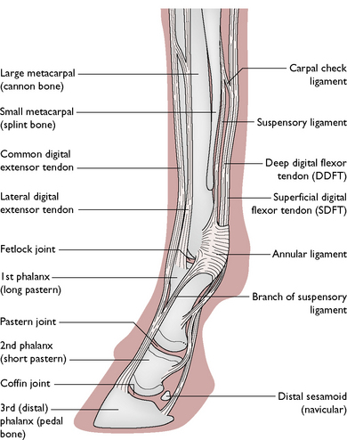
Important tendons found within the lower leg (Fig. 16.9) include:
The ligaments and tendons in the lower fore- and hindlimbs work in conjunction with a variety of muscles to form the suspensory apparatus (Fig. 16.10). The function of the suspensory apparatus is to support and suspend the limb and fetlock joint and prevent over-extension and collapse of the limb. It consists of the suspensory, intersesamoidean, collateral sesamoidean and distal sesamoidean ligaments, which are attached to the proximal sesamoid bones.
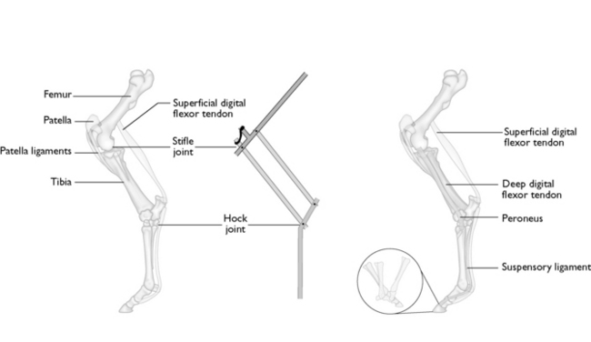
Fig. 16.10 The stay apparatus and suspensory apparatus of the hindlimb.
(With permission from Aspinall V 2006. The complete textbook of veterinary nursing. Butterworth-Heinemann, London, p 141.)
Stay updated, free articles. Join our Telegram channel

Full access? Get Clinical Tree


