Chapter 41The Hind Foot and Pastern
The contribution of the hind foot and pastern to hindlimb lameness is considerably less important than that of the digit to forelimb lameness. Extensive information regarding lameness of the foot and pastern in the forelimb is found in Chapter 26Chapter 27Chapter 28Chapter 29Chapter 30Chapter 31Chapter 32Chapter 33Chapter 34Chapter 35Chapter 82. This information is directly applicable to the hindlimb. During lameness examination, the hind foot and pastern are easily overlooked because they are in a potentially dangerous place to examine. Other regions of the hindlimb have been presumed to be more important, and, historically, little has been taught about the distal part of the limb. Without a commitment to performing diagnostic analgesic techniques in the distal aspect of the hindlimb, there is little way to discover whether the digit is the authentic source of pain unless the problem is severe or obvious. Subtle primary or compensatory lameness problems of this region are likely to go unnoticed daily. However, in certain sports horses, such as the draft horse, lameness of the hind foot and pastern is so common that this region cannot be overlooked. I am curious as to why infectious osteitis of the distal phalanx is more common in the hind feet. Of 26 affected limbs in 21 lame foals with infectious osteitis of the distal phalanx, most likely hematogenous in origin, 18 (69%) were hindlimbs.1 Infectious arthritis in foals occurs more commonly in hindlimb joints, specifically the femoropatellar and tarsocrural joints, than in forelimb joints, a finding that is equally difficult to explain.2
Anatomy and Innervation of the Hind Foot and Pastern
The bones are essentially the same as in the forelimb. The hind distal phalanx is narrower compared with the forelimb and has a steeper dorsal angle. The plantar surface is more concave, and the plantar processes are closer together.1 In external appearance, the hind foot usually is more upright than the fore foot, but abnormal wear and shoeing practices can produce a common pathological condition of a low, underrun heel (see Chapter 6). The hind middle phalanx is narrower and longer, and the hind proximal phalanx is slightly shorter than the corresponding bones in the forelimb.3 The ligaments, tendons, digital flexor tendon sheath (DFTS), and distal interphalangeal and proximal interphalangeal joints are the same as in the forelimb. Innervation of the foot is derived primarily from the medial and lateral plantar nerves, which originate from the tibial nerve (see Chapter 10). The medial and lateral plantar metatarsal nerves, which originate from the deep branch of the lateral plantar nerve, become superficial just distal to the “bell” of the second and fourth metatarsal bones. Unlike the palmar metacarpal nerves, the hindlimb medial and lateral plantar metatarsal nerves supply sensation to the pastern region and coronary band, a fact that can complicate interpretation of perineural analgesia in the hind digit.4
Examination, Clinical Signs, and Diagnosis
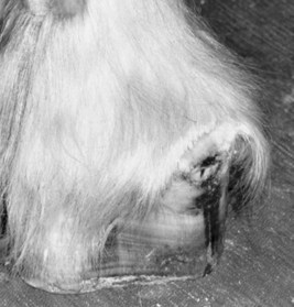
The clinical examination of the hind foot and pastern was described in Chapter 6, and a detailed description of the clinical investigation of the foot and shoeing is found in Chapter 27Chapter 28Chapter 30Chapter 31Chapter 32Chapter 33Chapter 34. A conscientious effort must be made during every lameness examination to evaluate the hind foot with the limb in both the standing and flexed positions. The type of shoeing and shoe wear are important in understanding potential lameness conditions of the foot, but perhaps more important, study of hind foot balance and shoeing can provide important clues in the diagnosis of lameness located more proximally in the limb. Dramatic abnormalities in balance and shoe additives, such as calks, grabs, trailers, and bars, can place added forces of shear and torsion on hindlimb bones and joints. Therapeutic recommendations cannot be made unless the clinician is aware of the type of shoes that are currently in place. Hoof imbalance has not been studied extensively in the hindlimb, but dorsal to plantar and medial to lateral hoof imbalance is a likely contributor to lameness in the proximal interphalangeal and metatarsophalangeal joints. Palpation for signs of inflammation, such as increased digital pulse amplitude, heat, swelling, and pain, must be performed but can be difficult in fractious horses. Palpation is difficult in the hindlimb because of the stay apparatus, which causes the digit to flex involuntarily. Palpation of the plantar pastern soft tissue structures is difficult, and subtle swelling and pain associated with the distal sesamoidean ligaments or other structures can be easily overlooked. In draft horses, signs of inflammation can be obscure (Figure 41-1). Examination with hoof testers is important, but normally horses show considerable sensitivity across the heel (see Chapter 6). Because the foot is incorporated in the rigid hoof capsule and swelling may not be apparent, even severe lameness conditions, such as abscesses or fractures, can go unnoticed. Commonly lameness of the hind foot is misinterpreted as originating high up in the limb, because clinical signs are not prominent and are easy to overlook.
Degree of lameness varies greatly. Most recognized problems of the hind foot cause obvious, often severe lameness, but occasionally subtle or insidious problems occur. Horses with the most common problems, such as a hoof abscess and penetrating wounds, may not bear weight. If they are bearing weight, horses usually are severely lame at the trot. Horses with severe hind foot lameness, particularly of the toe region, may walk with a shortened caudal phase of the stride, similar to horses with pain in the coxofemoral region (see Chapter 7). Horses with most lameness conditions typically have a shortened cranial phase of the stride at both the walk and trot. Observing a paradoxical shortened caudal phase of the stride at the walk and the more typically seen shortened cranial phase of the stride at the trot can be an indication of hind foot pain. The horse may bear weight only on the toe. Turning may accentuate pain. At the trot, horses with hind foot and pastern lameness travel like horses with pain that originates anywhere distal to the distal aspect of the crus, and no typical alteration in limb flight is observed. When the horse is viewed from behind, limb flight is characterized by moving the foot straight ahead or slightly medial to the line of expected limb flight and then stabbing laterally during the later portion of the stride, just before impact.
Stay updated, free articles. Join our Telegram channel

Full access? Get Clinical Tree


