Chapter 4 The Genus Staphylococcus
THE GENUS STAPHYLOCOCCUS
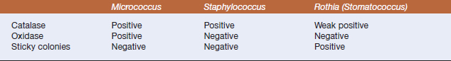
Staphylococci are the most commonly isolated Micrococcaceae from veterinary clinical specimens. They occur in pairs, in grapelike clusters, or singly, and colonies are generally white to off-white, with smooth surfaces and butyrous consistency; many strains of Staphylococcus aureus have a golden pigment from which the organism derives its name. Members of the genus Staphylococcus can usually be differentiated from micrococci based on cell morphology (the latter form tetrads and cells tend to be larger than those of the staphylococci) and pigment production on solid media. Rothia (Stomatococcus) species are found infrequently in veterinary specimens, and a weak catalase reaction, ovoid cell shape, and sticky colonies are useful phenotypic features (Table 4-1). Rothia species are further distinguished from other members of the Micrococcaceae bythe low G+C content of its DNA (30%-38%), the presence of teichoic acid in their cell walls, andthe ability to tolerate high levels of NaCl (15% to saturated).
There are 32 recognized species of staphylococci (Table 4-2), 13 of which are indigenous to humans, with the remainder associated with various nonprimate animals. They are aerobic or facultatively anaerobic, and are capable of generating energy by respiratory and fermentative pathways. Staphylococci are nutritionally fastidious, with complex nitrogen requirements; most species require several amino acids, vitamins (thiamine and niacin), and uracil (to grow anaerobically).In complex, nutritionally complete media, the organism has a generation time of approximately 20 minutes.
TABLE 4-2 Species of Staphylococcus Important in Veterinary Medicine
| Staphylococcus Species | Veterinary Importance |
|---|---|
| S. aureus | Wound infections in all animals; mastitis, skin infections; joint infections, especially in chickens; diarrhea in pet birds; vaginal infections in dogs and horses |
| S. aureus ssp. anaerobius | Isolated occasionally from ovine caseous lymphadenitis |
| S. epidermidis | Opportunistic pathogen; bovine mastitis, skin abscesses in other animals |
| S. warneri | Septicemia in lovebirds |
| S. saprophyticus | Possible opportunist in urinary tract infections |
| S. kloosii | Normal skin and mucous membrane flora in squirrels and opossums |
| S. intermedius | Skin and ear infections in dogs, occasional bovine mastitis; isolated occasionally from birds and horses |
| S. hyicus | Skin infections, arthritis in pigs; skin, milk of cattle; avian arthritis |
| S. chromogenes | Bovine milk, skin of pigs and cattle; normal skin and mucous membrane flora in cattle |
| S. sciuri | Normal skin and mucous membrane flora in squirrels |
| S. lentus | Normal skin and mucous membrane flora in sheep and goats |
| S. gallinarum | Normal skin and mucous membrane flora in turkeys |
| S. caprae | Normal skin and mucous membrane flora in goats |
| S. equorum, arlettae | Normal skin and mucous membrane flora in horses |
| S. felis | Otitis externa, cystitis, abscesses and wounds in cats only |
| S. auricularis, S. capitis, S. carnosus, S. caseolyticus, S. cohnii, S. haemolyticus, S. hominis, S. lugdenensis, S. muscae, S. pasteuri, S. piscifermentans, S. saccharolyticus, S. schlieferi, S. simulans, S. vitulus, S. xylosus | None known |
Eleven capsular polysaccharide serotypes have been described for S. aureus, and the most common in clinical isolates are types 5 and 8. The main capsular components are N-acetylaminouronic acids and N-acetylfucosamine. The genes for capsule production are in a single chromosomal operon.
Staphylococcal bacteriophages were among the first demonstrated, and this has led to the establishment of a bacteriophage typing method for epidemiologic studies. Most pathogenic strains belong to phage groups II and III, but all arecapable of causing disease.
Diseases and Pathogenesis
Intracellular survival and spread are mediated in large part by production of toxins. Membrane-active toxins, some of which are enzymes, pro-tect the organism against the host response and provide access to host-derived nutrients. Many of the approximately 30 extracellular proteins produced by S. aureus are plasmid encoded, providing an armada of potential virulence factors that varies from strain to strain (Table 4-3). A single bacterial product is rarely of overriding importance in development of disease. Alpha toxin subunits bind to cell membranes, oligomerize, and form pores, leading to necrosis. Prostaglandins and other inflammatory mediators are released from some target cells, and macrophage function is compromised. Systemic effects of alpha toxin are most pronounced on cardiac and central nervous system tissue. Most bovine isolates of S. aureus produce β-toxin, a sphingomyelinase that lyses susceptible cells by enzymatic degradation of phospholipids in the outer leaflet of the membrane. Its role in pathogenesis is less clearly understood, but it augments staphylococcal growth in vivo. Hyaluronidase is a putative virulence attribute that may contribute to spread of the organism in the host.
TABLE 4-3 Virulence Factors of Staphylococcus aureus
| Virulence Factor | Effects |
|---|---|
| Capsule | Inhibits phagocytosis; promotes adherence |
| Peptidoglycan | Leukocyte chemoattractant; decomplementation |
| Teichoic acid | Fibronectin binding |
| Protein A | Immunoglobulin binding |
| Toxins (α, β, others) | Antiphagocytic; cytotoxic |
| Exfoliative toxins | Serine proteases; split cellular bridges in stratum granulosum |
| Enterotoxins | Superantigens; nauseogenic, diarrheagenic |
| Toxic shock syndrome toxin | Superantigen; endothelial damage |
| Coagulase | Converts fibrinogen to fibrin |
| Hyaluronidase | Hydrolyzes hyaluronic acid in connective tissue |
| Lipase | Hydrolyzes lipids |
| Nuclease | Hydrolyzes deoxyribonucleic acid (DNA) |
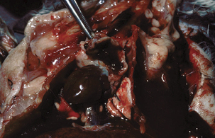
Staphylococcus aureus causes suppurative infections and septicemia (Figure 4-1) in all species, and a few examples are illustrative. Arthropod bite wounds in lambs and other animals may become infected, and these animals may also develop lameness and bacteremia. Abscesses form in kidney, liver, joints, and brain, and death is rapid. The organism may also be associated with ovine periorbital eczema.
< div class='tao-gold-member'>
Stay updated, free articles. Join our Telegram channel

Full access? Get Clinical Tree


