Chapter 9 The Genera Dermatophilus and Nocardia
THE GENUS DERMATOPHILUS
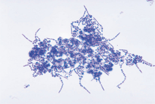
Members of the genus Dermatophilus are aerobic, gram-positive, branching, filamentous rods. They produce motile zoospores, and aerial mycelia are ordinarily absent (Figure 9-1). The substrate mycelium consists of long filaments that branch laterally at right angles. Septa are formed in transverse and horizontal planes and give rise to parallel rows of coccoid cells, often referred to as “railroad tracks.” These organisms are catalase positive and non–acid fast.

FIGURE 9-1 Clustering of Dermatophilus congolensis (Giemsa stain).
(Courtesy Public Health Image Library, PHIL #2986, Atlanta, date unknown, Centers for Disease Control and Prevention.)
Diseases, Epidemiology, and Pathogenesis
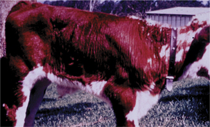
Dermatophilus congolensis is an obligate parasite of animals and affects many species, causinggeneralized exudative dermatitis in livestock. Dermatophilosis occurs in cattle in tropical and subtropical regions (Figure 9-2); disease in sheep is especially common in areas of high rainfall. Temperate breeds of cattle are more severely affected than tropical breeds. Economic losses derive from effects on production of beef, mutton, milk, hides, skins, and wool, and are especially notable in relation to cattle production in West and Central Africa and in the Caribbean, and sheep production in Australia.
The life cycle of the organism involves germination of cocci and production of hyphae that elongate through the epidermis and then undergo transverse and longitudinal division to produce filaments that release cocci. Motile zoospores develop from cocci and are released from wet crusts to establish new sites of infection on the same animal or new hosts.
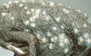
Dogs, rabbits, deer and other ungulates, foxes, seals, and pigs are affected by scabs and hair loss. Cats are often infected by way of puncture wounds, and the resulting abscesses involve the subcutis, muscles, and lymph nodes, with chronic draining fistulas. The organism is also associated with ulcerative lymphangitis in cattle and ulcerative and hyperkeratotic skin diseases in lizards and alligators (Figure 9-3), and has been isolated sporadically from various skin diseases in monkeys, ground squirrels, and polar bears. Several cases acquired from animals have occurred in humans. Dermatophilus chelonae is a newly characterized organism isolated from skin lesions in chelonids (turtles and tortoises).
Diagnosis
Diagnosis is based on the finding of morpholog-ically compatible organisms in stained smearsfrom clinical materials; conventional or fluorescent antibody stains may be useful. Gram-positive, branching segmented hyphae are seen, with hyphal elements larger and less regularly shaped than those of members of the genera Streptomyces and Nocardia. After 24 hours of incubation on solid media, D. congolensis produces tiny, grayish white round colonies that pit and adhere to the agar. Colonies may become orange after 2 to 5 days, and they are frequently β-hemolytic. Stains reveal segmenting, longitudinal and transverse filamentsand coccoid spores that are often deep purple and in packets. Septation of hyphal elements results in formation of zoospores that are motile by way of polar flagella. Differentiation is by standard phenotypic assays (Table 9-1).
TABLE 9-1 Differentiation of Dermatophilus species
| Dermatophilus Species | ||
|---|---|---|
| D. congolensis | D. chelonae | |
| Acid from glucose | Pos | Pos |
| Growth at 25°C | Less | More |
| Nitrate reduction | Neg | Wk pos |
| Double-zone hemolysis | Neg | Pos |
| Putrid odor | Neg | Pos |
| Capsule | Neg | Pos |
Neg, Negative; Pos, positive; Wk pos, weak positive.
Stay updated, free articles. Join our Telegram channel

Full access? Get Clinical Tree


