Chapter 6 The Genera Actinomyces and Arcanobacterium
THE GENUS ACTINOMYCES
The species most often associated with disease in domestic animals are Actinomyces bovis, Actinomyces viscosus, and Actinomyces suis. Other species are presented in Table 6-1.
TABLE 6-1 Animal-Pathogenic Actinomyces Species Other Than Actinomyces bovis, Actinomyces viscosus, and Actinomyces suis
| Actinomyces Species | Conditions |
|---|---|
| A. hordeovulneris | Canine pleuritis, peritonitis, arthritis, abscesses; associated with grass awns |
| A. howelli | Dental plaque of cattle; questionable pathogen |
| A. hyovaginalis | Porcine purulent vaginitis and other lesions; isolated from aborted swine fetuses |
| A. israelii | Actinomycosis in cattle and swine; important human pathogen |
| A. naeslundii | Isolated from porcine abortions |
| A. bowdenii | Recent isolate from dogs and cats; resembles A. viscosus |
Diseases and Pathogenesis
Actinomyces bovis causes actinomycosis, or lumpy jaw, in cattle (Figure 6-1), as well as occasional similar human infections. Bovine actinomycosis usually affects bony structures, and is most commonly seen in the mandible, or lower jaw. Pulmonary infections may occur in cattle and swine. The organism has also been associated with equine fistulous withers and similar conditions, sometimes in company with Brucella abortus or Brucella suis. Disease in other ruminants has also been reported, and dogs often develop actinomycotic spondylitis as a consequence of migration of foxtails from the upper respiratory tract. Actinomyces hordeovulneris is a newly described species that is more common than A. bovis in these canine infections. Actinomycosis can also occur concurrently with lung adenocarcinomas in dogs.
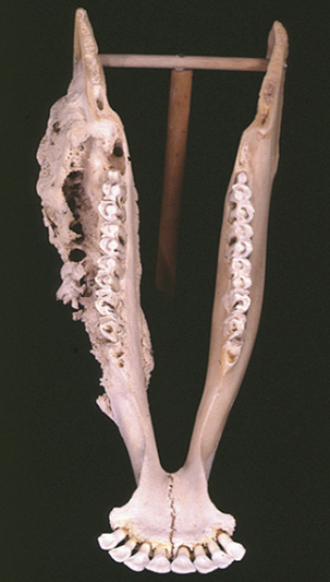
Bone honeycombed with pus-filled sinus tracts replaces normal bone (see Figure 6-1), and affected animals may lose teeth, become unable to chew, and develop mandibular fractures. Jaw lesions are often impressive in extent, but if vascular dissemination occurs, infection does not establish in other tissues. Circulating antibody developed as a result of infection has no impact on recovery, and it is likely that resistance is cell mediated.
Actinomyces spp. are prominent in the human oral cavity and are important factors in plaque development. Extensive study has revealed unique attributes that favor colonization and persistence in this environment. Fimbrial adhesins are perhaps the most important of these in that they enable adherence to receptors on tooth and mucosalsurfaces and interaction with other plaquebacteria. Sialidase may amplify the adhesion process by exposing host cell receptors. Diverse enzymatic activities of Actinomyces spp. enhance its own lifestyle and that of other oral bacteria, possibly exerting a positive selective effecton the actinomycetes. Such information is not available for the Actinomyces spp. associated primarily with animal diseases, but the same selective pressures may apply. Application of similar genetic systems and methods of approach to animal pathogenic Actinomyces spp. provides many opportunities, perhaps leading to better methods for prevention.
Diagnosis
Lesion-associated granules in A. viscosus infection contain the typical filamentous masses and diphtheroidal forms, but the clubs that are characteristic of A. bovis are absent (Figure 6-2). It produces flat, smooth-to-granular “molar tooth” colonies (Figure 6-3), which, at approximately2 mm in diameter, are perhaps twice as large as those of A. bovis. Microcolonies, after 24-hour incubation in an atmosphere enriched for CO2, have a dense center and filamentous fringe, and after 72 to 96 hours are circular, convex, smooth and white to cream. Actinomyces viscosus is catalase positive, unlike other members of the genus. The organism is relatively uncommon in purulent exudates, and cultural false negatives may result. Granules may be cultured in thioglycollate broth.
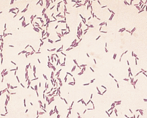
FIGURE 6-2 Gram stain of Actinomyces viscosus.
(Courtesy Public Health Image Library, PHIL #1256, William A. Clark, Atlanta, 1977, Centers for Disease Control and Prevention.)
Stay updated, free articles. Join our Telegram channel

Full access? Get Clinical Tree


