The Foot and Shoeing

Chapter 27 The Foot and Shoeing


image Foot Balance, Conformation, and Lameness


Andrew H. Parks


Athletic injury usually results from imposition of repetitive stresses that exceed the capacity of the tissues. The magnitude of stresses and hence the likelihood of injury frequently depend on balance and conformation. Therefore balance and conformation are extremely important in maintaining optimal limb function and limiting athletic injury.


Conformation describes shape—in this case, the shape of the distal aspect of the equine limb—and conveys the size and relative proportions of the limb. Balance embraces both the conformation and function of the hoof—conformation because it describes the shape of the hoof, and function because it describes the way the hoof relates to the skeletal structures of the limb and the ground at rest and at exercise. Balance is divided into geometric (static) balance and functional (dynamic) balance.


Balance and conformation are both three-dimensional concepts. Balance usually is divided into three planes: frontal (dorsal), sagittal, and transverse. Balance in the frontal plane is called mediolateral balance and in the sagittal plane is called dorsopalmar (plantar) balance.


To understand how balance and conformation affect stresses that cause injury, it is necessary to consider the function of the distal limb and then examine how it changes with conformation and balance. Therefore consideration must be given to the musculoskeletal system, the hoof and the ground, and the interfaces. There are some substantial differences between the front and hind feet. Because almost all research and documented clinical observation are related to the front feet, all discussion in this chapter refers to the front feet unless specifically stated otherwise.


The hoof is the interface between the musculoskeletal system and the ground. The hoof functions both as an extension of the distal phalanx, as a lever about the distal interphalangeal (DIP) joint, and as an entity in itself. As part of the integument the hoof behaves differently to the structures of the musculoskeletal system, both in its manner of constant growth and its biomechanical properties. As the hoof capsule is constantly worn at the ground surface, it is replaced by the germinal epithelium of the coronary band and the sole. In nature and in an appropriately trimmed foot, there is an approximate balance between growth and loss of the hoof capsule so that the growth rings are parallel.1-3 The exact mechanism by which hoof growth is regulated is unknown, but several factors are known to influence it: season, inflammation, nutrition, and topical irritants.4 Growth of the wall also is inversely related to pressure on the coronary band. Hoof wall growth proximal to a hoof wall resection or horizontal grooving of the hoof wall appears accelerated, whereas the immediately adjacent hoof wall growth may be retarded.5 This effect may be mediated by an effect on the vasculature of the coronary band.


During normal hoof growth the hoof wall migrates distally in relation to the distal phalanx by active separation and reformation of desmosomes as the primary epidermal lamellae move past the secondary epidermal lamellae.6 The distal growth of the hoof wall under normal loading patterns is approximately even around the circumference of the hoof, and the position of the coronary band in relation to the distal phalanx is static. However, in response to locally increased and decreased loads within the hoof wall, migration of the coronary band proximally and distally in relation to the distal phalanx is superimposed on the normal pattern of hoof wall migration, suggesting that a whole segment of the wall can displace distally and even proximally by movement within the lamellae.


The stiffness of the hoof wall changes radially, and the outer stratum medium is stiffer than the inner stratum medium but is much less stiff than bone throughout. The stiffness of the hoof wall at the toe and quarters is similar, but the difference in thickness indicates that the quarters are more flexible.7 The hoof wall sustains strains greater than bone, but under normal circumstances it operates within its elastic range at a fraction of its yield capacity.8 The stiffness of the hoof wall increases with increased strain rate.9 The hoof wall is viscoelastic. It responds to a rapidly applied force in an elastic deformation so that it returns to its original form rapidly; however, to a slowly applied force, it deforms in such a manner that when the force is removed, the hoof wall returns to its original form slowly.10 Because of its physical properties the hoof wall is more fracture resistant than bone,11 but because it is a much less stiff material than bone, it will bend and shear more readily. The biomechanical properties of the soft tissues between the hoof capsule and distal phalanx are less well understood, but the periosteum of the distal phalanx fails before the junction between the epithelial and dermal lamellae.8 The lamellar junction is much less stiff than the hoof wall12; dorsally the lamellae are oriented perpendicular to the tangent to the hoof wall, but at the quarters they are in a more palmar direction.13


Foot Function


At Rest


At rest a horse bears approximately 28% to 33% of its body weight on each forelimb. The exact roles of the wall, sole, and frog in weight bearing are undetermined. Weight bearing has traditionally been viewed with a horse on a flat, firm surface so that the weight-bearing surface is the full circumference of the wall and the immediately adjacent sole, although the weight is not evenly distributed around the perimeter of the foot. Studies on feral horses indicate that the toe and quarters are worn so that if the horse stood on a flat, firm surface, the weight would be transmitted through the wall at the heel and the junction of the toe and quarter biaxially, although there is some variation related to the terrain on which the horse has lived.14 The dorsal aspect of the toe and midquarters would not bear weight. Domestic horses that were allowed to wear the feet “naturally” at pasture and then stood on different surfaces showed remarkably different loading patterns.15 When stood on a firm surface, greatest contact was at the medial and lateral aspects of the heel and just medial and lateral to the dorsal aspect of the toe, comparable to feral horses. When stood on sand, the greatest contact was with the central aspect of the sole, and the total contact area was approximately four times greater for horses standing on sand than on a flat, firm surface.


Any part of the ground surface of the foot is potentially weight bearing. Each point of contact that bears weight transmits that force to the ground, although the pressure at each point varies. The sum of all the forces from all points of contact is called the ground reaction force (GRF). It is represented as a vector, with a magnitude and direction, and a location or point of force, which is also called the point of zero moment. In the stationary horse this force is almost vertical and located slightly medial to the dorsal third of the frog. Therefore the GRF is dorsal to the center of rotation of the DIP joint, with a resultant moment about the joint. This moment is opposed by an opposite moment created by tension in the deep digital flexor tendon (DDFT). The GRF acting through the phalanges creates a moment about the metacarpophalangeal joint that is opposed by tension in the digital flexor tendons and the suspensory ligament (SL).


At Exercise


Stride Phases


The stride is divided into flight and stance phases.16 The stance phase of the stride is further subdivided into initial contact, impact, support, and breakover. The foot should move in a sagittal plane parallel to the longitudinal axis of the horse.5 In an exercising horse the GRF changes in magnitude, point of force, and direction with and within the phases of the stride. The GRF is separated into components in three axes: x-axis (mediolateral), y-axis (dorsopalmar), and z-axis (vertical).


Support Phase

The support phase extends from the end of impact until the onset of breakover. At a walk the vertical GRF is biphasic, with peaks at either side of the middle of the stride, but at the trot there is a solitary peak approximately halfway through the stride.26,27 For most of the stance phase the GRF is slightly medial to the dorsal third of the frog.28 The force is absorbed and energy is stored by the digital flexor tendons and SL as the metacarpophalangeal joint extends29 so that the maximal GRF coincides with maximal extension of the metacarpophalangeal joint.17,30 At the walk, forces in the superficial digital flexor tendon (SDFT) and DDFT peak before the peak in the GRF, but the force in the accessory ligament of the DDFT (ALDDFT) peaks during the second half of the stride as the DIP and metacarpophalangeal joints extend.31,32 The GRF in the dorsopalmar direction is negative during the first half of the stride as the limb decelerates. The foot continues to slide forward after initial impact until arrested, at 6% of the stride duration in the forelimb33 and 23% in the hindlimb34 in trotters on a dirt track. The fore foot bounces more on impact, whereas the hind foot slides more.21 During the second half of the stride the horizontal GRF becomes positive as the limb accelerates to provide propulsion. The balance of propulsion and retardation is such that the forelimbs contribute more to retardation and the hindlimbs to propulsion.27 Faster gaits create higher GRFs and greater strains in the hoof wall,8 DDFT, SDFT, and SL.35 Under in vitro loading conditions, hoof wall strains increase with load as strain field epicenters develop around the circumference of the hoof, at the junction of the middle and distal thirds of the hoof, regardless of load.36


The distal phalanx is loaded during the stance phase. It was initially thought that the palmar processes rotate palmarly during loading,37 but recent finite element analysis indicates that the toe rotates distally and that the palmar processes move proximally; the findings of the finite element analysis, though not yet verified in vivo, are consistent with observations of hoof wall deformation and strains.38 The sole flattens and spreads as the heel expands,2,39 more so distally than proximally.8 At the same time, the dorsal hoof wall flattens and rotates palmarly to parallel movement of the distal phalanx. Frog contact with the ground during exercise appears to be variable.25,39 The role of frog pressure in hoof expansion is undetermined; there is evidence that indicates it is not involved, yet other evidence suggests that frog pressure is not the sole determinant of hoof expansion but may enhance it.25,40 Either way, the frog must function as an effective expansion point to permit movement of the sole ventrally and the heel abaxially. It is hypothesized that the digital cushion, in conjunction with the cartilages of the foot, participates in dissipating energy during impact through a hydrodynamic mechanism.41,42 The DIP joint passively flexes43 and the metacarpophalangeal joint extends. During the second half of the stride the DIP joint extends43 and the tension in the ALDDFT increases.32 Tension in the collateral sesamoidean and distal sesamoidean impar ligaments also increases,43 and pressure on the navicular bone increases.44 The metacarpophalangeal joint flexes. The point of action of the GRF moves toward the toe toward the end of the stance phase.16


Stride Characteristics


The time and motion characteristics of the stride are important in determining the animation qualities of the gait and the speed of the horse. Higher movement of the foot and greater flexion and extension of the joints represent greater animation. Long retraction with a high starting point is considered desirable, and longer strides are associated with greater speed. Maximum stride frequency is inversely related to speed index.46-48


The ground surface affects the angle of the hoof to the ground during the stride, the duration of the stride, and the absorption of impact energy. On a flat, firm surface the plane of the hoof is the same as that of the ground, but on a surface such as sand, the angle of the hoof with the ground increases gradually during the stance phase of the stride.35 This rotates the plane of the sole so that it is more perpendicular to the vector of the GRF, which appears to aid traction and propulsion. Ground footing has been divided into three types: dense hard surfaces, surfaces with friction damping such as sand, and structural damping surfaces such as wood chips.49 Friction damping occurs through displacement of small particles, whereas structural damping occurs through viscoelasticity of the particles. The duration of impact oscillations is related to the hardness of the surface. Harder surfaces are associated with a longer duration of impact oscillations than soft surfaces and less energy absorption.50 A loose cushion on the surface reduces the peak impact force.51 Racetracks with a hard surface result in faster race times, but horses are more likely to sustain injury associated with the increased energy of impact.51 Ground footing also affects stride and swing duration, which are both longer on an elastic, softer surface than a hard, firm surface.52 At a walk, strain in the DDFT and ALDDFT is lower on sand than on a hard, flat surface,35 such that it resembles the effect of a heel wedge.


Optimum Balance and Conformation


Optimum function should intuitively demand optimum conformation and balance. Practitioners have inherited many empirical notions that often are based on what types of conformation and balance do not work and cause problems. Generally these ideas predate modern motion analysis and therefore relate to conformation and static balance. Modern techniques that allow dynamic evaluation of function have supplemented and sometimes contradicted the geometric approach.


Static Balance and Conformation


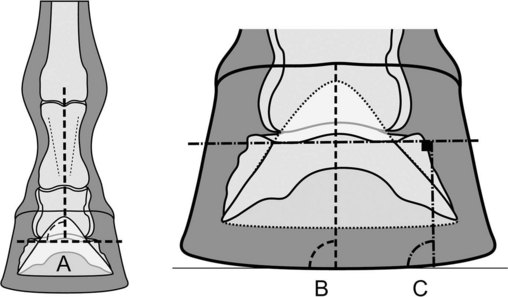
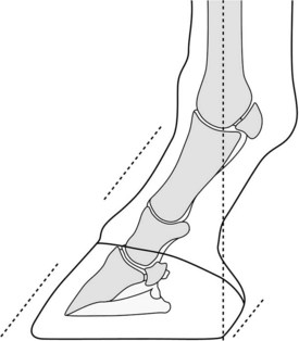
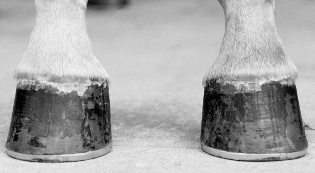
Viewed from the lateral aspect, the foot-pastern axis should be straight; that is, the dorsal hoof wall should be parallel to the dorsal surface of the pastern, and the angle of the heel should approximate that of the dorsal hoof wall (Figure 27-1). The angle of the dorsal hoof wall and the foot-pastern axis to the ground is variable, but it frequently is cited as 50 to 54 degrees in forelimbs and approximately 3 degrees steeper in hindlimbs.53 Other studies report higher or lower means and variable relationship between the angles of the dorsal hoof wall of the forelimbs and hindlimbs.54,55 In domestic horses the length of the hoof wall has been approximately linked to the weight of the horse (7.6 cm for 360 to 400 kg; 8.25 cm for 430 to 480 kg; 8.9 cm for 520 to 570 kg).53 In feral horses the toe length ranges from 6.7 to 8.9 cm14,54 but is independent of weight.54 In domestic horses with either trimmed or shod hooves, the length of the heel should be approximately one third that of the toe,2,55 but in feral horses it varies with the terrain.14 An imaginary line that bisects the third metacarpal bone (McIII) should intersect the ground at the most palmar aspect of the weight-bearing surface of the heel.56,57


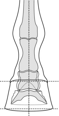
On a lateromedial radiographic image the dorsal hoof wall should be 14 to 20 mm thick depending on breed58-60 and parallel to the dorsal surface of the distal phalanx. The angle made by the distal solar border of the distal phalanx with the ground ranges from 2 to 10 degrees.58,60,61 The center of rotation of the DIP joint is palmar to the center of the ground surface of the foot. Viewed from the dorsal aspect, a line bisecting the metacarpal region should bisect the phalanges and foot, so that the foot is approximately symmetrical, including the mass of foot on either side of the line and the heights and angles of the walls.1,3,57,61 The medial quarter is frequently steeper so that the medial wall is shorter than the lateral wall.2,62,63 A line drawn between any two comparable points on the coronary band should be parallel to the ground, and a vertical line bisecting the McIII should be perpendicular to a line drawn across the coronary band or the ground surface of the foot62 (Figure 27-2).


On a dorsopalmar radiographic image the center of the DIP joint should be centered over the ground surface of the foot. Ideally, the articular surface of the distal phalanx should be parallel to the ground, but it is more important that the interphalangeal joint spaces be even.


Viewed from the ground surface the width and length of the hoof capsule of the fore foot should be approximately equal, although the hoof capsule may be slightly wider than it is long.54,62 The hind foot is invariably slightly longer than it is wide. The point of breakover is best assessed from the ground surface and should be located near the center of the toe. Dynamic studies of the location of the GRF during breakover show that it deviates laterally during breakover, returning toward the dorsopalmar axis of the limb before the foot leaves the ground20; this suggests that breakover normally occurs lateral to the dorsopalmar axis of the foot. The ideal location for breakover in the dorsopalmar axis is disputed. In a traditionally trimmed and shod horse, breakover is positioned where the line of the dorsal hoof wall intersects the ground, although it may ideally be located more palmarly. With the position of the hoof wall used as the reference point, breakover is between the dorsal margin of the hoof and the white line.64 Alternatively, breakover is 2.5 to 3.8 cm dorsal to the apex of the frog or 0.6 cm dorsal to the dorsal margin of the distal phalanx.14,65 The relationship of the longitudinal axis of the frog to the underlying distal phalanx is relatively constant compared with the rest of the ground surface of the hoof. The medial and lateral aspects of the ground surface of the foot are symmetrical about the central axis of the frog,3,57,64 although slight asymmetry, the lateral sole being about 5% wider than the medial sole, may be beneficial.62 The latter is compatible with an even coronary band and a steeper medial wall. The size of the foot should be proportional to the weight of the horse.54,55 The sole should be concave. The frog width should be at least 50% to 67% of frog length,55,62 and the weight-bearing surface of the heel should coincide with the widest part of the frog.55


Imbalance and Poor Conformation


Balance and conformation cannot be considered in isolation, because poor conformation predisposes a horse to developing imbalance, and an imbalanced foot may cause a horse to stand as if it had poor conformation.1 The effects of imbalance have been examined by experimentally inducing imbalance and by clinical observation. To understand the effects of imbalance it is simplest to consider the consequences of deliberately unbalancing the foot. Poor conformation cannot be readily altered under experimental conditions, and its effects must be assessed by comparison between horses with different conformations.


Mediolateral Imbalance


Mediolateral imbalance is caused by either poor trimming of a horse with good conformation or poor conformation causing excessive stress on one side of the foot so that it grows more slowly than the other side. Inappropriate shoe placement can promote imbalance. If a shoe is set too much to one side or the other, or if the shoe is rotated so that the shoe covers less of the medial or lateral aspects of one heel than the other, it alters the mediolateral stress on the hoof capsule.66 A single heel calk causes elevation of one side of the foot; the foot tilts and rotates and may contribute to interference.66


Mediolateral imbalance can be induced by applying a wedge pad to elevate the medial or lateral side of a foot or trimming a foot unevenly. The coronary band is no longer parallel to the ground or perpendicular to the sagittal axis of the limb. The horn tubules of the dorsal hoof wall are no longer oriented in the sagittal plane. Dorsopalmar radiographic images demonstrate that the DIP joint space is narrower on the side of hoof elevation, and the middle phalanx slides to the lower side.43 In addition, the condyle of the middle phalanx on the elevated side of the foot moves palmarly, in effect causing the distal phalanx to rotate on the middle phalanx so that the dorsal margin of the distal phalanx rotates away from the elevated side43 (Figure 27-3), but the horse has the appearance of being toed toward the elevated side.1


The GRF shifts toward the elevated wall.67 In foals, compressive strains were immediately increased in the lateral cortex of the McIII and decreased in the medial cortex by elevation of the lateral wall with a wedge.68


The immediate dynamic effects of mediolateral imbalance result in a greater frequency of mediolateral asymmetrical footfall, the lengthened side landing first.19 The location of the GRF displaces abaxially toward the lengthened side of the foot.19,69,70


Prolonged mediolateral imbalance affects the relationship between the hoof capsule and the distal phalanx, causes distortion of the hoof capsule, and alters hoof wall growth.71 If a foot is trimmed unevenly, so that one wall is longer than the other, the longer wall grows more slowly than the shorter wall. The longer wall develops a flare, and the shorter wall becomes underrun.1,71 In more severely affected horses the coronary band bulges abaxially to create a lip at the proximal margin of the hoof wall on the elongated side. The horse breaks over on the shorter side of the toe. It is my impression that the solar margin of the distal phalanx partially realigns with the ground surface, either because the wall actually migrates proximally or because distal movement of the hoof wall is inhibited, resulting in a net proximal displacement of the coronary band in relation to the distal phalanx. Ultimately, prolonged mediolateral imbalance causes remodeling of the phalanges because of redistributed stresses according to Wolff’s law.


Rotational deformities and angular deformities may mimic each other. In most rotational deformities, it appears as if the metacarpophalangeal joint is the most proximal joint to be rotated, but it is not uncommon for the carpus to appear rotated. With rotational deformities, the McIII is vertical and the observer can rotate around the limb until the metacarpal and phalangeal axes are correctly aligned unless the rotation occurs within the phalangeal axis. In horses with angular deformities that occur proximal to the fetlock, including base-wide or base-narrow conformation, the McIII may not be vertical and there may not be a viewpoint from which the metacarpal and phalangeal axes are correctly aligned.


Rotational deformities, such as toe-in and toe-out conformation, alter the position of the ground surface of the foot in relation to the midsagittal vertical axis of the limb. Toe-in conformation causes the foot to wing out during the flight phase of the stride,1 and if the limb does not deviate from a vertical axis, the foot lands on the lateral heel quarter and breaks over at the lateral toe. Toe-out conformation causes the foot to wing in during the flight phase of the stride. The foot lands most frequently on the lateral heel quarter, as with toe-in conformation (see Figure 4-13). Breakover is less consistently directed than breakover in horses with toe-in conformation. Angular deformities, including base-wide and base-narrow conformation, and varus and valgus deformities also alter the position of the ground surface of the foot in relation to the ideal vertical axis of the limb. If an angular deformity (e.g., varus) is severe enough that the distal aspect of the wall at the quarter is axially directed, it will become underrun. Rotational and angular deformities may occur in combination, complicating the picture.


Dorsopalmar Imbalance


Dorsopalmar imbalance has several causes. A broken-back foot-pastern axis may follow poor trimming, either leaving the toe too long or trimming the heel too short. A shod horse wears its heels against the shoe whereas the toe wears very little, so the hoof angle changes up to 3 degrees over 8 weeks.2,72 This change must be allowed for by trimming the toe slightly more than the heel, because even trimming around the foot causes a gradual decrease in the hoof angle.3 Using too small a shoe or leaving a shoe on too long causes the palmar ground contact to move dorsally, imposing greater stresses on the heel, which then is prone to collapse.63,66 Toe grabs and heel calks alter the foot axis and concentrate stress.66 Thoroughbred horses may have a genetic predisposition for a broken-back foot-pastern axis; galloping causes the angle of the dorsal hoof wall to decrease, and removing the horse from training causes it to increase.73


At rest, elevation of the heel causes the DIP and pastern joints to flex and the metacarpophalangeal joint to extend.74,75 The effect is greatest at the DIP joint.74,75 In addition, elevation of the heel increases DIP joint pressure and localizes the contact dorsally between the middle and distal phalanges; toe elevation localizes articular contact to the palmar aspect of the joint.76 In vitro, elevation of the heel with a wedge decreases the strain in the DDFT, the extensor branches of the SL, and the medial hoof wall77 and decreases the moment about the center of rotation of the DIP joint.78 The horizontal distance between the center of rotation of the DIP joint and the toe decreases with heel elevation. In addition, the horizontal distance between the toe and an imaginary vertical line bisecting the McIII dropped to the ground is decreased. The clinical consequence of heel elevation is decreased heel growth indicative of increased stress within the wall of the heel.


Lowering the heel or raising or lengthening the toe to create an acute hoof angle or long toe increases the likelihood of toe-first landing.19,22 Heel wedges increase the maximum flexion of the DIP joint during the support phase of the stride and decrease the maximal extension of the DIP joint during breakover.79 Elevating the heel increases the likelihood of heel-first landing.19 The overall impulse (force × time) on the foot is least when the foot-pastern axis is straight, indicating that a straight foot-pastern axis is least injurious to the foot.19 At a walk, elevating the heel decreases the strain in the DDFT and its AL32,78 with little effect on the SDFT and SL. The decreased strain in the DDFT is reflected in decreased pressure on the navicular bone.44 Elevating the toe results in a marked increase in strain in the ALDDFT and a lesser increase in strain in the DDFT at the end of the stride as a result of increased extension of the DIP joint.32 Strains in the SDFT and SL are either reduced or unchanged.32,78 Horses with small hoof wall angulation have a prolonged breakover, but the length of stride, duration of the stance, and swing phases are unchanged.19,22 When hind feet are trimmed with more acute hoof wall angulation, breakover is delayed but the timing of impact is unchanged as normal coordination is restored during the swing phase of the stride. There is an increase in overreach distance, the distance between the print of the front foot and the landing point of the hind foot.80 Heel wedges delay the dorsal shift in the GRF and decrease the maximum torque about the DIP joint during the second half of the stride. Toe wedges have an opposite effect.32,70 However, neither toe nor heel wedges alter the dorsopalmar position of the point of force during midstance of the stride, indicating that the heel is not unloaded. Both toe and heel wedges cause medial displacement of the point of force.70 Increasing the length of the toe prolongs breakover but does not alter stride length; however, it increases maximal flexion of the metacarpophalangeal joint during the swing phase.81,82


The position of breakover in the sagittal plane appears to influence the angle of the dorsal hoof wall and the distal phalanx. Moving the point of breakover palmarly from the most dorsal margin of the hoof wall increases the angle of the dorsal hoof wall and the ground and increases the alignment between the middle and distal phalanges.65,83 Whether this effect is related to the biomechanical properties of the dorsal hoof wall or relief of pain within the foot is undetermined. The effect of increased hoof angle on hoof wall strain is inconsistent. In an in vitro model, hoof wall strain did not change with increased hoof wall angle.77 In contrast, in an in vivo experiment, increased hoof wall angle increased hoof wall strain more at the lateral quarter than at the toe and not at all at the medial quarter.84


The effect of pastern length and the angle of the foot-pastern axis are less well established. The angle of the hoof-pastern axis to the ground is a feature of a horse’s conformation. It cannot be changed experimentally, but comparing horses with different conformations shows that the point of force in horses with a small hoof-pastern axis angle is more palmarly positioned than in horses with a larger axis angle.28


Prolonged dorsopalmar imbalance also has delayed effects because of the nature and growth of the hoof capsule. In barefoot horses, trimming the feet to reduce the dorsal wall angle by leaving the toe long causes the ground surface of the foot and the frog to become narrower, and the shape of the ground surface tends to skew away from a circular shape.85 As might be expected, the heel appears to grow much faster, but curiously, the wall at the toe also grows faster. Neither the area of the sole nor the length of the frog change. Interestingly, when the foot is trimmed with a short toe to increase the dorsal wall angle, neither the width nor the shape of the frog or hoof changes.85 Clinically, the same appears to occur in shod horses. In horses with extremely long toes the foot becomes “hoof bound.” The heel of long-toed horses is predisposed to become underrun because the heel bends dorsally. Both of these phenomena are seen in Tennessee Walking Horses or American Saddlebreds intentionally shod with long hooves.


Imbalance and Poor Conformation as a Cause of Lameness


Poor conformation and imbalance of the distal aspect of the limb are common, as is pain causing lameness that can be isolated to the distal aspect of the limb. However, demonstrating the correlation is not always straightforward. In some horses an obvious disease process and obvious imbalance coexist, and when the imbalance is treated the lameness improves. In other lame horses, imbalance is evident with no other clinical, radiological, or scintigraphic evidence of disease, and treating imbalance also improves the lameness. In yet other lame horses, there is evidence of imbalance, with or without other evidence of disease, but treating the imbalance does not improve the lameness. To my knowledge, only one study thoroughly investigated the effects of hoof balance on injury. The odds of catastrophic musculoskeletal injury and suspensory apparatus failure were lower when the lateral sole area was greater than the medial sole area.62 Suspensory apparatus failure was more likely the greater the difference between the angles of the dorsal hoof wall and the heels. McIII condylar fractures were less likely with a steeper toe angle.


Mediolateral imbalance is associated with a shift in the point of force of the GRF, distortion of the hoof capsule, induced asymmetry of the articulations of the distal limb, and rotation of the DIP joint. With increased compressive stresses the following problems are clinically presumed to follow imbalance: subsolar bruising, hemorrhage in the white line from laminar tearing, pain from shearing heel bulbs, quarter or heel cracks, thrush in narrow frogs, pedal osteitis, fractures of the palmar process of the distal phalanx, sidebone, synovitis, osteoarthritis, and more proximal fractures.1,57,63,66


The effects of dorsopalmar imbalance should be separated into the effects of broken-forward and broken-back foot-pastern axes. A broken-back foot-pastern axis increases the load on the palmar aspect of the foot during weight bearing and increases the stresses in the toe at breakover. It causes hyperextension of the DIP joint and increases the tension in DDFT and pressure on the navicular bone. Therefore it can be expected to be associated with heel bruising, lamellar tearing at the toe, osteitis of the palmar processes of the distal phalanx, navicular disease, tendonopathy at the insertion of the DDFT, and more proximal injuries of the tendons or suspensory apparatus.3,57,63,66 In the hindlimb a broken-back foot-pastern axis appears to be associated with tarsal and back pain.


A broken-forward foot-pastern axis (upright) appears to be less pernicious.3,57 It increases the load on the dorsal half of the foot and decreases tension in the DDFT. The principal findings are subsolar bruising distal to the dorsal distal margin of the distal phalanx and subsequent osteitis of the distal phalanx.57


An upright foot-pastern axis has traditionally been considered to predispose toward concussive injuries of the weight-bearing structures in the limb, whereas a foot-pastern axis with an acute angle to the ground predisposes toward strains and sprains of the flexor apparatus. Similarly, a long pastern has been considered to predispose toward strains and sprains.


Any angular deformity located more proximal in the limb that increases the mediolateral symmetrical loading of the foot can be expected to have effects similar to mediolateral imbalance. Rotational deformities do not seem to be a frequent cause of problems. Toe-out conformation is more likely to cause interference, but anecdotally, toe-in conformation is considered more likely to cause lameness.


Undoubtedly, technological advances in diagnostic imaging and pain localization will help identify other combinations of disease and structure. Epidemiological studies will confirm the relationships between the different features of poor conformation and imbalance and disease.


Clinical Identification of Hoof Imbalance


The horse must be observed while it is standing squarely to assess each limb in relation to the whole and in motion on a flat, level surface. Each foot must be examined on and off the ground.


Mediolateral Imbalance


Visual inspection should note the position of the entire limbs to identify angular or rotational deviations more proximally in the limb that may have repercussions for the foot (Figure 27-4). Visual inspection of the foot on the ground should note rotational deformities of the metacarpophalangeal joint and placement of the foot in relation to the sagittal axis of the limb. The hoof capsule should be inspected closely for asymmetry of the coronary band. This is frequently a strictly visual inspection, but graphing the height of corresponding medial and lateral points on the coronary band provides objectivity that can highlight an imbalance and provide a record for future comparison.55,88 The medial and lateral walls should be inspected for flares and evidence of an underrun heel, lipping at the coronary band, and even spacing between the growth rings.


The ground surface of the foot reflects changes elsewhere in the hoof capsule. The foot should be approximately symmetrical about the center of the frog. In mediolateral imbalance, the sole may appear wider on the side with a flare in the wall and narrower on the side with an underrun wall. Dorsal displacement of the ground surface of one heel bulb in relation to the other accompanies proximal displacement of the coronary band at the heel commonly associated with sheared heel. Wear of the shoe or wall at the toe indicates the point of breakover. Alternatively, the breakover point may be identified by lifting the antebrachium cranially, allowing the metacarpal region and pastern to hang passively, and then lowering the foot; the point of breakover is the first part of the foot to touch the ground57; however, this assumes that breakover occurs at the same point regardless of the gait and speed of the horse. Breakover frequently occurs slightly lateral to the center of the toe in horses with normal balance, but any marked asymmetry in breakover may indicate mediolateral imbalance. It also may follow angular or rotational deformities of the limb. Asymmetrical bruising adjacent to the wall at either quarter may signify excessive concussion caused by mediolateral imbalance or laminar tearing in a wall with a flare.


Examination of the distal aspect of the limb for rotational or angular conformation by viewing the limb on the ground from the dorsal aspect may be misleading because it is influenced by weight bearing. The ground surface of the foot automatically aligns with the surface of the ground regardless of the relative lengths of the medial and lateral hoof wall. This causes secondary rotation within the phalangeal axis. To circumvent this rotation, to find the point of breakover the limb can be examined off the ground by lifting the limb by holding it forward from under the carpus; the angulation or rotation within the distal limb is observed by sighting down the metacarpal region, pastern, and hoof.57


The traditional way to assess mediolateral balance is to sight across the ground surface of the foot with the leg off the ground, holding the proximal metacarpal region and allowing the digit to hang downward in the sagittal plane with the metacarpophalangeal and interphalangeal joints in passive extension. A line drawn across any two corresponding points on the circumference of the ground surface of the wall should be perpendicular to the axis of the limb as judged by the metacarpal region. If the limb is perfectly symmetrical about the axis of the limb, without angular or rotational deformities, and the observer is directly above the limb, this technique is probably satisfactory within the limits of the observer. I question the accuracy of this technique because most distal limbs are not symmetrical but have at least some element of rotation about the metacarpophalangeal joint. A smaller number of horses have true angular deformities at the metacarpophalangeal joint. For reliability, there must be consistency in the extension of the metacarpophalangeal joint and in the position of the observer. T-squares have been used to improve the reliability of this observation,4,57 but misalignment of the T-square with the axis of the metacarpal region decreases accuracy.


Dynamic mediolateral balance is assessed by observing the horse from in front and from behind at a walk and at a trot. Because the degree to which symmetry of landing can be detected is a function of the frequency of observation and speed of the horse, only more severe imbalances can be detected at a trot compared with a walk, and more subtle differences in timing remain undetected unless a video recorder or more sophisticated measuring equipment is used. During the flight phase of the stride, movement of the foot, phalangeal axis, and more proximal limb is observed in relation to the plane of travel to correlate with previously noted rotational and angular deformities.


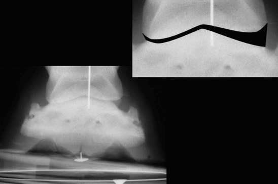
A dorsopalmar radiograph is the only means to assess the relationship between the hoof capsule and the phalanges (Figure 27-5). Overt imbalance can be detected on routine dorsopalmar radiographs, but detection of more subtle changes requires strict technique because apparent radiological imbalance can be readily induced artificially. Both feet must be weighted equally, because unilateral weight bearing induces mediolateral asymmetry and rotation of the interphalangeal joints.43 The foot must be allowed to assume its natural orientation to the rest of the limb, most reliably achieved if it is placed on a swiveling block. Deformation of the hoof capsule may be induced by rotation within the limb, which may change the angulation of the distal phalanx with the ground.89 The metacarpal region must be within 10 degrees of vertical in the frontal (dorsal) plane. The x-ray beam must be horizontal and centered on the midsagittal plane so that a wire marker centered on the dorsal hoof wall bisects the central sulcus of the frog on the radiographs. Neither toe-in nor toe-out conformation alters the radiological measurements of mediolateral balance if assessed in this manner.90,91 Interpretation of balance from dorsopalmar radiographs is usually based on examining either the relationship between the articular surface of the distal phalanx and the ground, which are ideally parallel, or the symmetry of the phalangeal joints, which should be of even width medially and laterally. When these appear to be in disagreement, I consider joint asymmetry to be more important because it is difficult to envisage a circumstance under which it is not harmful. In addition, interpretation of imbalance is more complex in horses in which the imbalance is chronic because the imbalance is associated with compensatory changes in hoof wall growth and also appears to be associated with movement of the hoof capsule in relation to the distal phalanx.


Although mediolateral imbalance can unquestionably cause lameness, the hoof capsule is not necessarily or even likely to be the site of pain that is associated with lameness. Rather the pain is associated with the effects of imbalance, that is, stress on the deeper structures of the hoof and the musculoskeletal structures of the distal aspect of the limb. Therefore it is not surprising that the lameness may improve with perineural or intraarticular analgesia of the distal aspect of the limb in a similar manner to osteoarthritis of the DIP joint or navicular disease.


Dorsopalmar Imbalance


The limb is examined for angulation at the carpus and metacarpophalangeal joint. The foot-pastern axis is visually inspected to determine whether the axis is straight or broken forward or back. This method provides only a rough guide. Visual examination can be improved by using a gauge, one limb of which is aligned with the pastern and one with the dorsal hoof wall. However, deciding exactly what landmarks to use on the pastern for alignment introduces irregularity. In addition, the axis changes as the horse shifts its weight or posture. Similarly, concavity of the dorsal hoof wall in the sagittal plane raises the issue of with what to align the pastern. If the dorsal hoof wall is concave, usually the top third of the hoof wall is the most closely aligned to the dorsal surface of the distal phalanx.


The length of the toe from the proximal aspect of the coronary band to the ground surface in the midsagittal plane of the hoof is readily measured with a tape and compared against reference values or previous measurements for the same horse. This information is greatly underused. The length and angle of the heel also are evaluated. Only the wall at the heel distal to the bulb should be evaluated, because inclusion of the heel bulb causes the angle to be underestimated. A heel that is angled more acutely to the ground than the dorsal hoof wall is longer than a heel that is parallel to the dorsal hoof wall. Interestingly, it is the angle of the distal phalanx to the ground, and not the angle of the heel, that correlates well with the force on the navicular bone.92


A long toe is associated with elongation and narrowing of the ground surface of the foot. The frog width decreases in a comparable manner with the width of the foot. The length of the frog should remain almost constant, so that an increase in the length of the ground surface of the foot is reflected in an increase in the distance between the apex of the frog and the most dorsal aspect of the toe or breakover point. The ground surface of the heel should be adjacent to the base of the frog. If the ground surface of the heel projects dorsally to the base of the frog, the heel is either too long, angled too acutely, or both. Hemorrhage in the white line at the toe caused by lamellar tearing may be a secondary indicator that the toe is too long.


In a horse with a broken-forward foot axis, a flexural deformity of the DIP joint must be distinguished from heel contraction secondary to pain. In a foot with a flexural deformity the heel and frog are more likely to be wide and the ground surface of the foot is triangular, resembling a hind foot. In contrast, a contracted foot has a narrow heel and frog.


The relationships between the individual phalanges and between the phalangeal axis and the hoof are determined radiologically. The phalanges are closest to alignment when the foot-pastern axis is straight. However, the proximal interphalangeal joint usually appears slightly extended (dorsiflexed), even with a straight foot-pastern axis.74 The dorsal hoof wall should be parallel to the distal phalanx. The angle of the solar margin of the distal phalanx with the ground, the thickness of the sole at the dorsal distal margin of the distal phalanx, and the distance from the dorsal margin of the distal phalanx to the toe should be evaluated. The center of rotation of the DIP joint is assessed in relation to the weight-bearing surface of the toe and heel. Normally, the center of rotation should be slightly palmar to the center of the ground surface of the foot. Dorsopalmar imbalance is likely if the solar margin angle is less than 2 degrees, the center of rotation of the DIP joint is markedly shifted toward the heel, and the horizontal distance between the toe and dorsal margin of the distal phalanx is elongated.


In hind feet, dorsoplantar imbalance associated with hyperextension of the DIP joint appears to take a slightly different form compared with the fore feet. The dorsal hoof wall is not necessarily long when viewed from the side. The toe frequently has been dubbed back, and it acquires a marked convexity. Viewed from the ground surface the concavity of the sole is exaggerated, and if the foot is shod, the frog lies between the branches of the shoe. This may be from descent of the frog, or proximal displacement of the heel. In my experience, these horses almost invariably are shod.

Stay updated, free articles. Join our Telegram channel

Jun 4, 2016 | Posted by in EQUINE MEDICINE | Comments Off on The Foot and Shoeing

Full access? Get Clinical Tree

Get Clinical Tree app for offline access