The European and Australasian Standardbreds

Chapter 109The European and Australasian Standardbreds


image The European Standardbred


Fabio Torre


Dimensions and Characteristics


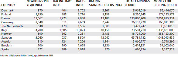
The European Standardbred (STB) is historically the result of different crosses between the classic American bloodlines and European families. The French STB is known for its stamina and aptitude for long distances. The American-derived families, however, have been selected for speed and racing at a fixed distance of 1 mile and are represented more widely in Italy and Sweden. Several countries (especially France, Italy, Germany, and the Scandinavian countries) have developed individual bloodlines and today are able to produce good-quality STB trotters. In Europe no races are available for pacers. In Table 109-1 the STB population and racing activity of different European countries are listed.


Europe has a large number of racetracks for STBs, and they are characterized by different circuit lengths, surfaces, and designs. Half-mile tracks have been replaced progressively by 1-km (image-
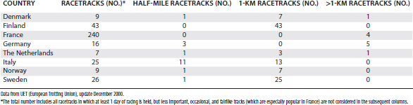
mile) tracks, and no 1-mile racetracks (with the exception of training centers) are present in Europe. The most characteristic and popular racetrack is the Plateau de Gravelle in Vincennes, Paris, where the Prix d’Amerique is held. The track surface is made of pressed charcoal powder; slopes and descents are present; and the circuit is egg-shaped, which is unique in Europe, where other racetracks have a regular design. Another famous French racetrack, Cagnes-sur-Mer in Nice, is well known for being 1200 m in length and for hosting the Criterium de Vitesse, one of the main European free-for-all races. In Sweden, Solvalla racetrack in Stockholm represents the heart of the Swedish STB racing, and in late May the multiple-heat race Elitlopp is held. In Norway, in addition to well-developed STB racing (the main racetrack being Bjerke near Oslo), races for Warmblood horses (a breed historically devoted to hard work but now genetically selected for speed) are still popular and meet the special interest of the local public.


A variety of track designs and purposes are present in Europe. For example, a large number of small country tracks are found in France, and local races are organized in a fairlike fashion, whereas the main racing activity is concentrated in Paris. In Italy, however, important races have a more even distribution among racetracks, Milan and Rome being the main places. In Table 109-2 the distribution of racing tracks in the main European countries is summarized.


Differences exist in availability of prize money among different countries, and this makes France and Italy the most attractive countries for trainers and owners. A number of well-known trainers started to move in the early 1980s toward France and Italy, especially from northern Europe, and this has enhanced the exchange of experiences between trainers and veterinarians.


Prize money distribution among horses of different ages has strongly influenced racing, especially in Italy, where 2- and 3-year-olds have the richest races. Higher purses for younger horses have also affected breeding programs and have stimulated an interest in many of the orthopedic problems affecting young horses, especially osteochondrosis, which has been studied widely in northern Europe.


Approaching Lameness Problems


Only STB trotters are allowed to race in Europe, and this makes the trainer’s work more challenging. Any possibility to switch gaits or to move an unnaturally fast gaited trotter to a potentially good pacer is precluded. When the fast gait is compromised by biomechanical problems and substantial interference between limbs (unlike in the pacer), corrections aimed at avoiding interference are attempted. Finding a solution for mechanical problems is not always possible, and corrective shoeing that alters the natural gait may cause compensatory lameness in different locations.


A series of mechanical limitations may be present at the beginning of training, and relatively soon they are followed by pain-related problems, usually affecting joints and less frequently digital flexor tendons and the suspensory ligament (SL). A less than 100% natural trotting athlete is more likely than a natural trotter to have its gait totally compromised, even by a mild subclinical lameness problem, and consequently gait limitations increase. However, anything that is attempted by the trainer to improve gait (corrective shoeing, special equipment that prevents turning the head and neck toward one side, or shifting the hindlimbs toward one shaft) forces the horse to a nonnatural fast gait and often results in lameness. In fact, when a nonnatural gait is forced, the end result is usually that a single limb (or a biped) is overloaded, and the uneven loading is exacerbated by the progression of training.


When young horses start training, they frequently have been subjected to basic lameness and radiological evaluations. This allows trainers the opportunity to treat conditions such as osteochondrosis or to be aware of other abnormalities. Prepurchase radiographic examinations and, when needed, preventative arthroscopic surgery (mostly for osteochondrosis of the tarsocrural joint and osteochondral fragments in the fetlock joints) are now practices that have received general acceptance. The reason to operate early is to have the horse rested before any training program is started. In yearlings eligible for autumn sales, it is important to perform surgery early to have presale radiographs without evidence of osteochondral fragments and to decrease effusion before the sale.


When moderate gait anomalies are present, experienced trainers usually give the horse time and keep going with a light exercise program instead of making radical changes. This allows, in many horses, a complete maturation of the equine athlete, and when the growth is complete and the muscular function well conditioned, the gait in many horses automatically improves without injuring the immature skeleton. Shoeing is also central in early training. Light plastic shoes are ideal to allow foot growth and expansion and to minimize trauma in the early phases of fast training.


Lameness Examination


Horses with acute, severe lameness should be allowed to rest. Radiographs are frequently diagnostic, revealing the most common severe musculoskeletal injuries affecting trained STBs, such as incomplete sagittal fractures of the proximal phalanx, fractures of the proximal sesamoid bones (PSBs), splint bone fractures, fractures of the third or radial carpal bones, fractures of a palmar process of the distal phalanx, fractures of the lateral condyle of the third metacarpal bone (McIII), slab fractures of the third tarsal bone, and stress fractures of the palmar aspect of the McIII or the plantar aspect of the third metatarsal bone (MtIII). Apical fractures of the PSBs are a common injury in young STBs, and the lateral PSB in the hindlimbs is the most common location.1


The conditions mentioned previously represent injuries of the racing STB requiring rest or surgical repair. More commonly the veterinarian is consulted for mild or obscure lameness, gait disturbances, or poor performance. In any case, a thorough history is mandatory before the lameness examination is initiated.


A basic lameness history must include the following:


Concerning conformation, the clinician should check the following:


Palpation


Many clinicians spend little time palpating a lame horse, a practice that I believe is a mistake. Areas of warmth (heat), especially in the hoof wall, must be detected, and regions of special interest include the fetlock joints, the metacarpal and metatarsal regions, carpus, hock, stifle, and back.


In the forelimb, the DIP joint capsule just above the coronary band is palpated to detect effusion. The character of the digital pulse is evaluated and compared between limbs.


Hoof tester examination can be considered part of palpation. When possible, feet are first tested without removing the shoes, and ideally the horse should be kept shod until any examination in movement is completed. When diagnostic analgesia is needed, hoof testing must precede palmar digital analgesia, and shoes may be removed temporarily if bar shoes or pads prevent accurate testing. These six points are tested in each horse: lateral and medial quarters, lateral and medial middle sole, and lateral and medial toe. Testing the frog rarely produces useful information, and squeezing the quarters from lateral to medial with hoof testers may cause pain unrelated to the primary lameness.2 Pain arising from the quarters, especially mild pain medially, should not be overrated because this region is frequently sensitive to hoof testers in normal horses. The contralateral foot may serve as a reference. In my experience the right medial quarter is the most common region to find pain elicited by hoof tester examination in STBs, and this is probably secondary to the counterclockwise direction of racing. This may be a clinically significant finding, but generally a painful response is considered more important when it arises from the toe or from the lateral side of the sole. When shoes are removed, a further evaluation of the lateral-to-medial balance is performed. The sole itself is observed, and when it appears flat and painful, this may correlate with type of shoes and padding that are used. Overzealous padding may add to, rather than relieve, pressure on the sole. A leather or rubber layer may allow sand to pack quickly under the pad and create pressure and secondary bruises. These horses are better managed with shoes in which the contact is limited to the hoof wall and no contact is made with the sole.


In the forelimb the fetlock joint is examined for effusion, alteration of the dorsal outline, and enlargement of the suspensory branches. The latter are palpated carefully with the joint flexed. Each branch is pressed gently axially, and alterations in consistency and pain response are noted. Range of motion of the fetlock joint is assessed. In a normal STB, the fetlock joint can be flexed up to 90 degrees (angle between the McIII and the proximal phalanx) without eliciting a painful response. With one hand holding the dorsal pastern region, the dorsal aspect of the fetlock joint can be palpated further by using the other hand to compress the dorsal joint capsule against the bony prominences of the sagittal ridge and condyles of the McIII. Horses with osteochondritis dissecans (OCD) or a hypertrophic synovial pad exhibit a painful response. The bony profile of the McIII must be followed with fingers to detect painful areas in the dorsal, lateral, and medial aspects. Palmar soft tissues are evaluated with the limb in a weight-bearing position and while being held off the ground. Each structure is palpated accurately to detect heat, pain, and edema in horses with acute lesions or fibrotic consistency and adhesions in those with chronic conditions. Fingers must be pressed firmly, deep in the proximal palmar metacarpal region, where pain originating from the proximal aspect of the SL is hard to detect. The carpus is better evaluated by holding the limb in a moderate degree of flexion. Careful digital palpation along the dorsal aspect should be performed. Of particular importance is the dorsomedial aspect where a thickened joint capsule and painful response correspond to the common finding of OA of the middle carpal joint. Palpation of the forelimb proximal to the carpus is rarely helpful. Elbow and shoulder lameness are rare, and bicipital bursitis has seldom been reported.


In the distal aspect of the hindlimb, palpation is similar to that described for the forelimb with the exception that foot lameness is less important. The metatarsophalangeal joint region is best evaluated while holding the limb in a semiflexed position. When the proximal metatarsal and distal tarsus regions are palpated, some false-positive painful reactions frequently are elicited, especially on the medial aspect. This pain frequently is overemphasized in horses with back pain, especially when the trainer thinks the horse has primary lameness in the tarsus. Effusion of the tarsocrural joint often indicates the likely presence of osteochondrosis (OC). The stifle must be palpated deeply and carefully because the joint is complex, but the structures that can be assessed are limited. The femorotibial joints seldom appear distended, but effusion of the medial femorotibial joint is an important abnormal clinical sign. More frequently, joint effusion is limited to the femoropatellar joint, but because the femoropatellar and medial femorotibial joints communicate, inflammation in one may result in effusion of both. With the exception of acute trauma, painful responses to palpation are rare in this area, even from the patellar ligaments that are identified easily. On the medial side, scar tissue and irregularities or enlargement of the medial patellar ligament may suggest previous desmotomy. When associated with femoropatellar effusion, the latter sign may suggest apical patellar fragmentation, and radiographic examination is indicated. The stifle can be flexed only in unison with most other hindlimb joints, so a painful response is not specific. With moderate flexion the medial collateral ligament can be stressed by the veterinarian pulling the tibia in a lateral direction with both hands and pressing the shoulder against the femur.


Palpation of the back is aimed at evaluating pain arising from joints (intervertebral, lumbosacral, sacroiliac, and sacrococcygeal), nerve roots, and muscles. The latter are the most likely origin of pain elicited by palpation, but other problems must be ruled out if associated clinical signs (atrophy, asymmetry) are present. Pain in the gluteal area may be secondary to many problems, including straight hindlimb conformation, hock or stifle pain, sore feet, gait imbalance, and stiff corrective harness equipment. Therefore pain in this area should not be treated as a primary problem unless a thorough clinical examination has ruled out other sources of pain.


Movement


After palpation, the horse is examined during movement. I commonly tranquilize each horse I examine for lameness. Tranquilization (10 mg of acetylpromazine maleate intravenously) improves the possibilities of handling the horse and lowers the risk of injuries to the veterinarian. Furthermore, the horse appears less stiff; mild lameness becomes somewhat more obvious; and the horse stands better for radiographic or ultrasonographic examination. The trainer must be consulted before injecting a tranquilizer because this practice may preclude racing because of doping regulations (the term used in Europe for a blood or urine drug test for a prohibited substance). Lameness is rarely detectable at the walk, but it is important to observe the way the horse lands with each foot to identify lateral-to-medial hoof imbalance. The horse is then trotted in a straight line on a firm surface, and the character of movements is observed. Abduction or circumduction of forelimbs is considered characteristic of carpal lameness because the horse appears to attempt to avoid flexion. In the hindlimb, stiffness has been related anecdotally to distal hock joint pain but in fact is not a specific sign. In horses with pain arising from the distal part of a hindlimb (frequently the metatarsophalangeal joint), the horse tends to moderately overflex the hock and stifle to shorten the weight-bearing phase of the stride.3 In horses with more severe lameness, drifting forward of the sound limb is observed, and a drop of the fetlock joint is easily seen. Abduction of the hindlimb is thought to be related to stifle lameness.


Flexion tests are used to supplement findings during movement and are similar to those used in other sports horses. Flexion of the carpus is accomplished by pulling the metacarpal region laterally, and the clinician’s elbow may act as a lever against the horse’s radius to stress the medial aspect. In young horses affected by carpitis, this maneuver frequently elicits pain.


Diagnostic Analgesia


Diagnostic analgesia can be performed in sequence from distal to proximal, or, to save time, selective analgesic techniques can be used. For example, European trotters with a positive response to forelimb lower limb flexion inconsistently respond to palmar digital analgesia; therefore analgesia of the metacarpophalangeal or DIP joints may be the first option. Perineural analgesia is preferred, however, to avoid minor risks of joint infections or to save the opportunity to provide intraarticular therapy immediately (when working in stables, trainers frequently are interested more in treatment than in diagnosis). I frequently start with low plantar analgesia in the hindlimbs because lameness of the digit is rare. In young STBs with forelimb lameness and a positive response to carpal flexion, I block the middle carpal joint first.


In horses with obscure lameness or when lameness is only apparent during fast exercise, examining the horse on the track may be useful. The horse is rigged in full harness and first examined trotting in a clockwise direction. Speed then is increased, and the horse is turned to train in a counterclockwise direction, the same as racing. Clinicians can drive the horse themselves, sit in a two-seat wagon, or observe the horse from a car or from a distance. In my opinion, watching the horse during exercise is important, especially when routine training and racing can be simulated, and I prefer the horse to be driven by its usual trainer. Having the horse fully equipped, mimicking the stress of racing, and observing the horse in turns are important advantages to this form of lameness examination.


Diagnostic analgesia is useful, but the clinician must be aware of related risks and make the trainer and owner aware as well. Diagnostic blocks should be avoided in horses suspected of having incomplete fractures. Radiographic examination should precede diagnostic analgesia in these horses. When examining a horse on the track after local analgesia, the trainer should be told to limit the speed as much as possible, avoiding any sudden stop or sharp turn. An experienced trainer normally is able to appreciate the benefits of a block quickly and without stressing the horse. Examination at speed is needed to make a diagnosis in horses with plantar process osteochondrosis fragmentation of the proximal phalanx and fragmentation of the distal border of the distal phalanx.


Diagnostic Imaging


Sophisticated equipment for diagnostic imaging is now available in most European equine clinics. Scintigraphy is available in Europe but is limited to large referral hospitals. Radiography remains the mainstay of equine diagnostic imaging, and the availability of excellent portable units has improved the radiographic examination under field conditions. Digital radiography offers advantages in terms of imaging processing, time saving, radiation protection, and easier image storage. Ultrasonographic examination is commonly performed.


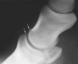
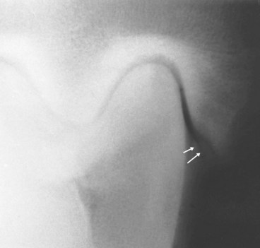
Common pathological findings in the distal limb of European trotters include osteophyte formation on the distal aspect of the middle phalanx, sometimes associated with modeling of the extensor process of the distal phalanx, as seen on a lateromedial image of the foot (Figure 109-1). This radiological pattern often is associated with heel growth proceeding faster than toe growth (club foot). When associated with a positive lower limb flexion test and lameness abolished using analgesia of the DIP joint, this radiological finding is important and indicates the presence of OA. Fragmentation of the extensor process of the distal phalanx has controversial clinical significance, but in my experience these fragments cause synovitis of the DIP joint and should be removed using arthroscopy.


The dorsoproximal-palmarodistal oblique radiographic image of the distal phalanx, unlike scintigraphy, rarely helps in diagnosing pain arising from stress remodeling of the distal phalanx; thus the diagnosis of pedal osteitis is limited to horses with chronic lameness characterized by substantial radiolucency.4 Fragmentation of the lateral and medial palmar processes of the distal phalanx, although controversial as a cause of acute lameness, may be an important source of pain best managed by shoeing.


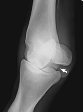
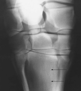
The flexed lateromedial image of the metacarpophalangeal or metatarsophalangeal joints is useful in evaluating lesions of the distal aspect of the McIII or the MtIII. In the hindlimb, the oblique images must be acquired in a proximal to distal direction (down-angled) to see better the area between the proximal phalanx and the base of the PSBs. Fragmentation of the proximal plantar aspect of the proximal phalanx represents a major cause of subtle hindlimb lameness (Figure 109-2).


A dorsopalmar image of the carpus helps to evaluate the proximal suspensory origin from the McIII, and focal or diffuse increased radiopacity of the McIII may be found (Figure 109-3). In yearlings and young horses, this image allows examination of the distal radius, and pathological modifications of the growth plate on the medial side (physitis) are a frequent cause of early lameness. The most common lesions of the carpus are seen with the dorsolateral-palmaromedial oblique image and the dorsal 35° proximal-dorsal distal oblique (skyline) image of the flexed carpus to highlight the distal row of carpal bones. The radiographs must be of excellent quality, and the appropriate projection must be obtained because false-negative radiographic images are frequent. Abnormal findings include areas of radiolucency in the dorsoproximal articular border of the radial fossa of the third carpal bone, radiolucent lines suggestive of fractures, and increased radiopacity of the third carpal bone.5 A moderate degree of increased radiopacity in the radial fossa of the third carpal bone is considered normal in racing horses, but more severe increased radiopacity associated with radiolucent areas represents a pathological finding.6,7


In the hindlimb the centrodistal and tarsometatarsal joints frequently appear normal radiologically even when distal hock joint pain is diagnosed clinically as the source of pain. Scintigraphy is an excellent tool for diagnosing OA of these joints.8 The tarsocrural joint is a predilection site for OC in STBs, and the presence of fragments associated with effusion may represent an indication for arthroscopic surgery. Lesions affecting the lateral trochlear ridge of the talus and medial malleolus of the tibia more frequently cause lameness and effusion than do those of the cranial aspect of the intermediate ridge of the tibia. In horses with effusion but without obvious fragmentation, a specific dorsal 10° to 15° lateral-plantaromedial oblique image is required to evaluate the axial aspect of the medial malleolus. Subtle osteochondral fragmentation or radiolucency easily can be overlooked (Figure 109-4).


Stay updated, free articles. Join our Telegram channel

Jun 4, 2016 | Posted by in EQUINE MEDICINE | Comments Off on The European and Australasian Standardbreds

Full access? Get Clinical Tree

Get Clinical Tree app for offline access