The Elbow, Brachium, and Shoulder

Chapter 40The Elbow, Brachium, and Shoulder


Anatomical Considerations


The elbow joint consists of the humerus, radius, and ulna. The distal aspect of the humerus develops from three ossification centers: the diaphysis, the distal epiphysis, and the epiphysis of the medial condyle. These close radiologically at 11 to 24 months of age. The radius and ulna have a single proximal epiphysis. The radial physis closes radiologically at 11 to 24 months of age, but the ulnar physis does not close until 24 to 36 months of age. Physeal closure occurs later in non-Thoroughbred (TB) breeds.


The cranial aspect of the olecranon of the ulna has articular and nonarticular components. The anconeal process and trochlear notch articulate with the humerus. At the distal part of the trochlear notch is a distinct ridge. Distal to this is a large, non–weight-bearing synovial fossa.


The elbow is a ginglymus joint, supported medially and laterally by collateral ligaments. The medial collateral ligament consists of a long superficial part and a deeper short part. The medial collateral ligament arises from an eminence on the medial epicondyle of the humerus. The deep part inserts on the radial tuberosity; the superficial part inserts on a more distal prominence, just distal to the interosseous space between the radius and ulna. The lateral collateral ligament arises from a depression in the lateral epicondyle of the humerus and inserts on the lateral tuberosity of the radius, just distal to the joint margin. The joint capsule is extremely thin caudally, where it forms a pouch in the olecranon fossa. Cranially the joint capsule is strengthened by oblique fibers and blends with the collateral ligaments medially and laterally.


The most readily palpable landmarks are the olecranon of the ulna and the lateral collateral ligament of the humeroradial joint. Minimal soft tissue covers the lateral aspect of the elbow, making it vulnerable to the effects of direct trauma and penetrating wounds.


The humerus is surrounded by muscles, which largely protect it from the effects of direct trauma. The deltoid tuberosity on the craniolateral aspect is usually readily palpable and is potentially vulnerable to the effects of direct trauma. The deltoid tuberosity is the most useful landmark for identifying the point for needle insertion for synoviocentesis of the intertubercular (bicipital) bursa. The proximal aspect of the humerus has several centers of ossification for the humeral head and the greater and lesser tubercles, which gradually fuse at 3 to 5 months of age. Radiological closure of the proximal humeral physis occurs at 24 to 36 months of age.


The scapula has four centers of ossification: the scapular cartilage, body of the scapula, cranial part of the glenoid cavity of the scapula, and supraglenoid tubercle and coracoid process. The ossification center for the cranial part of the glenoid cavity fuses directly with the body of the scapula, and this is complete radiologically by 5 months of age. The physis between the supraglenoid tubercle and coracoid process and the body of the scapula closes radiologically at 12 to 24 months of age—earlier in TB and TB crossbreeds than in ponies. These physeal lines remain weak links, and it is through these that fractures of the supraglenoid tubercle and cranial part of the glenoid cavity of the scapula tend to occur.


The scapula is attached by the serratus ventralis muscles to the axial skeleton. Other muscles involved in attachment of the thoracic limb to the trunk and neck are the four pectoral muscles, the brachiocephalicus, and omotransversarius.


The brachial plexus lies on the axial aspect of the scapula and is derived from the sixth, seventh, and eighth cervical nerve roots and the first thoracic nerve. These nerve roots are potentially vulnerable to trauma where they exit the cervical and cranial thoracic vertebrae, and these are relatively common sites for neuroma formation after trauma. The nerves of the brachial plexus are responsible for innervation of many of the principal muscles of the shoulder region.


The scapulohumeral or shoulder joint is unusual because of the absence of collateral ligaments. Stability therefore depends on muscular support on the medial and lateral aspects by the subscapularis, teres minor, infraspinatus, and supraspinatus. Cranial support is provided by the biceps brachii and supraspinatus, and caudal support is rendered by the long head of the triceps brachii. Overlying muscles make it impossible in mature horses to appreciate distention of the joint capsule by palpation. The joint capsule attaches closely to the margins of the scapulohumeral joint, and this, together with the surrounding muscles, restricts arthroscopic evaluation within the joint. The suprascapular nerve wraps around the cranial margin of the scapula proximal to the supraglenoid tubercle and provides innervation to the infraspinatus and supraspinatus muscles.


Considering its embryological development and function, the so-called intertubercular (bicipital) bursa would be more appropriately called a tendon sheath.1 The bursa surrounds the tendon of biceps brachii, which originates from the supraglenoid tubercle of the scapula. The craniodistal pull of the biceps brachii results in cranial and distal displacement of fractures of the supraglenoid tubercle. Communication was identified between the scapulohumeral joint capsule and the intertubercular bursa using contrast arthrography in 3 of 18 limbs (17%)2; thus in some horses intraarticular analgesia of the scapulohumeral joint has the potential to cause improvement in lameness caused by pain arising from the intertubercular bursa. The tendon of biceps brachii is partially cartilaginous proximally and passes over the smooth intertuberal groove of the humerus and then becomes predominantly muscular with a tendonous core.


The most important palpable landmarks in the shoulder region are the cranial and caudal eminences of the greater tubercle of the humerus. The notch between these eminences provides the portal for arthrocentesis of the scapulohumeral joint. The scapular spine is usually readily palpable except in exceptionally fat horses or heavily muscled individuals and becomes more prominent if the supraspinatus or infraspinatus muscles atrophy.


Diagnosis


Clinical Signs


In the adult horse lameness associated with the elbow or shoulder region is comparatively rare, except after direct trauma caused by a fall, collision with a solid object such as a gatepost, or collision with another horse. In immature athletic horses, stress fractures of the scapula, humerus, and radius; osseous cystlike lesions; and osteochondrosis are quite common.


Lameness associated with the shoulder or elbow region is usually sudden in onset and generally moderate to severe. After trauma to the shoulder or elbow, or in association with severe lameness, the horse tends to stand with its weight inclined toward the contralateral limb, not fully load bearing on the lame limb. The horse may resent turning on the limb. Muscle atrophy in the shoulder region is not specific for lameness associated with the proximal aspect of the limb but is often more severe than if the pain arises farther distally. Rapid loss of the bulk of supraspinatus and infraspinatus muscles alone is likely to reflect damage to the suprascapular nerve, whereas involvement of additional muscles is more likely to reflect a brachial plexus injury. Swelling in the elbow or shoulder region usually reflects direct trauma but may be seen with subluxation or luxation of the shoulder or elbow joint. Patchy sweating is sometimes seen with a lesion of the brachial plexus. Pain elicited by deep palpation is relatively unusual and is generally associated with direct trauma. Care should always be taken to compare the reaction with that to palpation of the contralateral limb. The reaction to manipulation of the proximal limb joints should also be interpreted with care because many normal horses resent extreme flexion, extension, or abduction.


Many normal horses show some resentment of firm palpation of the brachiocephalicus muscles at the base of the neck. These muscles often become sore with a more distal source of pain causing lameness. Primary muscle pain causing lameness does sometimes occur and is associated with a more marked pain reaction on palpation and muscle spasm. It may cause a lameness most evident when the horse is ridden at the walk, characterized by lifting of the head and neck as the ipsilateral limb is protracted. Lesions of the tendon of biceps brachii are also sometimes associated with lameness that is worse at walk than trot.


If lameness is mild, then the character of the lameness is nonspecific; but if lameness is moderate to severe, it is often characterized by a shortened cranial phase of the stride, a reduced height of the arc of foot flight, and a marked head lift and nod. imageThese gait characteristics are evident at the walk and the trot. Observation of the moving horse from the front and the side is particularly useful. The horse may pivot on the lame limb when turning. The gait characteristics of shoulder slip (see page 473) are more easily identified by observing the horse walking toward you. Lameness is frequently accentuated with the lame limb on the outside of a circle. In horses with proximal limb lameness, especially associated with muscle fibrosis, lameness may be evident only when the horse is ridden, performing specific movements. Manipulative tests of the proximal limb joints are rather nonspecific and frequently unrewarding.


Lameness may fluctuate in degree under different circumstances and within an examination period; therefore it is important to observe the horse for a sufficient length of time before proceeding with local analgesic techniques. If lameness is apparent only when the horse is ridden, the horse may be sensitive to the diagonal on which the rider sits.


Lameness caused by trauma resulting in only bruising usually improves rapidly, within a few days. Persistence of lameness merits further investigation.


Local Analgesia


It is important to recognize the potential effect of a median nerve block, performed in the proximal antebrachium, on elbow pain. Elbow lameness may be substantially improved, presumably because of local diffusion of the local anesthetic solution. Techniques for intraarticular analgesia of the elbow and shoulder and intrathecal analgesia of the intertubercular bursa are described in detail in Chapter 10. Intraarticular analgesia of the elbow and shoulder joints usually improves but rarely eliminates pain associated with either joint. The elbow and shoulder joints are relatively large; therefore use of at least 10 mL of local anesthetic solution (mepivacaine, 2%) is recommended for the elbow joint and 20 mL for the shoulder joint. Retrieving synovial fluid from each joint is usually possible. Absence of resistance to injection is not a guarantee that the needle is in an intraarticular location. Walking the horse after the block facilitates circulation of the local anesthetic solution throughout the joint. Although improvement in lameness may be seen rapidly, within 10 to 15 minutes after injection, at least 1 hour should elapse after the block before the result is considered negative. The block generally is effective for up to 2 hours. The block usually has no influence over pain associated with periarticular structures.


When performing intraarticular analgesia of the shoulder, it is important to recognize that there is communication with the intertubercular bursa in some horses. In some horses instability of shoulder, so-called shoulder slip, appears transiently (for up to 2 hours) after injection of local anesthetic solution. This prohibits interpretation of the nerve block. The cause is presumably diffusion of local anesthetic solution to nerves innervating the muscles responsible for maintaining stability of the shoulder. Positive-contrast arthrography has shown that injection of volumes greater than 20 mL pose a danger of pooling at the site of injection or leakage from the joint capsule.. An experimental study demonstrated that deposition of mepivacaine over the suprascapular nerve could result in transient shoulder slip, but this may be the result of diffusion to affect the brachial plexus (see page 473).3


Intrathecal analgesia of the intertubercular bursa may improve lameness associated with lesions of the tendon of biceps brachii, the bursa itself, or the humeral tubercles, but accurate intrathecal injection may not be reliable.4 The bursa is large, and use of 20 mL of local anesthetic solution is recommended.


False-negative results to both intraarticular analgesia of the shoulder and elbow joints and intrathecal analgesia of the intertubercular bursa may occur, and radiological, scintigraphic, and ultrasonographic evaluation of these areas is indicated if clinical signs are suggestive of shoulder or elbow region pain. Many other potential sources of pain in the proximal limb cannot be desensitized by local analgesic techniques. If the response to distal limb analgesia and the blocks described previously is negative, nuclear scintigraphic evaluation may be warranted.


Imaging


Radiography and Radiology


Radiographic examination of the elbow requires a minimum of mediolateral and craniocaudal images.5 The limb should be pulled forward sufficiently to avoid the pectoral muscle mass to evaluate the distal aspect of the humerus properly. Additional oblique images may be of value in selected horses. Routine radiographic examination of the scapulohumeral joint should include mediolateral and craniomedial-caudolateral oblique images. In some horses cranioproximal-craniodistal oblique images of the humeral tubercles are useful. Examination of the entire length of the humerus can be difficult, because often when such examination is indicated, the horse has pain and is reluctant to allow the limb to be adequately protracted. Accurate evaluation of the entire scapula is also not easy because of superimposition over the thoracic vertebrae and the contralateral limb.


Fast-speed, rare earth screens and appropriate film are essential if using conventional radiography. Use of a grid will greatly enhance image quality, especially in the shoulder region. High exposure factors are required (e.g., 100 kV, 100 mAs, for a mediolateral image of the shoulder). Underexposure will result in lesions being missed.


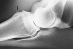
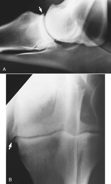
It is important to recognize that the cranial articular margin of the proximal radius has several lips that should not be confused with osteophytes (Figure 40-1). The cranial tuberosity of the proximal aspect of the radius may appear roughened in slightly oblique mediolateral views.


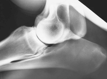
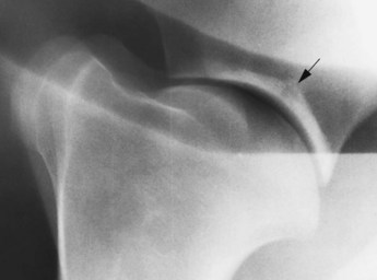
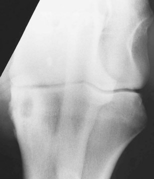
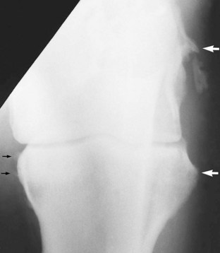
In the scapulohumeral joint a small circular radiolucent region is sometimes seen in the subchondral bone in the middle of the glenoid cavity of the scapula (Figure 40-2). A radiolucent edge effect is often seen in the proximal humerus, the result of superimposition of the lateral rim of the glenoid cavity of the scapula.


Ultrasonography


Diagnostic ultrasonography is invaluable for assessing muscle structure in the shoulder region. Normal muscle has a homogeneous echogenicity. Identification of hyperechogenic regions indicative of muscle necrosis, fibrosis, or mineralization is usually associated with lameness.


Evaluation of the elbow joint itself is limited in the weight-bearing position because of the difficulty in getting access medially. Examination of the lateral collateral ligament of the humeroradial joint is straightforward. For evaluation of the medial aspect, the limb should be pulled forward, but unless the medial collateral ligament is under tension, its echogenicity may lack homogeneity.


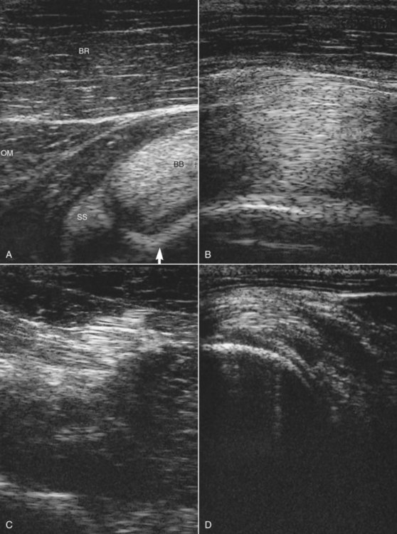
In the shoulder region ultrasonography is important for assessing the intertubercular bursa, humeral tubercles, tendon of biceps brachii (Figure 40-3, A and B), tendons of insertion of the supraspinatus and infraspinatus muscles (Figure 40-3, C and D), and the infraspinatus bursa (Figure 40-3, B).6-10 Care should be taken to ensure that the horse is fully load bearing on the limb, because hypoechogenic artifacts can be created, especially in the tendon of biceps brachii, unless the musculature is under tension. The medial and lateral lobes of the tendon of biceps brachii and the isthmus between them should each be evaluated individually, because getting the entire structure into focus simultaneously is difficult.


Differential Diagnosis


Elbow


Osseous Cystlike Lesions


Osseous cystlike lesions occur most commonly medially in the proximal radial epiphysis (Figure 40-5) and usually result in acute-onset, relatively severe lameness in immature athletic horses.


Lameness may vary greatly in degree, within and between examinations, and is usually substantially improved by intraarticular analgesia. In addition to a well-defined osseous cystlike lesion, there is often periosteal new bone on the proximal medial metaphyseal region of the radius. Intraarticular medication with hyaluronan or corticosteroids (triamcinolone acetonide or methylprednisolone acetate) has resulted in successful resolution of lameness.1,11 In some horses the osseous cystlike lesions have resolved radiologically. Surgical treatment by curettage of the cyst using an extraarticular approach has been successful in some horses, but fracture through the cyst has been a recognized complication.12 Recurrent lameness after surgical treatment is also possible.13 An ill-defined osseous cystlike lesion is occasionally seen in older horses with OA of the elbow with no known previous history of lameness; it is likely that the cystlike lesion had been present asymptomatically before the development of pain associated with OA.


Less commonly, large, less well-defined osseous cystlike lesions have been identified in the distal aspect of the humerus in young Thoroughbreds being prepared for the yearling sales or just after entering training.1 Lameness is acute in onset, persists despite box rest, and is generally not influenced by intraarticular analgesia of the elbow. Nuclear scintigraphic examination reveals a region of IRU in the distal aspect of the humerus, more centrally located than that associated with a stress fracture (see page 463). Careful radiological examination reveals a less well-defined osseous cystlike lesion. Conservative management has resulted in persistent lameness, and the results of surgical treatment have been disappointing. Young horses with smaller osseous cystlike lesions in the distal medial humerus have been identified and have responded to conservative management.12


Collateral Ligament Injury


Injury to the collateral ligaments of the elbow is not common and usually results from a traumatic injury such as a fall. Damage to the lateral collateral ligament has been identified most frequently. Severe injuries may also be associated with injury to the joint capsule, and osteoarthritis may ensue. Lameness is acute in onset. Subtle soft tissue swelling may be appreciated in the elbow region, and manipulation of the elbow may induce pain. Lameness may be partially improved by intraarticular analgesia of the elbow. If damage is restricted to extraarticular structures, then the response may be negative. Nuclear scintigraphic examination may be helpful in these horses. IRU occurs at the sites of ligament attachment on the distal aspect of the humerus and proximal aspect of the radius.


Definitive diagnosis requires ultrasonographic examination and identification of disruption of the normally linear pattern of echoes within the ligament. Sometimes periosteal new bone or avulsion fractures can be identified at the region of ligamentous attachment.16 Radiographic examination should also be performed to identify any concurrent pathological condition of the bone, such as enthesophyte formation, an avulsion fracture, or secondary OA (Figure 40-6).


Horses with minor lesions have responded well to a period of box rest and controlled walking exercise, but those with lesions that have been associated with entheseous new bone formation often have had persistent lameness and have developed OA.


Enthesopathy of the Biceps Brachii


Tearing of the attachment of the biceps brachii from the cranioproximal aspect of the radius may be associated with a traumatic injury such as a fall, but frequently history does not suggest the cause. Lameness is sudden in onset and often not associated with any localizing signs, except pain on manipulation of the elbow in horses with acute lameness. The lameness has no particular characteristics. The response to local analgesic techniques is negative. Radiographic examination may reveal periosteal new bone on the cranioproximal aspect of the humerus in horses with chronic lameness17 (Figure 40-7). Sometimes an adjacent mineralized fragment is present, an avulsion fracture or dystrophic mineralization. Some new bone formation on the cranioproximal aspect of the radius can be seen in normal horses, reflecting previous injury; therefore care should be taken in interpreting its current clinical significance. Nuclear scintigraphic examination is helpful for acute lameness before periosteal new bone develops and in horses with chronic lameness. Ultrasonographic evaluation has not been helpful. Treatment is by rest. The prognosis is guarded to fair.


Jun 4, 2016 | Posted by in EQUINE MEDICINE | Comments Off on The Elbow, Brachium, and Shoulder

Full access? Get Clinical Tree

Get Clinical Tree app for offline access