The Ear


The Ear


The ear (organum vestibulocochleare [auris]) evolved as an organ of balance and hearing in vertebrates. The most primitive part is the inner ear (auris interna), which in all vertebrates consists of a membranous labyrinth within a bony labyrinth and functions for both balance and hearing. Fish have only an inner ear, whereas amphibians and reptiles developed an additional chamber, the middle ear (auris media), formed by a tympanic cavity (cavum tympani) that is an extension of the pharynx. The tympanic cavity connects with the pharynx via the auditory tube, (formerly eustachian or pharyngotympanic tube), which is closed to the outside by a tympanic membrane, or eardrum. A portion of the hyomandibular bone from the second branchial arch develops into a sound-conducting ossicle, the columella, which transmits vibrations from the tympanic membrane, across the air-filled tympanic cavity, to the inner ear. In mammals two more bones, the malleus and incus, are added to the middle ear, and the columella becomes a stapes, making a chain of three bones from the tympanic membrane to the inner ear. Although lizards and birds have an external ear (auris externa) it consists only of an auditory meatus and a short canal. To protect the entrance to the canal and help direct sound into it, lizards sometimes have specialized scales, and birds have specialized feathers and a cutaneous muscle for pulling a skin flap across the opening. Only mammals (except cetaceans and a few others) have a well-defined external ear formed by a cartilaginous auricle (auricula) or pinna covered by skin and moved by muscles.


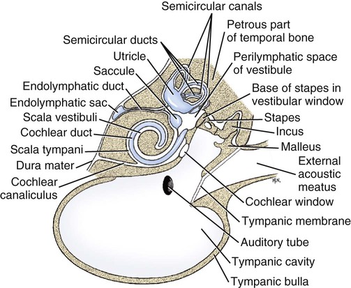
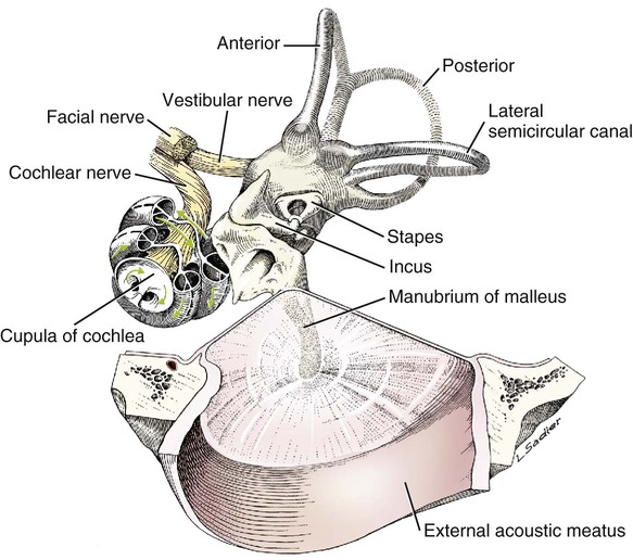
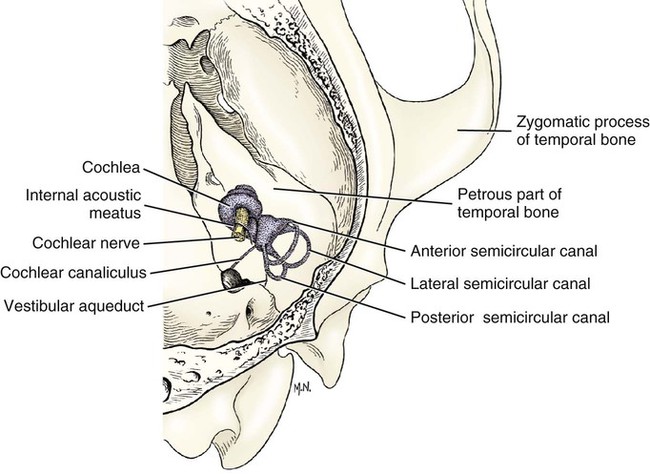
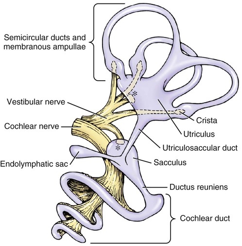
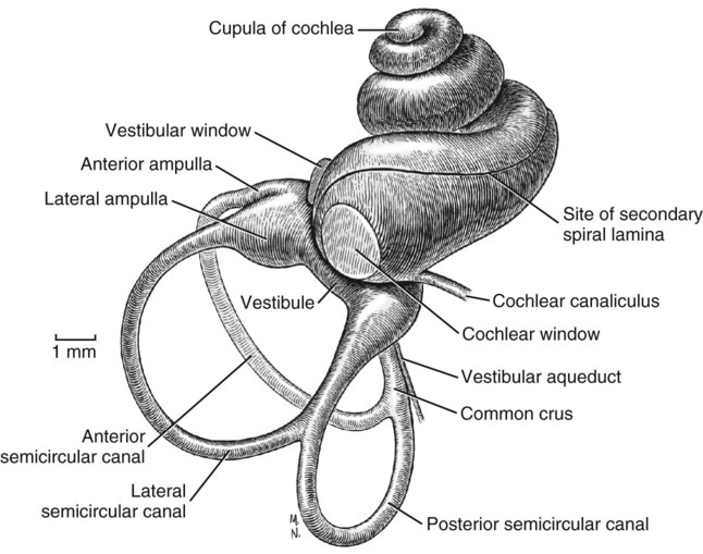
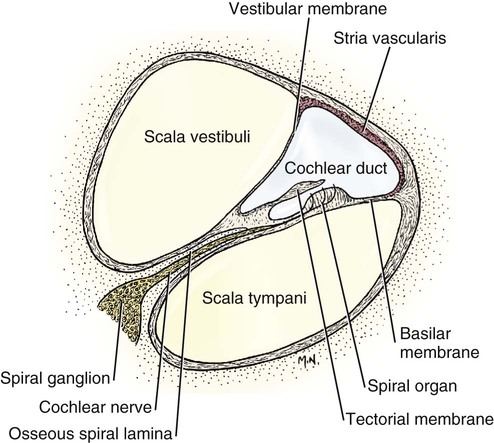
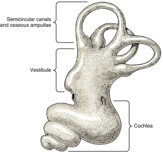
The three major components of the dog ear will be considered here in developmental sequence, which is similar to their phylogenetic evolution: (1) the inner ear, (2) the middle ear, and (3) the external ear (Fig. 20-1).


The receptor for special proprioception, the vestibular system, develops in conjunction with the receptor for the auditory system (special somatic afferent system). They are derived from ectoderm but are contained in a mesodermally derived structure. Together these receptors are the components of the inner ear. The ectodermal component arises as a proliferation of ectodermal epithelial cells on the surface of the embryo adjacent to the developing rhombencephalon. This structure is the otic placode, which subsequently invaginates to form an otic pit and otic vesicle (otocyst) that breaks away from its attachment to the surface ectoderm. This saccular structure undergoes extensive modification of its shape but always retains its fluid-filled lumen (endolymph) and surrounding thin epithelial wall as it becomes the membranous labyrinth of the inner ear. Special modifications of its epithelial surface at predetermined sites form the receptor organs for the vestibular and auditory systems.


Corresponding developmental modifications occur in the surrounding paraxial mesoderm to provide a supporting capsule for the membranous labyrinth. This fluid-filled (perilymph) ossified structure is the bony labyrinth contained within the developing petrous portion of the temporal bone (Fig. 20-1).


These membranous and bony labyrinths are formed adjacent to the first and second branchial arches and their corresponding first pharyngeal pouch and first branchial groove. The first branchial groove gives rise to the external ear canal. The first pharyngeal pouch forms the auditory tube and the mucosa of the middle-ear cavity. The intervening tissue forms the tympanum. The ear ossicles are derived from the neural crest of branchial arches 1 (malleus and incus) and 2 (stapes). These ossicles become components of the middle ear associated laterally with the tympanum (malleus) and medially with the vestibular window of the bony labyrinth of the inner ear (stapes).


The Inner Ear


Bony Labyrinth


Anatomically, the bony labyrinth in the petrous part of the temporal bone consists of three continuous fluid-filled portions. These areas are the large vestibule, and the three semicircular canals and the cochlea, which arise from the vestibule. All three continuous bony components contain perilymph, a fluid similar to cerebrospinal fluid (CSF), from which it may be at least partly derived.


Vestibule


The vestibule is an irregular, oval space, approximately 3 mm in diameter, that communicates with the cochlea rostrally and with the semicircular canals caudally. The walls of the vestibule are marked by depressions and ridges that correspond to the various portions of the enclosed membranous labyrinth. The medial wall contains two depressions: Caudodorsal is the elliptical recess, which contains the utricle, and rostroventral to it is the spherical recess for the saccule. The vestibular crus separates the two recesses. Several groups of small openings that accommodate the nerves of this region occur near the recesses. These tiny groups of foramina are called maculae cribrosae. In the vestibule are two openings: the more dorsal vestibular window in which is inserted the foot plate of the stapes and the more ventrorostral cochlear window, which is closed by a membrane and is located at the end of the cochlea where perilymph vibrations can be dampened into the tympanic cavity.


Semicircular Canals


There are three semicircular canals, an anterior, a posterior, and a lateral canal (Figs. 20-2 and 20-3). They lie caudal and slightly dorsal to the vestibule. Each canal describes approximately two thirds of a circle in a single plane, and each is approximately at a 90-degree angle to the other two. The segment of the canal that communicates with the vestibule is called the crus. Each canal has two crura that communicate with the vestibule (with the exception of the common crus, to be noted later). One crus of each canal has a dilation, the osseous ampulla (ampullae osseae) near the junction with the vestibule. The lumen diameter of the canals averages roughly 0.5 mm, the ampulla being approximately twice as large.


The anterior canal of one ear is roughly parallel with the posterior canal of the opposite ear. The lateral canal of each side occupies a nearly horizontal plane. The anterior canal is the longest. The arc it forms measures approximately 6 mm across at the widest part. The lateral canal forms an arc that measures approximately 4.5 mm, while the arc of the posterior semicircular canal is the smallest, measuring only 3.5 mm in medium-sized dogs. These measurements vary with the size of the dog. The common crus is formed by the nonampullated ends of the posterior and anterior canals. In sculptured specimens the anterior semicircular canal is seen to surround the floccular fossa, a small but deep depression on the medial side of the petrous part of the temporal bone. This depression is occupied by the paraflocculus of the cerebellum. The ampullated end of the posterior canal and the nonampullated end of the lateral canal are united for a short distance caudal to the vestibule.


Cochlea


The cochlea is the bony shell that surrounds the cochlear duct in a spiral of three and one-quarter turns around a central hollow core of bone, the modiolus, which contains the cochlear nerve and blood vessels. The cochlea points ventrorostrally and slightly laterally within the promontory of the petrous part of the temporal bone (Fig. 20-4). The osseous spiral lamina that winds around the modiolus, much like the thread of a screw, nearly bisects the lumen of the cochlea into two portions, called the scala tympani and scala vestibuli (Fig. 20-5). The osseous spiral lamina begins within the vestibule and ends at the apex in a free hooklike process, the hamulus. The scala vestibuli communicates with the vestibule, and hence the perilymph within it is acted on by the vibrations of the base of the stapes in the vestibular window. The cochlear window (see Figs. 20-1 and 20-3) is an opening situated near the rostral end of the vestibule by which the scala tympani communicates with the tympanic cavity. A secondary tympanic membrane closes this cochlear window. The membranous cochlear duct, formerly scala media, completes the separation of the two scalae. The scalae communicate at the apex of the modiolus by a small opening, the helicotrema, formed at the free border of the hamulus. The basal turn of the cochlea is approximately 4 mm in diameter and lies close to the medial side of the vestibule. The total height of the cochlea measures approximately 7 mm. Longitudinal modiolar canals and a spiral modiolar canal serve for the distribution of both blood vessels and nerves to the cochlea. The source of perilymph may depend on its location. One source is CSF from the subarachnoid space that gains entrance to the scala tympani of the cochlea via the perilymphatic duct (ductus perilymphaticus) (not part of the membranous labyrinth) in the small cochlear canaliculus (canaliculus cochleae). This small canal courses directly ventrad from a point on the ventral wall of the scala tympani near its origin to communicate with the subarachnoid space (see Fig. 20-1). The other source is as an ultrafiltrate from the cochlear blood vessels in the modiolus into the scala vestibuli.


Membranous Labyrinth


The ectodermally derived membranous labyrinth consists of four fluid-filled compartments, all of which communicate. These compartments are contained within the components of the bony labyrinth and include the saccule and utriculus within the bony vestibule connected by the utriculosaccular duct, the three semicircular ducts within the bony semicircular canals that connect to the utriculus and a cochlear duct within the bony cochlea that is connected to the saccule by the ductus reuniens. The endolymphatic duct is an extension from the utriculosaccular duct through the bony vestibular aqueduct to the intracranial dura where the duct expands into a endolymphatic sac. The endolymph contained within the membranous labyrinth is thought to be derived from the blood vessels and epithelium of the stria vascularis along the peripheral wall of the cochlear duct and is absorbed back into the blood through the blood vessels surrounding the endolymphatic sac. The three semicircular ducts are the anterior (vertical), posterior (vertical), and lateral (horizontal). Each semicircular duct is oriented at right angles to the others. Thus rotation of the head around any plane causes endolymph to flow within one or more of the ducts. Each semicircular duct connects at both ends with the utriculus.


Crista Ampullaris


The crista ampullaris is the receptor organ associated with each semicircular duct. At one end of each membranous semicircular duct is a dilation called the ampulla. On one side of the membranous ampulla, a proliferation of connective tissue forms a transverse ridge called the crista ampullaris. It is lined on its internal surface by columnar neuroepithelial cells. On the surface of the crista is a gelatinous structure that is composed of a protein-polysaccharide material called the cupula, which extends across the lumen of the ampulla. This neuroepithelium is composed of two basic cell types: hair cells and supporting cells. The dendritic zones of the vestibular neurons are in synaptic contact with the base of the hair cells. These hair cells have on their luminal surface 40 to 80 hairs, or modified microvilli (stereocilia), and a single modified cilium (kinocilium). These structures project into the overlying cupula. Movement of fluid in the semicircular ducts causes deflection of the cupula, which is oriented transversely to the direction of flow of the endolymph. This deflection bends the stereocilia, which is the source of the stimulus by way of the hair cells to the dendritic zone of the vestibular neuron that is in synaptic relationship with the plasmalemma of the hair cell. In one end of each semicircular duct is one membranous ampulla with its crista ampullaris. Because the three semicircular ducts are all at right angles to each other, movement of the head in any plane or angular rotation affects a crista ampullaris and stimulates vestibular neurons. These cristae function in dynamic equilibrium.


Macula


The macula is the receptor organ found in the utriculus and saccule, which are located in the bony vestibule. These maculae are on one surface of each of these saclike structures. Each macula is an oval-shaped plaque in which the membranous labyrinth has proliferated. The surface of the macula consists of columnar epithelial cells. This neuroepithelium is composed of hair cells and supporting cells. Covering the neuroepithelium is a gelatinous material, the statoconiorum (otolithic) membrane. On the surface of this membrane are calcareous crystalline bodies known as statoconia (otoliths). Similar to the hair cells of the cristae, the macular hair cells have projections of their luminal cell membranes—stereocilia and kinocilia—into the overlying statoconiorum membrane. Movement of the statoconia away from these cells is the initiating factor in bending the stereocilia to stimulate an impulse in the dendritic zones of the vestibular neurons that are in synaptic relationship with the base of the hair cells. The macula of the saccule is oriented in a vertical direction (sagittal plane), whereas the macula of the utriculus is in a horizontal direction (dorsal plane). Thus gravitational forces continually affect the position of the statoconia relative to the hair cells. These structures are responsible for the sensation of the static position of the head and linear acceleration or deceleration. They function in static equilibrium. The macula of the utriculus may be more important as a receptor for sensing changes in head posture, whereas the macula of the saccule may be more sensitive to vibrational stimuli and loud sounds.


Vestibular Nerve


The dendritic zone of the vestibular portion of cranial nerve VIII is in a synaptic relationship with the hair cells of each crista ampullaris and the macula utriculi and macula sacculi. The axons course through the internal acoustic meatus with those of the cochlear nerve. The cell bodies of these bipolar-type sensory neurons are inserted along the course of the axons within the petrous portion of the temporal bone, where they form the vestibular ganglion (Giene and Kuder 1983). The vestibular nerve axons pass to the lateral surface of the rostral medulla where they enter the medulla and terminate in telodendria at one of two sites. The majority terminate in the vestibular nuclei in the medulla and pons. A few course directly into the cerebellum by way of the caudal cerebellar peduncle.


Cochlear Duct-Spiral Organ


The most highly developed and differentiated portion of the membranous labyrinth is the cochlear duct (Figs. 20-5 and 20-6). This duct has a triangular shape with its base, the stria vascularis adjacent to the peripheral wall of the cochlea. A thin vestibular membrane forms the roof of the cochlear duct and a thicker basilar membrane forms the floor of the duct. The spiral organ (organum spirale), formerly organ of Corti, is a collection of hair cells and supporting cells that rests on the basilar membrane. These structures are involved in the transduction and transmission of sound impulses via the cochlear nerve to the brain. Special somatic afferent axons in the cochlear nerve make synaptic contact with these hair cells. Their neuronal cell bodies are centrally located in the attachment of the spiral lamina to the modiolus. The axons leave the inner ear through the internal acoustic meatus accompanied by the vestibular nerve and synapse in the cochlear nuclei on the lateral side of the medulla oblongata. The basilar membrane separates the endolymph of this duct from the perilymph of the scala tympani, which is a part of the cochlea. The thin vestibular membrane separates the endolymph of the cochlear duct from the perilymph in the scala vestibuli of the cochlea. Thus the membranous cochlear duct filled with endolymph is enclosed within the bony cochlea (scala tympani-scala vestibuli), which is filled with perilymph. These fluids have different chemical compositions and are not in open communication with each other. Likewise the membranous semicircular ducts containing endolymph are enclosed within the semicircular canals that contain perilymph. The distinction between the osseous (Fig. 20-7) and the membranous labyrinth (see Fig. 20-6) is sometimes blurred in textbooks by the carefree use of canal for duct and cochlea for cochlear duct. To understand the structural and functional relationships of the labyrinths to each other, the terminology must be kept clear and consistent.


Hearing depends on the ability of sound waves in the external gaseous environment reaching the tympanic membrane via a patent external acoustic meatus. Here the oscillations in the air are converted to oscillations in the three auditory ossicles in the tympanic cavity. At the vestibular window the bony oscillations are converted to oscillations of the perilymphatic fluid, which in turn affects the spiral organ by oscillations of the basilar membrane in the cochlear duct. Any interruption of this pathway can result in loss of hearing. Most deafness is related to disorders of the spiral organ. Foss and Flottorp (1974) found that hearing in puppies first occurred as a functional modality at 14 days after birth (on average), which coincides with the opening of the eyelids. Congenital inherited sensorineural deafness is very common in many breeds of dogs and is usually present shortly after birth. Johnsson et al (1973) studied deafness and pathology of the cochlear in Dalmatian dogs. Rouse et al (1984) considered abnormal otoconia and calcification of the labyrinth in deaf Dalmatian dogs. Albinotic and abiotrophic forms are recognized and can be diagnosed in puppies using brainstem auditory response testing (de Lahunta and Glass, 2009.)


Shambaugh (1923) injected the vessels of the dog labyrinth and found that the arterial supply was from a single labyrinthine artery that entered through the internal acoustic meatus. In some specimens a small artery came along the cochlear canaliculus and was distributed to a small area of periosteum along the scala tympani. The first branch to be given off from the labyrinthine artery was the anterior vestibular artery, a vessel that supplied the macula of the utriculus and the cristae of the lateral and anterior semicircular ducts. The second branch of the labyrinthine artery supplied the crista of the posterior duct, the posterior crura of the posterior and horizontal canals and ducts and the crus commune. The venous drainage from the labyrinth was collected by two trunks, the larger one leaving along the cochlear canaliculus collected all of the blood from the cochlea as well as most of the blood from the capillaries supplied by the anterior and posterior vestibular arteries. The lesser vein leaving along the vestibular aqueduct drained the remainder of the labyrinth.

< div class='tao-gold-member'>

Only gold members can continue reading. Log In or Register to continue

Stay updated, free articles. Join our Telegram channel

Jul 18, 2016 | Posted by in PHARMACOLOGY, TOXICOLOGY & THERAPEUTICS | Comments Off on The Ear

Full access? Get Clinical Tree

Get Clinical Tree app for offline access