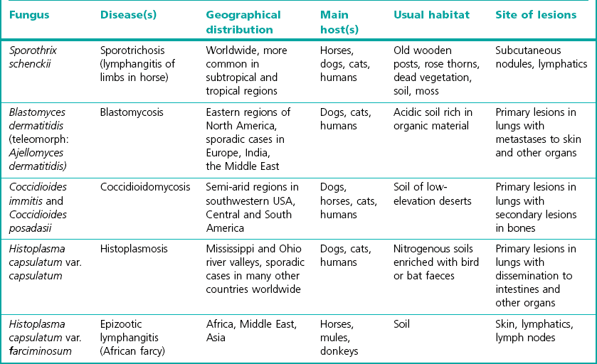
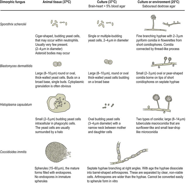
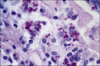
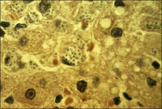
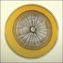
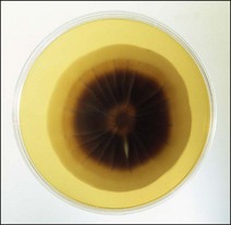
Chapter 41 The dimorphic fungi present two growth forms, a mould when growing saprophytically in the environment or when on culture media at 25–30°C, and a yeast or yeast-like form in animal tissues or when cultured on enriched media at 37°C. The mould or mycelial phase tends to be the more stable of the two. These fungi cause deep or systemic mycoses in animals and humans. Table 41.1 indicates the fungi included in this group and gives the main hosts and disease, the reservoir of infection and the geographical distribution. Three varieties of Histoplasma capsulatum are recognized, var. capsulatum (chiefly a New World pathogen), var. duboisii (an African human pathogen) and var. farciminosum (an Old World equine pathogen). However, phylogenetic studies have identified at least eight clades suggesting that the three varieties are phylogenetically meaningless (Kasuga et al. 2003). Teleomorphs have been identified for some of these fungi. The teleomorph of B. dermatitidis is Ajellomyces dermatitidis and the teleomorph of H. capsulatum is Ajellomyces capsulatus (phylum Ascomycota). A summary of the diagnostic tests used to identify the dimorphic fungi is given in Box 41.1. All of the dimorphic fungi can cause disease in humans and should be treated with respect. Cultures of Coccidioides immitis in particular, represent a major biohazard for laboratory personnel because the arthrospores, produced on media at 25–37°C, can easily form an infective aerosol. The culture of this dimorphic fungus should either be avoided or appropriate precautions must be taken. These include the use of a biological safety cabinet when handling any material suspected of containing the organism, especially cultures. Cultures on slopes in screw-capped bottles are recommended and if culture plates are used these must be taped. The cultures of C. immitis should be covered with sterile water or saline before introducing an inoculation needle to prevent dispersion of the arthrospores. All microscopic preparations must be done in a biohazard cabinet. Cultures should be autoclaved as soon as the final diagnosis of C. immitis has been made. Isolates of Coccidioides immitis can be divided into ‘Californian’ and ‘non-Californian’ isolates, a second species has been proposed Coccidioides posadasii to distinguish the latter (Fisher et al. 2002). Table 37.2 (see Chapter 37) and Figure 41.1 indicate the microscopic morphology of the dimorphic fungi in animal tissue and in cultures at 25°C and 37°C. Histopathological sections are most useful for demonstrating the yeast forms in animal tissues (Figs 41.2 and 41.3). At 25°C growth is visible in three to five days. Colonies are white to cream at first, becoming wrinkled with delicate aerial hyphae and then later turning dark and leathery (Figs 41.4 and 41.5). At 37°C colonies are yeast-like, smooth, soft and cream to tan in colour. Growth occurs in about three to five days.
The dimorphic fungi
Laboratory Diagnosis
Safety aspects
Direct microscopy
Colonial morphology
Sporothrix schenckii
< div class='tao-gold-member'>
![]()
Stay updated, free articles. Join our Telegram channel

Full access? Get Clinical Tree


The dimorphic fungi
Only gold members can continue reading. Log In or Register to continue
