The Digestive System

CHAPTER 36 The Digestive System*


Prenatal and Neonatal Development


The signals that regulate the differentiation and development of the fetal gastrointestinal tract (GIT) are not well understood. It is likely that factors exist in the amniotic fluid that may play a role in this process. Gut peptides are found in the fetal human GIT within the first trimester and in the amniotic fluid by the second trimester. In porcine and ovine fetuses, GIT development was impaired when the swallowing of amniotic fluid was prevented. Compared with controls, these animals showed a decrease in the weight of their small intestine, pancreas, and liver and a generalized thinning of the gut wall throughout the length of the GIT. Other factors, including cortisol, insulin, and numerous other hormones, present in both the fetal and maternal circulation also may have significant effects on GIT development. Although there have been very few studies examining the prenatal development of the GIT in dogs and cats, some brush-border enzymatic activity and nutrient transport activity are developed by parturition.


At birth, the GIT undergoes perhaps the most drastic change in function of any organ system except the lungs. During the first 24 hours, the canine small intestine nearly doubles in weight. At this time, the GIT must take over from the placenta the huge task of transferring nutrients from the outside world to the neonatal circulation. However, the intestinal wall of kittens grows little during the first week after birth. Because neonatal puppies and kittens have very small energy and glucose reserves, failure to make this accommodation becomes a serious if not fatal problem in a matter of hours.


The normal neonatal GIT is fully capable of digestion and absorption of its primary substrate, mother’s milk. Many of the brush-border enzymes found in the mature GIT are present to facilitate the final stages of digestion and thus absorption. The activity of these enzymes increases markedly just before parturition; therefore premature animals may experience digestive difficulties.


The neonatal GIT is not well suited for ingesta other than milk. Newborn puppies lack certain pancreatic enzymes, and the muscularis layer of their small intestine is about 50% thinner than that found in adult dogs. Some of the brush-border enzymes, particularly the α-glycosidases, are not well developed, and this can cause problems if sugars such as sucrose or maltose are used in homemade milk replacers. Brush-border enzymes of the mature GIT are present, but their activity increases as the puppy ages. Suckling induces hypertrophic and hyperplastic mucosal cells in the neonatal dog.


Although the neonatal GIT may have difficulty handling foods other than milk, it is highly specialized for milk digestion and absorption. Not surprisingly, the changes that occur in the developing GIT are well matched to changes in the composition and volume of the milk with which it is presented. The first milk, colostrum, is rich in protein, immunoglobulins, hormones, and other factors that promote hypertrophy and hyperplasia of the neonatal GIT. Puppies fed milk replacer instead of colostrum experience a much smaller increase in intestinal mass during the first 24 hours of life.


The ability to internalize large molecules such as proteins persists up to 2 weeks after birth in many species. In puppies, this may help compensate for inadequate activities of pancreatic proteases that are secreted in only small amounts during the first 1 to 2 weeks of life. Neonatal puppies also secrete very little pancreatic lipase, but this is compensated for by secretion of gastric lipase. As the concentration of fat in milk increases over time, the secretion of pancreatic lipase also increases.


Gastric capacity can be approximated as 100 to 250 ml/kg body weight in adult dogs with puppies having a greater range in relative size. The entire neonatal GIT is colonized with bacteria at day 1; subsequentially the relative proportions of various groups of bacteria change, with anaerobic groups increasing in numbers.


From the second week to the seventh week of life, there is a multifold decrease in milk intake as a percentage of body weight. Similarly there is a threefold increase in solid food intake as a percentage of body weight from the third week to the seventh week. By 3 weeks of age, the puppy’s GIT will have undergone considerable changes. The thickness of the gut wall will have nearly doubled primarily because of hypertrophy of the tunica muscularis. This will facilitate the passage of solid ingesta along the lumen of the gut. The pancreas will have developed adequate capacity to produce digestive enzymes and antibacterial factors. The introduction of solid food provides both a source and a substrate for bacterial growth, and these factors take over from those found in milk to help establish normal GI microflora.


In later life, the ability to change digestive function varies among species in accordance with natural variation in the diet. The cat, which is an obligate predator, is less able to vary its pancreatic and GIT enzyme activity than is the more omnivorous dog. Dogs also possess a tremendous capacity to adapt their GIT function over an enormous range of energy requirements. For most dogs, the largest demand on the GIT for processing nutrients occurs during growth. A weaned puppy may require 2 to 3 times the energy per unit body weight of a sedentary adult dog.*


The Oral Cavity


Examination of the oral cavity should be consistent and systematic and include inspection and palpation of the gingiva, teeth, tongue, lingual frenulum, floor of the mouth, buccal surface, and hard and soft palates. Gentle opening and palpation of the oral cavity should not be painful or cause prompt withdrawal of the head. The breath of a growing, healthy dog or cat is not unpleasant. Alterations in the odor of the breath usually indicate a disease state, food the animal has eaten, or medication it has received. Offensive, foul-smelling breath may be caused by oral lesions, necrotic respiratory disease, or alimentary tract disease associated with belching.


The ingestive and masticatory functions of the oral cavity depend largely on the ability of the oral cavity to form a closed, hollow compartment, requiring labial and palatal competence. Cleft palate, cleft lip, or other causes of altered competence of the oral cavity should be identified during the oral examination.


Stomatitis


Any infectious, physical, or chemical agent or traumatic insult that significantly alters the replication, maturation, or exfoliation of the healthy mucosa favors the occurrence of stomatitis.


Feline rhinotracheitis virus (FRV) and feline calicivirus (FCV) cause most of the oral lesions seen in young cats. Buccal, lingual, and nasal ulcers frequently accompany these viral respiratory infections, although ulcers are generally more severe in FCV than in FRV infections. Feline leukemia virus and feline immunodeficiency virus may also be associated with oral lesions as a persistent glossitis, periodontitis, palatitis, and/or gingivitis, presumably because of virus-induced immunosuppression.


Foreign objects frequently cause traumatic insult to the oral mucosa of young dogs and cats, probably owing to the animals’ curious natures and normal developmental chewing habits. The animal generally presents in varying phases of recovery, with secondary bacterial infection and scar tissue often camouflaging the foreign object(s). Removal of the foreign object(s) usually effects a cure. Intermittent problems from migrating grass awns or porcupine quills may be eliminated only by thorough dissection of irritated tissue. Traumatic injury to the tongue, palate, or lips is common, and the original wound is often aggravated by secondary bacterial infections. Oral burns from electrical cords are especially common in young dogs and cats that chew through the insulation of the cords. Pulmonary edema, seizures, and cardiac arrhythmias frequently accompany these electrical shock injuries.


In the immune-mediated diseases (i.e., pemphigus diseases, bullous pemphigoid, and lupus erythematosus), it is unusual in either the dog or cat for oral lesions to occur without skin involvement, especially at other mucocutaneous junctions, such as eyelids, nostrils, anus, vagina, and prepuce. Pemphigus foliaceus, the most common of the pemphigus diseases, rarely affects the oral cavity. Any of these immune-mediated diseases may first appear in the dog or cat at 4 to 6 months of age and typically follow the same lesion(s) development and patterns as seen in the adult. Their treatment follows the same regimens as used for the adult.


Lymphoplasmacytic stomatitis commonly affects young to middle-aged cats, but a cat as young as 4 months of age may be affected. Calicivirus has been isolated from some affected cats. The definitive cause of the oral lesions is unknown, but an immunologic basis has been suspected. Abyssinian and Somali are commonly affected cat breeds. Lymphoplasmacytic stomatitis typically follows the same lesion(s) development (i.e., small erythemic papules to severe mucosal proliferation of gingiva and/or palatal arches) as seen in the adult cat. Oral lesion(s) treatment follows the same regimens as used for the adult.


Mineral and vitamin deficiencies, heavy-metal poisoning, and coagulation abnormalities may also cause oral lesions. Although rarely seen, deficiencies in many of the B-complex vitamins and possibly in zinc may contribute to the appearance of oral lesions in growing, malnourished dogs or cats. Silver-gray Collies affected with cyclic hematopoiesis often develop a recurrent stomatitis that coincides with absolute neutropenia. Proliferative eosinophilic granulomas have been seen in the oral cavity of Siberian Huskies and in many breeds of young cats (i.e., in kittens as young as 8 weeks of age).


The definitive cause of a stomatitis cannot always be determined by history, physical examination, cytologic examination, bacterial and fungal culture, or tissue biopsy. In many cases, bacterial cultures reveal the normal mixed flora of the oral cavity, often making antimicrobial etiologies unreliable. In these cases, therapy to treat the symptoms is often efficacious. Daily flushing of the mouth with chlorhexidine (diluted to 0.1% to 0.2% solution in tepid water) followed by rinsing the mouth with copious amounts of fresh water will help cleanse the affected areas. Soft, palatable food may need to be provided for several days during the initial healing phase. Ampicillin, amoxicillin, or cephalosporins may be administered daily in an oral liquid form for both a local antimicrobial effect and subsequent systemic effect after absorption. Antimicrobial therapy may be indicated for up to 3 weeks.


Tonsillitis


Small-breed young dogs are most commonly affected with primary tonsillitis. Anorexia, lethargy, dysphagia, coughing, gagging, and fever are the usual complaints. The absence of tonsillar swelling does not necessarily preclude the diagnosis of tonsillitis, especially in cats. Improvement of the animal, observed with antimicrobial therapy, generally precedes resolution of the inflammation, although tonsillar swelling may continue to be present. Recurrence is common, with exacerbations lasting several days. Tonsillectomy is rarely required in recurrent tonsillitis unless the tonsillar enlargement obstructs the pharynx. Most dogs can be effectively treated medically and appear to “outgrow” the problem. Medical treatment of tonsillitis includes using broad-spectrum antimicrobial agents and supportive care as discussed for stomatitis. Bacterial culture of inflamed tonsillar tissue usually produces β-hemolytic streptococci, coliforms, or other bacteria of the normal oral flora; therefore identification of antimicrobial etiologies may be unreliable.


Secondary tonsillitis may be associated with systemic disease or local predisposing anatomic or pathologic factors. The presence of tonsillitis does not indicate the site of primary disease. Any persistent irritation or inflammatory process of the oral cavity or pharynx (e.g., abscesses, gingivitis, lodged foreign object(s), or persistent vomiting or regurgitation) may induce a secondary tonsillitis. The gagging, regurgitation, and vomiting of foam in cases of tonsillitis necessitate differentiation from megaesophagus, pyloric disorders, cricopharyngeal achalasia, productive cough, retropharyngeal abscesses, and lodged foreign object(s) in the pharynx, larynx, or esophagus. Treatment should be directed toward eliminating the underlying secondary cause of the tonsillitis.


The Salivary Glands


Congenital enlargement of the parotid salivary glands has been reported in dogs. The dogs typically present with hypersalivation (drooling) and are treated effectively with parotid duct ligation.


Sialadenitis may be caused by bite wounds, lacerations, blunt trauma, or extension from cellulitis and abscesses of the head and neck. Sudden swelling in the region of a salivary gland, fever, inappetence, and pain on opening the mouth are usually evident on physical examination. Diagnosis of sialadenitis is usually obvious, but cytologic examination confirms salivary tissue involvement. Treatment should be aimed at the underlying cause. Ways of providing symptomatic relief include the administration of antimicrobial agents, application of warm compresses, replacement fluid therapy, drainage if abscess is present, and frequent feeding of small amounts of a nutritionally complete, soft, palatable food.


A sialocele is a collection of saliva in tissue. Any age, sex, and breed of dog or cat can be affected. Sialocele formation most commonly involves the sublingual and mandibular salivary glands, where blockage of a duct or rupture of the gland causes extravasation of saliva into the surrounding connective tissue. The saliva may gravitate to the sublingual area (often referred to as a ranula), intermandibular space, mediastinum, or pharyngeal area (Figure 36-1). Swelling may develop suddenly but is more likely to be in the form of a slowly developing, fluctuant sac. Diagnosis of sialocele is based on physical appearance of the cervical swelling and on cytologic evaluation. Needle aspiration of the swelling yields a viscous, mucoid fluid that is usually clear or brown. Total excision of the affected gland, which is usually the sublingual or mandibular gland, and drainage of the sialocele provide the only long-term therapy. Removal of an elliptic portion of the wall of a ranula allows direct drainage of saliva into the oral cavity. The surgical techniques for the management of salivary gland disorders are described elsewhere.
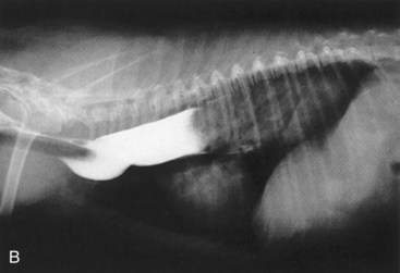


The Oropharynx


The events of swallowing can be divided into oral, pharyngeal, and cricopharyngeal stages. Dysphagia is uncommon in dogs and rare in cats. Dysphagia is usually recognized in puppies and kittens shortly after weaning.


Oral dysphagias may result from disturbances to various motor and sensory tracts and peripheral nerves. Branches of cranial nerves V, IX, and X provide sensory innervation for swallowing, and cranial nerves V, VII, IX, X, XI, and XII deliver motor innervation via the nucleus ambiguus and the respiratory centers. Signs of oral dysphagia may include difficulty in prehension of food and lapping water, excessive chewing or chomping, hypersalivation, and diminished or absent gag reflex. Evidence of denervation of the tongue may exist, and food may drop from the animal’s mouth or be retained in the buccal cavity.


Pharyngeal dysphagias are less consistent, and related signs are more difficult to localize. The most common signs are coughing with repeated unsuccessful attempts at swallowing and laryngotracheal aspiration of swallowed material. Often the puppy or kitten will regurgitate masticated food several hours after ingestion, or food or liquid material will be misdirected into the nasopharynx, leading to nasal discharge.


Cricopharyngeal dysphagias typically manifest as problems of asynchrony (i.e., incoordination of pharyngeal contraction and cricopharyngeal sphincter relaxation) or as achalasia (i.e., failure of cricopharyngeal sphincter relaxation). Signs of cricopharyngeal dysphagias can vary, depending on whether there is an achalasia or asynchrony.


Differentiation of oral, pharyngeal, and cricopharyngeal dysphagias is important. Myotomy of the muscles of the cricopharyngeal sphincter gives dramatic improvement in cricopharyngeal achalasia. However, oral and pharyngeal dysphagias are generally made worse by the myotomy procedures. Oral and pharyngeal dysphagias are best managed by treating any underlying medical illness, changing the consistency of the animal’s usual diet, and feeding the animal in an elevated position. If conservative therapy is not helpful and the owner does not wish to continue feeding by one of the enteral methods, cricopharyngeal myotomy may be considered. The owner should be aware that cricopharyngeal myotomy may make the animal’s condition worse.


The Esophagus


An important species difference between the dog and cat is in the musculature of the esophageal body. The entire length of the canine esophageal body is composed of striated muscle, whereas the distal one third to one half of the feline esophagus is composed of smooth muscle.


Vascular ring anomalies are congenital malformations of the great vessel system that interfere with esophageal function. These anomalies produce an extramural obstruction of the esophagus at the base of the heart and rarely affect the trachea or cardiovascular system. The most frequently encountered vascular ring anomaly in the young dog or cat is persistent right aortic arch. The incidence of vascular ring anomalies is higher in young dogs than in young cats. Irish Setters, Boston Terriers, and German Shepherd Dogs are the most commonly affected dog breeds.


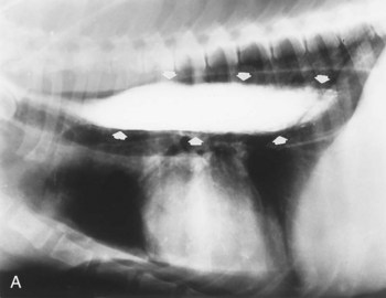
Signs caused by the vascular ring anomalies result from esophageal entrapment and obstruction, with subsequent precordial megaesophagus. When weaning to solid food, puppies and kittens will repeatedly regurgitate. Although regurgitation is usually associated with eating when solid food is first fed, it will occur at variable times after eating as the precordial megaesophagus worsens. Occasionally signs in the puppy or kitten are associated with ingestion of maternal milk. Affected animals are obviously malnourished and underweight. The diagnosis of a vascular ring anomaly-induced megaesophagus can usually be confirmed by a positive contrast esophagogram (Figure 36-2).


Treatment is early surgical correction by ligation and transection of the stricturing ligament or vessel and complete mobilization of the esophagus from the connective tissue in the area of entrapment. Prognosis is guarded pending complete postoperative recovery because of frequent complicating factors (e.g., existing malnourishment, weakened state of the animal, aspiration pneumonia, and persistent megaesophagus despite surgical correction). Most animals show substantial improvement if they survive the immediate postoperative period. Persistent megaesophagus occurs if the esophagus develops sacculations, an extensive dilation that fills the anterior region of the mediastinum, or dilation of the esophagus caudal to the vascular ring anomaly.


Esophagitis is unusual in the young dog and cat and usually occurs because of traumatic insult, ingestion of a chemical or thermal irritant, or gastric acid reflex. Thermal injuries may occur when eager eaters bolt hot food or when puppies or kittens are fed hot gruel. Gastric acid reflux is uncommon as a primary event but can occur any time the lower esophageal sphincter pressure is compromised, such as during anesthesia or severe blunt trauma directly to the thorax or abdomen. Esophagitis can be limited to mucosal damage or may extend into the submucosa and musculature. If only the mucosa is involved, the esophagitis is usually mild and self-limiting. However, esophagitis with submucosal and musculature involvement often leads to severe ulceration with subsequent perforation, fibrotic stricture formation, persistence of inflammation, and/or disturbed motor activity (acquired megaesophagus).


Signs of esophagitis are regurgitation and dysphagia. Esophagoscopy may be required to make a definitive diagnosis by visualization of mucosal changes; questionable cases may be confirmed by histopathology. Treatment of esophagitis is aimed at eliminating the underlying cause and symptomatic management of the esophagus and the systemic effects that the esophagitis created. Means of providing symptomatic relief include esophageal rest by feeding through a gastrostomy tube or by frequent small feedings of nutritionally complete, soft, bland food and administration of antimicrobial agents, H2-receptor antagonists, metoclopramide, and sucralfate slurry (crush 1 gm of sucralfate and mix with 10 ml of water; give 5 ml of slurry 4 to 6 times daily).


Idiopathic megaesophagus is characterized by motor disturbances of the esophagus that result in abnormal or unsuccessful transport of ingesta between the pharynx and stomach.


Both congenital and acquired forms of idiopathic megaesophagus occur in young dogs and cats. There is evidence that congenital idiopathic megaesophagus is inherited in both the dog (e.g., Wire Fox Terrier and Miniature Schnauzer) and cat. The incidence is highest in Great Danes, German Shepherd Dogs, Irish Setters, Labrador Retrievers, and Chinese Shar-Peis. Esophageal dysfunction in Chinese Shar-Peis may result from segmental hypomotility and esophageal redundancy. In the cat, Siamese and Siamese-related breeds have the highest incidence.


Acquired idiopathic megaesophagus may occur spontaneously in any young dog or cat. In most instances, the underlying cause is undetermined; however, megaesophagus may occur in several systemic diseases that affect the nervous system or skeletal muscles. These diseases include myasthenia gravis, polymyositis, toxoplasmosis, canine distemper, hypothyroidism, hypoadrenocorticism, and myotonia with myopathy. Other potential diseases that could contribute to megaesophagus are systemic lupus erythematosus, tick paralysis, botulism, tetanus, lead poisoning, ganglioradiculitis, dysautonomia, anticholinesterase compounds, and polyneuritis. Megaesophagus has also been associated in cats with pyloric dysfunction and feline dysautonomia, often referred to as the Key-Gaskell syndrome. Canine or feline dysautonomia, the main features of which are dilated pupils, dry mucous membranes, megaesophagus, constipation, and dysuria and urinary incontinence with urinary bladder distention, is a dysfunction of the autonomic nervous system. The cause of dysautonomia is currently unknown.


The onset of signs associated with congenital idiopathic megaesophagus usually begins around the time of weaning. Signs may include effortless regurgitation of esophageal contents, weight loss, polyphagia, weakness, dehydration, impaired skeletal mineralization, ballooning of the cervical esophagus that is synchronized with respiration, and recurrent laryngotracheal aspiration that often leads to recurrent pneumonia. Oral fetor may be present owing to stagnation of fermenting ingesta retained in the dilated esophagus. Regurgitation may be seen immediately on feeding or up to 12 or more hours later. The time interval between eating and regurgitation can be related to the degree of dilation or to the general activity of the animal. Usually both liquids and solids are poorly tolerated.


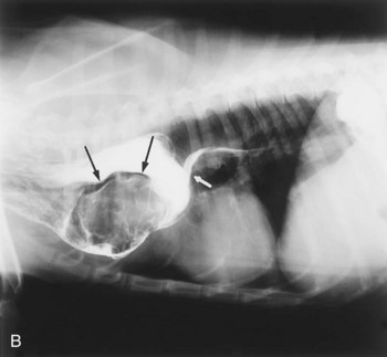
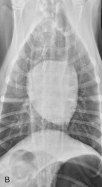
Survey thoracic radiographs consistently reveal a dilated esophagus (Figure 36-3). Evidence of aspiration pneumonia is frequently present and consistent with the signs of dysphagia and regurgitation.


When the esophagus is not observed visually on survey radiographs, a contrast esophagogram is required. A barium contrast study better defines the degree of esophageal dilation, lack of function, and extent of involvement (Figure 36-4). The study helps rule out congenital vascular ring anomalies or other causes of localized obstruction that might contribute to megaesophagus and outlines the funnel shape of the caudal region of the esophagus to rule out invasive processes that may cause irregular or asymmetric narrowing.
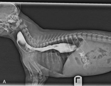
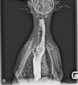


Treatment of congenital idiopathic megaesophagus consisting of proper dietary management in terms of frequent, elevated feedings with foods of appropriate consistency for the particular animal (some handle bulky foods well; others better tolerate gruels) generally results in spontaneous improvement in a number of animals. This had led investigators to believe that congenital idiopathic megaesophagus may be caused by delayed neurologic development of esophageal innervation. With elevated feedings and minimal distention of the esophagus, normal esophageal motor function may develop. If stasis of esophageal contents is allowed, however, gradual overdistention and atony result, contributing to persistent megaesophagus. The earlier the dysfunction is recognized and the dietary management instituted, the better the prognosis. Puppies and kittens diagnosed at weaning and managed appropriately have a better prognosis than those whose condition is recognized later, at around 4 to 6 months of age. Once severe megaesophagus occurs, complete recovery is unlikely. Aspiration pneumonia and malnourishment limit the longevity of these animals.


Disorders of the Gastroesophageal Junction


Disorders affecting the function of the gastroesophageal junction are uncommon. The disorders more commonly associated with its altered function are hiatal hernia with reflux esophagitis and gastroesophageal intussusception. Hiatal hernia is the result of a congenital or acquired defect of the phrenoesophageal ligament that allows displacement of the gastroesophageal junction forward into the thoracic cavity. Hiatal hernia may also be associated with an upper respiratory obstruction, especially common in young Chinese Shar-Peis. Gastric contents reflux through the incompetent junction into the caudal region of the esophagus, where they produce varying degrees of irritation to the esophageal mucosa. Signs may include regurgitation with possible hematemesis, dysphagia, altered breathing pattern, and weight loss. Diagnosis requires a high index of suspicion followed by a barium contrast study performed under fluoroscopic observation.


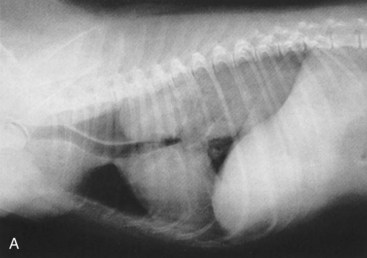
Gastroesophageal intussusception or invagination involves the telescoping of all or part of the stomach into the esophageal lumen (Figure 36-5). Occasionally the spleen and pancreas are also included. The cause of this condition is not understood, but an incompetent gastroesophageal junction must be suspected. Gastroesophageal intussusception generally occurs in puppies of large breeds and in kittens, particularly those with congenital megaesophagus. A puppy or kitten will be presented to the veterinarian with sudden onset of difficulty in breathing, impending shock, and a history of vomiting. The sudden onset of signs and radiographic appearance of the mass are the keys to the diagnosis. Treatment is surgical reduction of the intussusception and a gastropexy.


Stay updated, free articles. Join our Telegram channel

Sep 11, 2016 | Posted by in SMALL ANIMAL | Comments Off on The Digestive System

Full access? Get Clinical Tree

Get Clinical Tree app for offline access