The Cervical Spine and Soft Tissues of the Neck

Chapter 53The Cervical Spine and Soft Tissues of the Neck


This chapter discusses disorders of the neck that may give rise to lameness or poor performance or result in an abnormal neck shape, abnormal neck posture at rest or while moving, or neck stiffness. Transient neurological conditions, conditions caused by trauma resulting in other injuries, and gait abnormalities that may be confused with lameness are considered, but those associated with compression of the cervical spinal cord are discussed in Chapter 60.


Anatomy


The neck consists of seven cervical vertebrae, which articulate both by intercentral articulations and by synovial articulations, which have large joint capsules to accommodate the degree of movement between adjacent vertebrae. Interposed between the vertebral bodies are intervertebral fibrocartilages to which is attached the dorsal longitudinal ligament, which lies on the floor of the vertebral canal. The ligamentum flavum connects the arches of adjacent vertebrae. The atlas (the first cervical vertebra) and the axis (the second cervical vertebra) have a unique shape and specialized joints. The atlanto-occipital joint is a ginglymus joint, which permits flexion and extension and also a small amount of lateral oblique movement. The atlantoaxial joint is a trochoid or pivot joint; the atlas and head rotate on the axis. The ligament of the dens is strong and fan shaped and extends from the dorsal surface of the odontoid peg (dens) to the ventral arch of the axis. The ligamentum nuchae extends from the occiput to the withers and consists of funicular and lamellar parts. The lamellar part separates the two lateral muscle groups. The atlantal bursa is interposed between the funicular part of the ligamentum nuchae and the dorsal arch of the atlas; a second bursa may exist between the ligament and the spine of the axis. The muscles of the neck can be divided into lateral and ventral groups. The neck has eight cervical nerves, the first of which emerges through the intervertebral foramen of the atlas, the second through that of the axis, and the eighth between the seventh cervical vertebra and the first thoracic vertebra. The sixth to eighth cervical nerves contribute to the brachial plexus.


Clinical Examination


It is important to recognize that head and neck carriage depends in part on conformation: the way in which the neck comes out of the shoulder and the shape of the neck. The shape of the neck is also influenced by the way in which the horse works. If a horse carries the head and neck high, with the head somewhat extended, the ventral strap muscles tend to be abnormally well developed, resulting in a ewe-neck conformation. Many horses naturally bend more easily to the right than to the left or vice versa, and the muscles on the side of the neck, especially dorsocranially, are developed asymmetrically. Such asymmetry is particularly obvious if the neck is viewed from above by the rider. If a horse is excessively thin, then the cervical vertebrae become prominent and the caudodorsal neck region becomes dorsally concave, whereas in a fit, well-muscled horse that works regularly on the bit, this region is dorsally convex. Most stallions and many native pony breeds have a prominent dorsal convexity to the neck region, resulting in a cresty appearance. A horse that is excessively fat tends to lay down plaques of fat throughout the body, including the neck region, and this can be misinterpreted as abnormal neck swelling.


If a horse is particularly thick through the jaw, that is, has a large mandible, it is physically difficult to work on the bit (i.e., flexing at the poll so that the front of the head is in approximately the vertical position). Although neck pain can cause a reluctance to work on the bit, more common causes include rider-associated or training problems, mouth pain, forelimb or hindlimb lameness, and back pain. Some horses strongly resist the rider’s aids to work on the bit, despite the absence of pain. The use of artificial aids such as draw or running reins, which give the rider a mechanical advantage, may help to break a vicious cycle and encourage the horse to become more submissive and compliant. Similarly, work on the lunge line using a chambon (a device that runs from the girth via a headpiece to the bit rings) can encourage the horse to work in a correct outline and develop fitness and strength of the appropriate musculature. Working the horse in trot over appropriately spaced trotting poles can also help to encourage a horse to work in a correct outline, with a round and supple back.


A rider may complain of neck stiffness or difficulties in getting a horse to bend correctly in a circle. Although this may be caused by neck pain, neck stiffness may be a protective mechanism by the horse to avoid pain associated with lameness, especially forelimb lameness. A horse with left forelimb lameness, for example, may be reluctant to bend properly to the left, and when unrestrained by a rider on the lunge, on the left rein may hold the neck and head slightly to the right, giving the appearance of looking out of the circle. Thus load distribution is altered and lameness minimized. Such lameness actually may not be evident during riding, although this may be the only circumstance under which the rider recognizes the problem. The lameness may be more obvious on the lunge or even in hand in straight lines. When a horse has an abnormal neck and/or head posture, a comprehensive clinical evaluation of the entire horse should be performed. Neck pain or abnormal posture may reflect a primary lesion elsewhere (e.g., central or peripheral vestibular disease, fracture of the spinous processes of the cranial thoracic vertebrae, a mediastinal or thoracic abscess, or a systemic disease such as tetanus).


Detailed examination of the neck should include assessment of the neck conformation, the shape and posture at rest, and the position of the head relative to the neck and trunk. The veterinarian should note any patchy sweating or change in hair color reflecting intermittent sweating that may suggest local nerve damage. Look carefully at the musculature to identify any localized atrophy. Palpate the right and left sides of the neck to assess symmetry and the presence of abnormal swellings or depressions and to identify any neck muscle pain, tension, or fasciculation. Deep palpation should be performed on the left and right sides of the neck to identify pain.


A series of nine equidistant acupoints (acupuncture points) exist along an arc on the crest of the neck.1 The most cranial is in the depression just cranial to the wing of the atlas and just caudal to the ear. The most caudal point is a few centimeters dorsocranial to the dorsocranial aspect of the scapula. Six intervertebral acupoints also exist between the vertebrae. An abnormal response to firm palpation of these points may reflect neck pain.


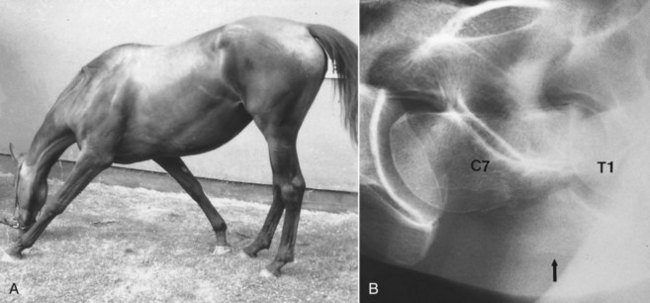
Neck flexibility should be assessed from side to side and up and down. This can be done by manually manipulating the neck, but many normal horses resist this. Holding a bowl of food by the horse’s shoulder to assess lateral flexibility is helpful. Ideally the horse should be positioned against a wall, so that the horse cannot swing its hindquarters away from the examiner during this assessment. The clinician should try to differentiate between the horse properly flexing the neck and twisting the head on the neck. Compare flexibility to the left and to the right. To assess extension of the neck, the veterinarian should evaluate the ease with which the horse can stretch to eat from above head height. Observing the horse grazing is helpful to assess ventral mobility of the neck. Especially with lesions in the caudal neck region a horse may have to straddle the forelimbs excessively to lower the head to the ground to graze (Figure 53-1, A). The horse should also be observed moving in small circles to the left and the right, and loose on the lunge.


Assessing skin sensation and local reflexes, such as the cervicofacial and the thoracolaryngeal reflexes, and comparing carefully the right and left sides may be useful. The consistency and patency of the jugular veins should be evaluated.


The horse should be observed moving in hand and on the lunge, and if necessary should be ridden, to assess neck posture and the presence of neurological gait abnormalities, restriction in forelimb gait, or lameness. The clinician should note how any gait abnormality is influenced by the positions of the head and neck. Forelimb lameness occasionally is associated with a primary cervical lesion, usually, but not invariably, together with other clinical signs referable to the neck.2


Imaging Considerations


Radiography and Radiology


Comprehensive radiographic examination of the neck requires at least five exposures, assuming that large cassettes or imaging plates are used, including the poll, cranial, midneck and caudal neck regions and the base of the neck to evaluate the first and second thoracic vertebrae.3 Lateral-lateral images are obtained easily in the standing position, but ventrodorsal images are best obtained with the horse in dorsal recumbency under general anesthesia, except in small ponies and foals. Positioning of the neck is important, because any rotation of the head and neck makes evaluation difficult, in particular the synovial articulations. Relatively large exposures are required for the more caudal neck regions, so radiation safety is important, and the cassette should be supported in a holder, not held by hand. A grid is useful, especially in the caudal neck region, to reduce scattered radiation. Obtaining exposures from left to right and right to left may be useful. Lateral oblique images of the cervical vertebrae may give additional information.


A number of variations of the normal radiological appearance of the cervical vertebrae should not be mistaken for lesions. A spur on the dorsocaudal aspect of the second cervical vertebra may project into the vertebral canal. The ventral processes of the sixth cervical vertebra and occasionally other vertebrae have small separate centers of ossification. The ventral lamina on the sixth cervical vertebra may be transposed onto the ventral aspect of the seventh cervical vertebra, unilaterally or bilaterally. The seventh cervical vertebra has a small spinous process, which may be superimposed over the synovial articulation between the sixth and seventh cervical vertebrae and should not be confused with periarticular new bone. In older horses small spondylitic spurs may be seen on the ventral aspect of the vertebral bodies. Modeling of the dorsal synovial articulations between the fifth and sixth and between the sixth and seventh cervical vertebrae is common in middle-aged and older horses3-5 (Figure 53-2).


image

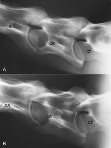
Fig. 53-2 A, Lateral radiographic image of the caudal cervical vertebrae of normal 4-year-old Thoroughbred. The synovial articulations between the fifth (C5) and sixth (C6) cervical and sixth and seventh (C7) cervical vertebrae are outlined smoothly. The intervertebral foramina are distinct. Compare with part B and Figure 53-8. B, Lateral radiographic image of the caudal cervical vertebrae of 9-year-old clinically normal horse. The synovial articulations are enlarged between the fifth (C5) and sixth (C6) cervical vertebrae and particularly between the sixth and seventh (C7) cervical vertebrae.


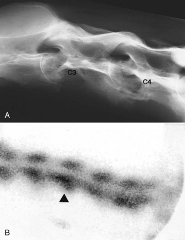
Major radiological abnormalities such as fusion of two adjacent vertebrae can be present subclinically, in part because of the great mobility between adjacent vertebrae (Figure 53-3, A). The clinical significance of such lesions may also be determined by the athletic demands placed on the horse.


Thermography


Thermographic examination of the neck is discussed in detail elsewhere (see Chapters 25 and 95). However, I have found thermography of limited usefulness, except for identifying acute superficial muscle injuries.


Clinical Conditions


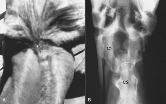
Occipito-Atlantoaxial Malformation


Occipito-atlantoaxial malformation (OAAM) is a congenital abnormality,13 and although it can occur in any breed,14 OAAM appears to be a heritable condition in Arabian horses15 (see Chapter 123). Clinical signs are usually recognizable within the first few weeks of life and include an abnormal neck shape in the poll region, with prominence on the left or right sides or both, and/or scoliosis (Figure 53-4, A). These signs are best appreciated when viewed from above. Usually no associated soft tissue swelling or pain exists, although an abnormal clicking sound may be audible because of subluxation of the atlantoaxial joint. The horse may have an abnormal limitation of movement in the poll region. The gait should be assessed carefully for neurological abnormalities; however, in many horses no neurological gait deficits are apparent.


Jun 4, 2016 | Posted by in EQUINE MEDICINE | Comments Off on The Cervical Spine and Soft Tissues of the Neck

Full access? Get Clinical Tree

Get Clinical Tree app for offline access