The Cardiovascular System

CHAPTER 32 The Cardiovascular System


Despite decades of advances in cardiac ultrasound and ready access to veterinary cardiologists across the country, the most often used, feasible, and economic means for identifying puppies and kittens with heart disease is by physical examination.


Abnormal or arrested development of the embryologic heart or great vessels is an important cause of morbidity and mortality in young animals. Although congenital heart disease (CHD) is usually considered present at birth, these lesions are not necessarily static from conception through adulthood. Some lesions live in accord with the fetal circulation but undergo profound physiologic modification by the circulatory alterations that occur at birth.


Fetal Circulation


In utero the heart feeds the systemic circulation in parallel, with crossover proximal and distal to the ventricles via the foramen ovale and ductus arteriosus (DA), respectively. Oxygen-rich blood from the ductus venosus and left hepatic vein is preferentially directed across the foramen ovale into the left ventricle.


At birth and through the early neonatal period, circulatory changes occur that transform the circulation into two separate circuits functioning in series. Pulmonary vascular resistance decreases as the fluid medium surrounding the pulmonary vessels is replaced by air, and the pulmonary vasculature dilates following exposure to oxygen. The placental circulation is removed, which increases systemic vascular resistance. Left ventricular systolic pressure in newborn puppies has been measured at 35 to 50 mm Hg, whereas right ventricular systolic pressure was 23 to 40 mm Hg. The decreasing pulmonary resistance and increased systemic resistance prevents right to left shunting through the DA. Increased oxygen tension and presumably prostaglandin inhibition produce muscular constriction of the ductus within the first few hours of life. Closure of the DA and reduction in pulmonary vascular resistance increase venous return to the left heart. The increased preload increases left atrial pressure and forces the septum primum (or valve of the foramen ovale) against the septum secundum, functionally closing the foramen ovale and producing a circulatory system in series.


Development of the Cardiovascular System


Right ventricular myocardial mass is equal to or greater than left ventricular myocardial mass at birth, presumably because fetal right ventricular blood flow is approximately twice that of the left ventricle. However, as pulmonary vascular resistance decreases and systemic vascular resistance increases, the left ventricular mass/right ventricular mass ratio changes from 0.80 at 1 day of age to approximately 1.0 at day 3. By 3 to 7 days of age left ventricular systolic pressure rapidly increases to 75 to 90 mm Hg, and by 3 to 4 weeks it is 120 mm Hg. Right ventricular systolic pressure remains at 20 to 30 mm Hg throughout. Postnatally the heart grows rapidly, with an average increase in weight of 7.7% daily from 1 to 17 days of age, predominantly as a result of left ventricular growth.


Compared with adults, puppies and kittens have lower blood pressure, stroke volume, and peripheral vascular resistance with greater heart rate, cardiac output, plasma volume, and central venous pressure at birth. These parameters progressively approach adult values during the first 7 months of life. The autonomic innervation of the heart and vessels is also incomplete in newborn animals, providing them with little baroreflex response to changes in homeostasis. In the early neonatal period, ventricular myocytes are different ultrastructurally compared with adults and may explain the significant reduction in the length-tension relationship of neonatal cats compared with adults. This combination of factors seems clinically important wherein young animals may have a limited ability to compensate for circulatory stresses, including hyperthermia, acid-base shifts, and hemorrhage.


Screening for Heart Disease


History and Physical Examination


CHD is most often identified by auscultation of a heart murmur at the time of immunization in historically asymptomatic animals. Less commonly, puppies and kittens are presented for evaluation of clinical signs, including stunted growth, coughing or tachypnea, abdominal distention, weakness, cyanosis, or syncope that may be attributable to cardiac disease.


A well-performed physical examination begins with an observation period enabling characterization of the pet’s general appearance and size, attitude, respiratory rate, and effort. The oral, conjunctival, and genital mucous membranes are inspected for evidence of cyanosis or poor capillary refill time. Cyanosis is seen when the deoxygenated hemoglobin concentration exceeds 5 g/dl and often signifies arterial hypoxemia, as seen with severe pulmonary disease or right-to-left shunting of blood (i.e., tetralogy of Fallot). Differential cyanosis, as occurs in right-to-left shunting PDA, is characterized by normal oral and conjunctival mucous membranes and cyanotic genital mucous membranes. Following examination of the mucous membranes, the jugular veins should be inspected for distention or abnormal pulsation that may accompany right heart abnormalities, like severe tricuspid insufficiency or PS. Normally the jugular veins are flaccid, collapse quickly after manual compression, and pulsations do not traverse greater than one third the height of the neck in a standing animal.


Following inspection of the head and neck, examination of the heart usually begins with localization of the cardiac impulse, or apical beat, and identification of palpable thrills or vibrations. Auscultation of the heart is initiated over the apical beat, and careful attention is given to identification of the heart sounds and the presence or absence of heart murmurs and arrhythmias. Abnormal heart sounds and murmurs are often focal and confined to the heart base or right hemithorax; therefore careful and complete auscultation of all areas of the heart is a necessity. Important considerations for successful auscultation include using a familiar and comfortable stethoscope, auscultating in a quiet environment, having a thorough understanding of the physiologic and pathologic genesis of cardiac sounds, and using a combination of practice and patience.


Heart sounds are brief auditory vibrations that can be defined by their intensity, frequency, and quality. Heart murmurs are prolonged auditory vibrations that occur as blood flows turbulently through stenotic or insufficient cardiac valves or through abnormal communications between the cardiac chambers. Murmurs may also occur subsequent to alterations in blood viscosity (i.e., anemia) or vessel diameter (the larger the vessel, the more likely blood flow is turbulent). The timing, intensity, configuration, location (defined as the point of maximal intensity), and radiation characteristics often provide valuable insight into the pathogenesis and significance of heart murmurs.


Electrocardiography


Electrocardiographic evaluation of animals with CHD enables characterization of the cardiac rhythm and identification of conduction abnormalities and patterns of chamber enlargement. With normal right heart dominance at birth, the mean QRS vector is directed cranially, ventrally, and to the right in newborn puppies. Presumably as the left ventricular mass increases over the first 12 weeks of life, the mean electrical axis shifts leftward and caudally as is typically seen in adult dogs. Compared with age-matched normal puppies, animals with severe PS have a pathological right ventricular hypertrophy pattern at birth, and they never develop leftward deviation of the mean electrical axis. Additional cardiac lesions that may contribute to a right ventricular enlargement pattern (right axis deviation; S waves in leads I, II, III, and aVF; increased S wave amplitude in leads I, II, CV6LL, and CV6LU) include tetralogy of Fallot, tricuspid valve dysplasia (TVD), ASD, large VSD, or diseases producing or accompanied by pulmonary hypertension. SAS, left-to-right shunting PDA, mitral valve dysplasia (MVD)/insufficiency, VSDs, and other conditions that result in left ventricular hypertrophy may produce increased amplitude of the R wave (leads II, III, aVF, CV6LL, and CV6LU) and widening of the QRS duration with an otherwise normal mean electrical axis.


Puppies and kittens normally display sinus rhythm without respiratory variations in the heart rate. However, similar to “normalization” of the mean electrical axis over the first several weeks of life, puppies establish vagal reflexes during the first 8 weeks of life that may contribute to sinus arrhythmia, wandering pacemaker, and atropine-responsive second-degree AV block. Pathologic arrhythmias, including supraventricular and ventricular premature contractions (VPCs), often accompany CHD that is capable of producing moderate to severe atrial enlargement (i.e., AV valve dysplasia) or ventricular concentric hypertrophy with compromised myocardial perfusion (i.e., SAS). Life-threatening arrhythmias may also occur as a consequence of accessory pathways (APs) or well-studied, but as of yet unknown, substrates.


Radiography


Radiographs yield valuable insight into pulmonary vascular and parenchymal changes that may accompany CHD. Although radiographs also provide a means to assess the cardiac silhouette, interpretation of specific changes associated with CHD is often inaccurate. Detection of chamber enlargement can be plagued by overinterpretation of normal right heart predominance in young animals or shifting of the cardiac apex in diseased conditions, leading to an erroneous diagnosis. A study assessing normal 3-month-old puppies found the mean ± standard deviation vertebral heart size on a lateral radiograph was 10.0 ± 0.5 vertebral bodies. This value is similar to normal adult dogs, and it did not vary significantly as puppies matured from 3 to 36 months. Although moderate to severe cardiomegaly associated with diseases that produce volume overload is relatively easy to document via radiography, concentric hypertrophy accompanying pressure overload may be difficult to detect without ultrasound.


Assessment of the pulmonary vasculature helps with identification of over- or undercirculation and impending left heart failure. Normally only a small proportion of the pulmonary capillaries are perfused. Left-to-right shunting lesions (i.e., PDA) produce pulmonary vascular overcirculation and recruitment of additional pulmonary capillary beds. Radiographically this may be identified as engorgement of pulmonary arteries and veins with widespread hypervascularity extending to the lung periphery. Right-to-left shunting defects (i.e., tetralogy of Fallot) bypass the pulmonary circulation, yielding small pulmonary arteries and veins with a relatively radiolucent interstitium.


Classification of Congenital Heart Disease


The descriptions in the following sections will classify conditions based on the classic location of murmurs associated with the specific congenital lesion. Although exam findings in many animals do not fit the classical descriptions, and complex lesions may further cloud recognition of disease entities, it is believed this classification may prove the most useful to veterinarians examining young dogs and cats on a daily basis.


Lesions Producing Left Basilar Murmurs


An audible systolic murmur with its point of maximal intensity (PMI) over the left heart base is most commonly produced by aortic stenosis, PS, tetralogy of Fallot, or an ASD. Low-grade innocent and physiologic murmurs also frequently display their PMI over the left heart base and will need to be differentiated from their more pathologic counterparts. Auscultation of a variable-intensity, left basilar (axillary), continuous murmur is most consistent with left-to-right shunting PDA.


Aortic Stenosis


SAS has likely replaced PDA as the most common congenital cardiac lesion in areas with a high population of large breed dogs. Although any breed of dog may be affected, evidence suggests Newfoundlands, Rottweilers, Boxers, Golden Retrievers, German Shepherd Dogs, and Bloodhounds have an increased relative risk for SAS. Valvular and supravalvular aortic stenosis are seen infrequently or rarely in dogs, and although uncommon, cats have been described with all three forms of fixed LVOT obstruction. SAS has been most thoroughly investigated in Newfoundlands, for whom breeding studies suggest an autosomal dominant mode of transmission with modifying genes or a polygenic mechanism. These same studies found that SAS may not be present at birth but instead begins to develop during the first 4 to 8 weeks of life. Because SAS lesions may progress as animals grow, it is not uncommon for the severity of the lesion and the associated murmur and cardiac changes to worsen from birth until young adulthood. Detection of mild disease is difficult; therefore ultimately genetic counseling is difficult.


The pathophysiologic consequences of aortic stenosis include left ventricular and in some instances left atrial hypertrophy, myocardial fibrosis, and aortic arch abnormalities. The hypertrophy and fibrosis produce a stiff, noncompliant left ventricle that ultimately necessitates higher atrial pressures to fill. Elevated left atrial, and hence pulmonary venous and pulmonary capillary hydrostatic, pressures may contribute to the development of pulmonary edema.


Puppies with mild to moderate SAS are often reported to be clinically normal at the time of immunization. Owners with severely affected dogs may report exercise intolerance, syncope, or a history consistent with left heart failure. In some cases sudden death occurs without premonitory signs. Auscultatory abnormalities are usually present in cases of moderate to severe SAS because the intensity and duration of the murmur tend to increase and lengthen, respectively, with more severe disease. Obstruction to blood flow through the LVOT produces an ejection, left basilar systolic murmur. The murmur tends to radiate up the carotid arteries and to the right so it can frequently be auscultated in the ventral cervical region, and in some cases the PMI is at the right heart base. Because SAS may progress through early adulthood, the murmur can increase in intensity between sequential physical examinations. Femoral pulse alterations, with palpably reduced and late rising pulse amplitudes, are often found in dogs with moderate to severe obstruction.


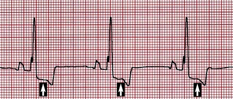
Although nonspecific, electrocardiographic changes that may accompany SAS include increased R wave amplitude and QRS duration consistent with left ventricular enlargement, as well as ST segment and T wave alterations suggestive of myocardial ischemia (Figure 32-1). VPCs may be evident on baseline electrocardiography (ECG), and studies of Holter examinations suggest the overall number and grade of VPCs display a modest correlation with pressure gradient. Ventricular tachycardia and ultimately fibrillation are the presumed mechanism for sudden death in dogs with SAS.


Radiography is limited in the assessment of ventricular enlargement in dogs with SAS because it produces concentric rather than eccentric hypertrophy. Nonetheless assessment of the cardiac silhouette still proves useful in dogs with moderate to severe SAS for detection of poststenotic dilation of the aorta or left atrial enlargement.


Echocardiography provides a reliable method for identification of moderate to severe SAS. Common findings include left ventricular concentric hypertrophy, a subvalvular fibrous ring or band narrowing the LVOT, and poststenotic dilation of the aorta. Hyperechogenicity of the left ventricular endocardial surface and papillary muscles may be seen in dogs with myocardial ischemia and replacement fibrosis.


Although the exact velocity cutoff to distinguish mild, moderate, and severe SAS is uncertain, it is generally accepted that gradients more than 100 mm Hg represent severe disease with a high likelihood of complications. Currently available diagnostic methods seem incapable of identifying dogs with grade 1 lesions. Even dogs with grade 2 lesions may be difficult to accurately assess purely based on the LVOT velocities because of the likelihood of breed and examination condition variations.


Although many dogs with mild to moderate SAS live normal lifespans devoid of clinical consequences, the natural history and responses to therapy of severe SAS are disappointing. Commonly reported outcomes of dogs with severe SAS include exercise intolerance, syncope, sudden death (most commonly in the first 3 years of life), and the development of congestive heart failure and/or endocarditis. In general, dogs with mild to moderate disease do not require specific therapy and can often exercise normally. Antibiotic prophylaxis is recommended for surgical procedures in all dogs with SAS because they appear to be at increased risk for developing endocarditis; however, the effectiveness of this therapy is uncertain. Exercise restriction is prudent for dogs with moderate to severe SAS, and β-blockers (i.e., atenolol) are frequently administered in an effort to combat arrhythmogenesis, reduce myocardial oxygen demands, and limit tachycardia. Currently the survival benefit imparted by β-blockers is unknown. Additional antiarrhythmic medications may be necessary to treat VPCs, ventricular tachycardia, or atrial fibrillation if they complicate the clinical situation. Similarly standard therapy for left heart failure, including diuretics, angiotensin-converting enzyme inhibitors, and possibly positive inotropic agents, are indicated for animals that develop pulmonary edema.


Pulmonic Stenosis


Valvular PS is the most common congenital right ventricular outflow tract (RVOT) obstruction in dogs and is the third most common congenital cardiac defect encountered. PS is less commonly identified in cats, and both species have been reported to infrequently display sub- and supravalvular forms of RVOT obstruction. Breeds with an increased relative risk for PS include English Bulldogs, Terrier breeds, Miniature Schnauzers, Chihuahuas, and Samoyeds. PS has been identified as hereditary in breeding studies of Beagles with a spectrum of pulmonary valve abnormalities. Grade 1 lesions display mild leaflet thickening with commissural fusion producing a central orifice. More severe cases of PS tend to be grade 2 lesions with moderate to severe thickening of the valve leaflets with fusion or hypoplasia producing RVOT obstruction. Fibrous thickening at the base of the valve may accompany valvular dysplasia. Boxers and Bulldogs have been identified with coronary arterial abnormalities that contribute to a unique form of subvalvular PS that is difficult to address surgically.


Physical examination of animals with PS often reveals a variable-intensity, left basilar, systolic, ejection murmur in a reportedly healthy puppy. Compared with murmurs of SAS, PS murmurs do not radiate as extensively to the right cranial thorax or up the carotid arteries. The femoral arterial pulse is usually normal. Jugular distention or abnormal pulsation should increase suspicion of right heart failure or concurrent cardiac defects (i.e., TVD). Many dogs with PS are reportedly asymptomatic during the first year of life when the murmur is initially detected. However, clinical signs, including exercise intolerance, syncope, and signs consistent with right heart failure often complicate severe cases of PS and have been reported in 34% to 83% of dogs evaluated.


In cases of moderate to severe PS, the ECG almost always displays criteria for right ventricular enlargement (Figure 32-2). The P waves are usually normal. Ventricular arrhythmias appear to be less common than in dogs with SAS.
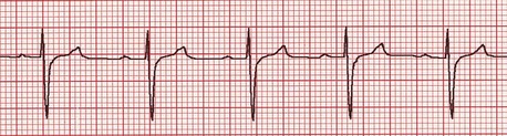


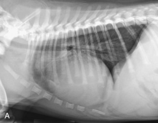
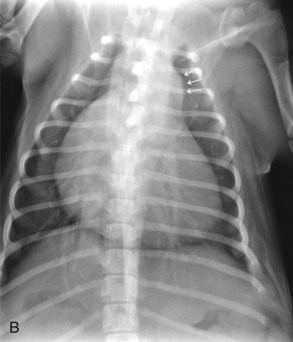
Radiography usually reveals moderate cardiomegaly with a right heart enlargement pattern (Figure 32-3). Poststenotic dilation of the main pulmonary artery produces loss of the cranial cardiac silhouette on the lateral view and dilation at approximately the 2 o’clock position on the dorsoventral (DV) view.


Echocardiography provides a method for definitive diagnosis and assessment of severity of PS, as well as identification of concurrent cardiac defects. Of the four cardiac valves, the pulmonary valve morphology tends to be the most difficult to clearly assess via transthoracic echocardiography. Therefore in some instances it is challenging to distinguish valvular PS from discrete subvalvular obstruction. Mild PS is usually categorized as a pressure gradient less than 50 mm Hg, whereas the cutoff for severe disease varies with peak gradients described more than 80 or 100 mm Hg. Contrast echocardiography may identify right-to-left shunting at the atrial level as RV diastolic dysfunction, and tricuspid insufficiency contributes to right atrial hypertension.


Although precise criteria for establishing a prognosis for dogs and cats with PS are lacking, it is generally accepted that dogs with uncomplicated mild to moderate PS often live normal, comfortable lives and do not require specific therapy. Serial echocardiographic examinations should be performed to determine whether the stenosis, right ventricular hypertrophy, tricuspid insufficiency, or right atrial dilation progresses. In animals with severe PS, clinical experience suggests they are more likely to develop exercise intolerance, right heart failure, or die suddenly. Surgical intervention is generally recommended even in asymptomatic dogs with severe PS.


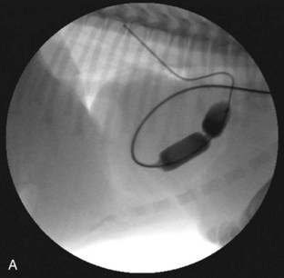
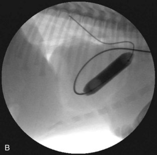
Several techniques have been described and are available for the treatment of PS. Valvular PS with a normally developed pulmonary annulus is most often treated by percutaneous balloon valvuloplasty (Figure 32-4). The goals of intervention include improving survival and resolving clinical signs in symptomatic animals. Surgical success, defined as a reduction of the pressure gradient into the mild category, or in markedly severe cases as a reduction in the pressure gradient of more than 50%, is achievable in up to 80% of dogs. Valvuloplasty has been reported to reduce clinical signs and mortality compared with animals not undergoing surgery.


Stay updated, free articles. Join our Telegram channel

Sep 11, 2016 | Posted by in SMALL ANIMAL | Comments Off on The Cardiovascular System

Full access? Get Clinical Tree

Get Clinical Tree app for offline access